Transcription
Electronic Supplementary Material (ESI) for Molecular BioSystems.This journal is The Royal Society of Chemistry 2016Supplementary InformationHigh-throughput production of a stable isotope-labeled peptide libraryfor targeted proteomics using a wheat germ cell-free synthesis systemNobuaki Takemori1*, Ayako Takemori1, Yuki Tanaka2, Jun Ishizaki3, Hitoshi Hasegawa3,Atsushi Shiraishi4, and Yuichi Ohashi41. Division of Proteomics Research, Proteo-Science Center, Ehime University, Ehime,Japan2. Advanced Research Support Center, Ehime University, Ehime, Japan3. Department of Hematology, Clinical Immunology and Infectious Diseases, EhimeUniversity Graduate School of Medicine, Ehime, Japan4. Department of Ophthalmology, Ehime University Graduate School of Medicine,Ehime, Japan*Correspondence: Nobuaki Takemori, Ph.D.Proteo-Science Center, Ehime University, Toon City, Ehime 791-0295, JapanTel: 81-89-960-5499. E-mail: takemori@m.ehime-u.ac.jp.Contents Supplementary Methods Supplementary Figures (3 figures)Fig. S1: A workflow diagram for the high-throughput synthesis of QconCATs.Fig. S2: Representative SDS-PAGE images of synthesized QconCATs.Fig. S3: SDS-PAGE analysis of aggregated QconCAT proteins during in vitrotranslation. Supplementary Tables (7 tables; see “Supplementary Table.xlsx”)
Supplementary MethodsCell-free protein synthesis and purificationArtificial genes encoding self-designed QconCAT sequences (Table S3) were synthesizedby commercial gene synthesis services (Biomatik USA, Wilmington, DE, USA). Asplit-primer PCR30 was performed to produce DNA templates for in vitro transcription.The first PCR was performed using a 10-nM primer set consisting of the gene-specificsense primer (Table S6) and an antisense primer (5ʹ- GCGGCCAACTTACTTCTGAC-3ʹ).Subsequently, the template DNA for in vitro transcription was amplified by the nextPCR using the second primer mix containing 100 nM of the SPu primer (5ʹGCGTAGCATTTAGGTGACACT-3ʹ), 1 nM of the deSP6E02 primer TCGAGGCTGCACGTGGCTATTCCTTTACCACCACCGCCGAGAAG-3ʹ), and 100 nM of the antisense primer.Cell-free synthesis of His-tagged QconCAT proteins was performed using thebilayer reaction method with a wheat germ extract WEPRO8240H (CellFree Sciences,Matsuyama, Japan) as described previously9,31. After in vitro translation, QconCATproteins were purified using a Ni-Sepharose High Performance resin (GE Healthcare,Pittsburgh, PA, USA). First, the synthesized protein solution (total 220 μL) was mixedwith 400 μL of equilibration buffer (20 mM phosphate buffer, 0.3 M NaCl, 10 mMimidazole, 6 M urea, pH 7.5) and then incubated with 10 μL of resin (50% slurry) for 1 hat room temperature. The resin was washed twice with 400 μL of wash buffer (20 mMphosphate buffer, 0.3 M NaCl, 50 mM imidazole, 6 M urea, pH 7.5). The binding proteinwas eluted with 30 μL of elution buffer (20 mM phosphate buffer, 0.3 M NaCl, 500 mMimidazole, 6M urea, pH 7.5). Purified QconCAT solution (0.7 μL) was solubilized with 10μL of the LDS lysis buffer containing 50 mM DTT and separated by NuPAGE Bis-Tris gels(Life Technologies, Carlsbad, CA, USA). After gel electrophoresis, protein bands werevisualized with Bio-Safe CBB staining (Bio-Rad, Hercules, CA, USA).In gel tryptic digestion of synthesized QconCAT proteinsProtein bands ( 500 ng total protein) were excised from the gel, and each gel piecewas destained with 50% (v/v) acetonitrile/100 mM NH4HCO3 (pH 8.9). Synthesizedproteins in the gel pieces were reduced by soaking with 10 μL of 40 mM DTT solutionfor 1.5 h at 37ºC and were subsequently alkylated by soaking with 10 μL of 250 mMacrylamide solution for 30 min at room temperature. Individual gel pieces weredehydrated with 1 mL of acetonitrile and then rehydrated with 2 μL of 0.1 μg/μLsequencing-grade modified trypsin (Promega, Madison, WI, USA), followed by
incubation in 50 μL of 100 mM NH4HCO3 at 37ºC for 16 h. Each peptide was extractedfrom the gel piece three times with 50 μL of 50% (v/v) acetonitrile/5% (v/v)trifluoroacetic acid. Peptide samples were concentrated using a centrifugal vacuumconcentrator, and then reconstituted with 10 μL of 0.1% (v/v) TFA solution. For MS/MSanalysis, 1 μL of peptide solution was injected onto LC.In-solution digestion of human serum proteinsHuman serum samples were obtained from three healthy donors after obtaininginformed consent in accordance with the procedures approved by the human ethicscommittee of the Ehime University. Major serum proteins were depleted using a MARSspin cartridges Human-14 (Agilent Technologies, Santa Clara, CA, USA).Immunodepleted serum samples (90 μg total protein) in 120 μL lysis buffer (8 M Ureain 150 mM Tris-HCl buffer, pH 8.8) were digested with Lys-C and trypsin as describedpreviously.32 After peptide purification with a C18 STAGE tip, the isolated peptides werecompletely dried using a vacuum centrifuge. The dried samples were resuspended in180 μL of 0.1% TFA and dispensed into 1 L aliquots. The obtained aliquots were mixedwith an internal standard (1 μL) containing tryptic digests of SI-labeled QconCAT andsubjected to SRM analysis.Mass spectrometry (MS/MS and SRM)All MS analyses were performed using a QTRAP 5500 hybrid triple quadrupole/linearion trap MS (AB SCIEX, Framingham, MA, USA) coupled with an Eksigent nanoLC systemvia a cHiPLC-nanoflex module (AB SCIEX). The peptides were ionized by a nano-ESIsource using PicoTip emitter with an internal diameter of 10 μm (New Objective,Woburn, MA, USA). The ion spray voltage was maintained at 2,300 V, and the positiveion mode was used for all experiments. The temperature of the interface heater washeld at 150 C. For MS/MS analysis, we used information-dependent acquisitionworkflow. Each acquisition cycle started with an enhanced mass spectrum (EMS) scan,followed by an enhanced resolution scan to determine the ion charge states andenhanced product ion (EPI) scans for 1–3 of the most intense peaks (charge states: 2to 4) detected from the EMS scan. The mass range was set to m/z 400–1,000 for EMSand m/z 100–1,000 for EPI. The scan rate was set to 10,000 Da/s for EMS and EPI. TheLC parameters used for each MS analysis are summarized in Table S7.MS/MS data analysis and protein identification were performed using the Analystver.1.5 and the ProteinPilot ver.4.0 (AB SCIEX), respectively. The database search wasperformed against the IPI human database version 20100624 using the following
parameters: sample type, SILAC (Lys 8, Arg 10); cys alkylation, acrylamide; digestion,trypsin; and search effort, rapid ID. The Skyline software33 was used to build SRMassays for the targeted peptides. The MS/MS search results from the ProteinPilotprogram were used to generate a spectral library with a peptide probability cut-off of0.9. Up to five of the most intense fragment ions (y or b ions) for doubly or triplycharged precursor ions were selected based on library data, and a set of transitionswith a dot product score 0.8 was then chosen for inclusion in the final assay. At leastthree transitions were selected per targeted peptide. The resolution at quadrupoles 1and 3 was set to unit mass and the dwell time was set to 20 msec. We manuallychecked for the presence of interference in the selected transitions in the case ofhuman serum analysis. The presence of interferences in each transition was evaluatedon the basis of comparison of the SRM chromatograms for human serum and referencepeptides. A set of at least three transitions with no apparent interference (dot productscore 0.8) was finally selected.References30.T. Sawasaki, T. Ogasawara, R. Morishita and Y. Endo, Proc Natl Acad Sci U S A,2002, 99, 14652-14657.31.32.33.T. Sawasaki, Y. Hasegawa, M. Tsuchimochi, N. Kamura, T. Ogasawara, T. Kuroitaand Y. Endo, FEBS Lett, 2002, 514, 102-105.N. Takemori, A. Takemori, J. Ishizaki and H. Hasegawa, J Chromatogr B AnalytTechnol Biomed Life Sci, 2014, 967, 36-40.B. MacLean, D. M. Tomazela, N. Shulman, M. Chambers, G. L. Finney, B. Frewen,R. Kern, D. L. Tabb, D. C. Liebler and M. J. MacCoss, Bioinformatics, 2010, 26,966-968.
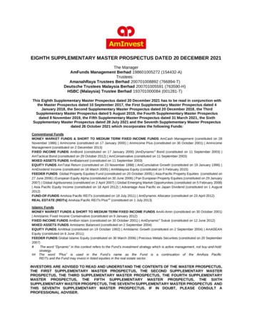
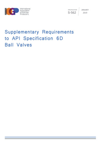
Figure S1. A workflow diagram for the high-throughput synthesis of QconCATs. PCRamplification of cDNA encoding for a targeted QconCAT was performed using asplit-primer PCR method.30 PCR products were used as the templates for in vitroprotein synthesis using WG-CFS. QT, a peptide tag sequence for the quantitation ofsynthesized QconCATs. His, his-tag sequence.1. Artificial QconCAT gene design and synthesis ( 1 month)2. PCR-based template DNA construction (7 hours)Gene specific primer1st PCRQconCAT1st PCR ProductPCRQconCATAntisense primer2nd PCRSPU1st PCR ProductdeSP6-E02-His-QTQconCATAntisense primerPCRSP6 E02HisQTQconCAT3. In vitro synthesis of QconCATs using WG-CFS (1 day)4. His-tag purification of synthesized QconCATs (2 hours)
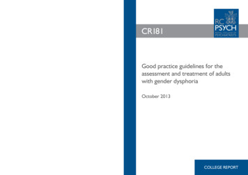
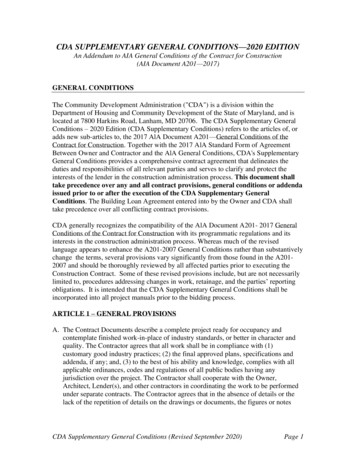
Figure S2. Representative SDS-PAGE images of synthesized QconCATs. SynthesizedQconCATs (ID: 7 162) were separated on acrylamide gels (NuPAGE 4-12% Bis-Tris gel)and visualized with CBB. Lane M: Molecular weight marker (BioRad Precision PlusProtein Unstained standards). Lane number: ID numbers of QconCATs.
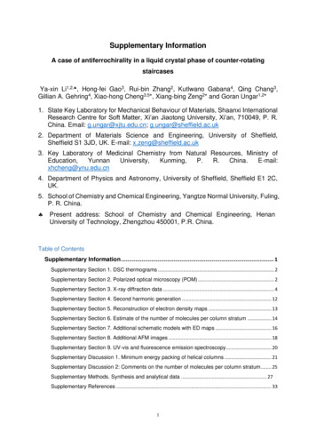
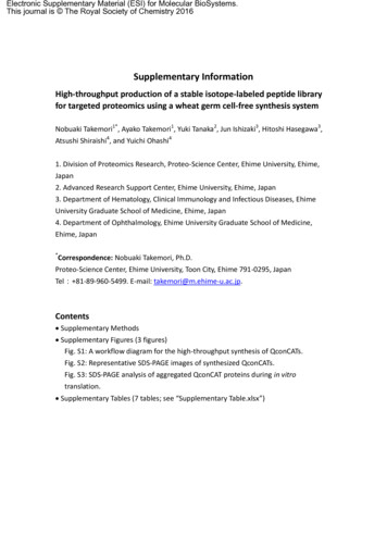
Figure S3. SDS-PAGE analysis of aggregated QconCAT proteins during in vitro translation.Aggregated QconCAT proteins were precipitated by centrifugation and solubilized withSDS lysis buffer. Resolubilized proteins were separated on a self-made 15%bis-acrylamide gel and visualized with CBB. Lane M: Molecular weight marker (BioRadPrecision Plus Protein Unstained M74808286kDa M 107 115 139 2015***
by commercial gene synthesis services (Biomatik USA, Wilmington, DE, USA). A split-primer PCR30 was performed to produce DNA templates for in vitro transcription. The first PCR was performed using a 10-nM primer set consisting of the gene-specific sense primer (Table S6) and an antisense primer (5ʹ- GCGGCCAACTTACTTCTGAC-3ʹ).