Transcription
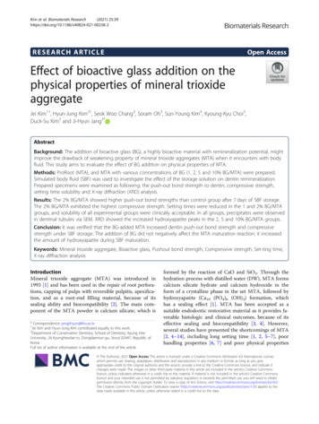
Kim et al. Biomaterials Research(2021) EARCH ARTICLEOpen AccessEffect of bioactive glass addition on thephysical properties of mineral trioxideaggregateJei Kim1†, Hyun-Jung Kim2†, Seok Woo Chang3, Soram Oh3, Sun-Young Kim4, Kyoung-Kyu Choi3,Duck-Su Kim3 and Ji-Hyun Jang3*AbstractBackground: The addition of bioactive glass (BG), a highly bioactive material with remineralization potential, mightimprove the drawback of weakening property of mineral trioxide aggregates (MTA) when it encounters with bodyfluid. This study aims to evaluate the effect of BG addition on physical properties of MTA.Methods: ProRoot (MTA), and MTA with various concentrations of BG (1, 2, 5 and 10% BG/MTA) were prepared.Simulated body fluid (SBF) was used to investigate the effect of the storage solution on dentin remineralization.Prepared specimens were examined as following; the push-out bond strength to dentin, compressive strength,setting time solubility and X-ray diffraction (XRD) analysis.Results: The 2% BG/MTA showed higher push-out bond strengths than control group after 7 days of SBF storage.The 2% BG/MTA exhibited the highest compressive strength. Setting times were reduced in the 1 and 2% BG/MTAgroups, and solubility of all experimental groups were clinically acceptable. In all groups, precipitates were observedin dentinal tubules via SEM. XRD showed the increased hydroxyapatite peaks in the 2, 5 and 10% BG/MTA groups.Conclusion: It was verified that the BG-added MTA increased dentin push-out bond strength and compressivestrength under SBF storage. The addition of BG did not negatively affect the MTA maturation reaction; it increasedthe amount of hydroxyapatite during SBF maturation.Keywords: Mineral trioxide aggregate, Bioactive glass, Pushout bond strength, Compressive strength, Set-ting time,X-ray diffraction analysisIntroductionMineral trioxide aggregate (MTA) was introduced in1993 [1] and has been used in the repair of root perforations, capping of pulps with reversible pulpitis, apexification, and as a root-end filling material, because of itssealing ability and biocompatibility [2]. The main component of the MTA powder is calcium silicate, which is* Correspondence: jangjihyun@khu.ac.kr†Jei Kim and Hyun-Jung Kim contributed equally to this work.3Department of Conservative Dentistry, School of Dentistry, Kyung HeeUniversity, 26 Kyungheedae-ro, Dongdaemun-gu, Seoul 02447, Republic ofKoreaFull list of author information is available at the end of the articleformed by the reaction of CaO and SiO2. Through thehydration process with distilled water (DW), MTA formscalcium silicate hydrate and calcium hydroxide in theform of a crystalline phase in the set MTA, followed byhydroxyapatite (Ca10 (PO4)6 (OH)2) formation, whichhas a sealing effect [1]. MTA has been accepted as asuitable endodontic restorative material as it provides favorable histologic and clinical outcomes, because of itseffective sealing and biocompatibility [3, 4]. However,several studies have presented the shortcomings of MTA[2, 4–14], including long setting time [1, 2, 5–7], poorhandling properties [6, 7] and poor physical properties The Author(s). 2021 Open Access This article is licensed under a Creative Commons Attribution 4.0 International License,which permits use, sharing, adaptation, distribution and reproduction in any medium or format, as long as you giveappropriate credit to the original author(s) and the source, provide a link to the Creative Commons licence, and indicate ifchanges were made. The images or other third party material in this article are included in the article's Creative Commonslicence, unless indicated otherwise in a credit line to the material. If material is not included in the article's Creative Commonslicence and your intended use is not permitted by statutory regulation or exceeds the permitted use, you will need to obtainpermission directly from the copyright holder. To view a copy of this licence, visit http://creativecommons.org/licenses/by/4.0/.The Creative Commons Public Domain Dedication waiver ) applies to thedata made available in this article, unless otherwise stated in a credit line to the data.
Kim et al. Biomaterials Research(2021) 25:39when interacting with body fluids (BF) [8, 9]. Methodssuch as adding additives (calcium chloride [10], sodiumhydrogen phosphate [12], methylcellulose [10], bioactiveglass (BG) [11] or changing the hydration liquid to calcium chloride [7], calcium lactate gluconate [7], andelastin-like polypeptide [6] have been attempted to overcome the disadvantages of MTA; these methods havedemonstrated improvements in some properties ofMTA.In most clinical application environments of MTA,more than one surface of the MTA material is in contactwith the periodontal or pulpal tissue. When the unsetMTA is contaminated with tissue fluid (e.g., blood andinterstitial fluid from dental pulp and periodontal tissues),the mechanical properties of MTA are adversely affected[15]. Previous investigations reported that blood contamination has detrimental effects on the resistance of displacement [8], microhardness, compressive strength, andsurface microstructure [9]; further, it is associated withchanges in the setting reaction of MTA with lower formation of calcium hydroxide crystalline structures [16]. Thus,additives and alternative liquid solutions to improve theproperties of MTA need to be addressed in clinically applicable conditions.In 1969, Dr. Larry Hench introduced BG for the firsttime; it was an alternative to nearly inert implant materials [17]. It is a highly bioactive material with silicon dioxide as its main component. When it encounters waterfrom BF or simulated body fluid (SBF), BG immediatelyundergoes ionic dissolution and glass degradation [17].It releases silica ions to form a silica-rich layer on thesurface. Outside this layer, calcium and phosphoric acidfrom the BF form a layer of calcium phosphate, whichbecomes hydroxyapatite when it crystallizes [18]. Further, BG can bond to mineralized and soft tissue [19];this leads to a superior surface area with a higher dissolution rate and faster apatite formation. In addition, itimproves the mechanical properties of such compositesfor natural bones [18].Owing to the ability of BG to form apatite, several investigations have been conducted in the recent years touse BG as a remineralization additive in dental restoration materials owing to its ability to form apatite [11,20]. Jang et al. [21] reported that dental composite resinsincluding BG remineralize surrounding dentin; Kimet al. [22] suggested that a glass ionomer including BGhad the same effect. Thus far, there have been a fewstudies on the addition of BG to MTA; among them, thephysical properties of MTA supplemented with BG,which are required for the evaluation of endodontic restorative materials, have been rarely investigated. Thisstudy aims to evaluate the effect of BG addition on thephysical properties of MTA, which includes push-outPage 2 of 11bond strength to dentin, compressive strength, settingtime, solubility, and X-ray diffraction (XRD) analysis.Materials and methodsMaterial preparationFor specimen preparation, DW was used as the liquidsolution in all experimental groups. The group nameswere labeled according to the type of powder used toprepare the MTA specimens. White ProRoot MTA(Dentsply Sirona, Tulsa, USA) was used as the controland labeled as MTA. The major constituents of MTApowder were tricalcium silicate (Ca3SiO5), dicalcium silicate (Ca2 SiO4), tricalcium aluminate (Ca3Al2O6), bismuth oxide (Bi2O3) and calcium sulfate (CaSO4). TheMTA with BG supplemented groups (BG/MTA groups)were prepared using the MTA and 63S BG (BondingChemical, USA) at four different concentrations: 1, 2, 5,and 10 wt%; these were labeled as 1% BG/MTA, 2% BG/MTA, 5% BG/MTA, and 10% BG/MTA, respectively.The composition of 63S BG was 63% SiO2, 31% CaO,and 6% P2O5, and its particle size was less than 20 μm.The L/P ratio of DW to the powder was 0.3 throughoutthe experiments, which was set according to the manufacturer’s recommendations.Preparation of storage solutions27 mM HCO3- Tris SBF with pH 7.4 was used to investigate the effect of the storage solution on dentinremineralization. The SBF was prepared as described byTas et al [23] The composition of Tris SBF is listed inTable 1.Push-out bond strengthExtracted caries-free human third molars were selectedfor this study. The included teeth were obtained frompatients whose teeth were indicated for extraction, andinformed consent was obtained. The experimentalprotocol using human teeth was reviewed and approvedby the Kyung Hee University Institutional Review Board(KHU-1808-1), and all methods were performed in accordance with the Declaration of Helsinki guidelines andTable 1 Compositions of 27 mM HCO3- Tris SBFaCompositionAmount (g/L)NaCl6.547NaHCO32.268KCl0.373Na2HPO4 2H2O0.178MgCl2 6H2O0.305CaCl2 2H2O0.368Na2SO40.071(CH2OH)3CNH26.057SBF Simulated body fluida
Kim et al. Biomaterials Research(2021) 25:39regulations. The midcoronal portion of the dentin of thetooth was horizontally cut into 2-mm-thick sectionsusing a water-cooled high-speed diamond bur (n 10).A cylindrical cavity (1.5 mm diameter) was prepared ineach dentin disc with a depth-cutting bur (Microcopy,Kennesaw, GA). The dentin discs were soaked sequentially in 17% ethylene diamine tetraacetic acid (EDTA)and 2.5% sodium hypochlorite for 1 min each, washedwith phosphate buffered saline, and dried. Each matrixwas mixed with DW and carefully loaded into the cavitywithout compaction pressure. The specimens werestored at 37 C under 95% humidity for 48 h [24] andstored in SBF for 3, 7, and 14 days in a 37 C cabinet[13]. The SBF solution was replaced every 2 days to prevent autogenous precipitation.After each storage period, the push-out bond strengthwas measured using a universal testing machine (AGSX; Shimadzu, Tokyo, Japan) at a 1.0 mm/min (diameter1 mm) crosshead speed. The maximum load applied tothe specimens in the cavity before dislodgement was recorded in Newtons. The push-out bond strength valuesin megapascals were calculated by dividing the maximum load by the total contact area of the cavity inmm2 [25].FE-SEM analysisThe two remaining samples from each group after thepush-out bond strength test (7 days of SBF storage) wereplaced individually in plastic containers containing 15 mlof SBF (pH 7.4), and they were then stored for an additional 14 days at 37 C to analyze the mixture-dentininterface [13]. The SBF solution was replaced every 2days to prevent autogenous precipitation.After 14 days of storage in SBF, the specimens werewashed in DW and processed for FE-SEM observations.The specimens were cut along the center to expose theinternal interface between the MTA and the dentin wallusing a high-speed diamond bur under water irrigation.The exposed internal surfaces were serially polished with600 grit SiC papers. The smear particles were removedby soaking in a 17% EDTA solution for 1 min. The specimens were dried at ambient temperature for 24 h. Theinterfaces were then observed under a FE-SEM (HITACHI S-4700, at an accelerating voltage of 10 kV, Tokyo,Japan) after Pt sputter coating.Compressive strengthThe compressive strength was determined using the ISO9917-1 method [26, 27]. Each material was mixed andplaced in an acrylic mold (4.0 mm inner diameter and6.0 mm height) [5]. After the placement, the completeassembly was transferred to a cabinet maintained at37 C under 95% humidity for a day for complete setting.The specimens were removed from the molds andPage 3 of 11checked visually for air voids or chipped edges. All defective specimens were discarded.The specimens were stored in SBF solution for 7 daysand maintained at 37 C [28–30]. The SBF solution wasreplaced every 2 days to prevent autogenous precipitation.The specimens were tested using the universal testingmachine at a crosshead speed of 1 mm/min. The maximum load required to fracture each specimen was measured, and the compressive strength (C) was calculatedin megapascals usingC ¼ 4P πD2 ;where P denotes the maximum load (N), and D denotethe mean diameter of the specimen (mm).Etting timeThe setting time was determined according to the International Organization for Standardization (ISO) 9917–1method using a Gillomore apparatus (Heungjin Co. Ltd.,Gyeonggi-do, Korea) [26, 27]. A total of three specimenswere prepared in a circular acrylic mold (inner diameter 10 mm; height 5 mm). The assembly was placedin a cabinet at 37 C and 95% humidity.The final setting time was measured using a 1-lb heavyneedle with a cylindrical tip (diameter 1.06 mm) of theGillmore apparatus. After mixing for 90 s, the indenterwas carefully loaded vertically onto the surface of thespecimens and allowed to remain there for 5 s. The indentations were repeated at 30 s intervals until the needle failed to make a visible indentation in the testspecimen to determine the approximate setting time.This process was repeated starting the indentation at 30s before the approximate setting time was determined,and indentations were made at 10 s intervals. This testwas repeated five times for each specimen.SolubilityThe solubility was assessed according to the ISO 6876standard [27, 31]. A total of eight specimens were evaluated to examine their solubility. Each material wasmixed and placed in split-ring molds (internal diameter 20 1 mm and 1.5 0.1 mm). The filled moldswere placed in a cabinet maintained at 37 C and 95%humidity for a day. The specimens were removed fromthe molds and the weight before the DW storage of eachspecimen was determined to the nearest 0.001 g using aprecision scale (AX200, SHIMADZU, Japan; precision 0.0001 g).The specimens were placed in a shallow dish, and50 1 mL of DW was added. The dish was covered andplaced in a cabinet maintained at 37 C for a day. Then,the specimens were removed and washed with 2–3 mLof fresh DW on a shallow dish. The water in the dish
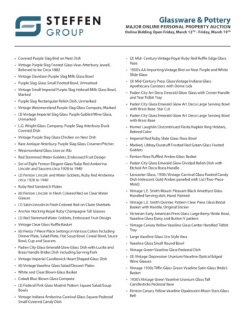
Kim et al. Biomaterials Research(2021) 25:39was evaporated without boiling and dried to a constantmass at 110 2 C. The dishes were weighed after cooling. The differences between this weight and the originaldish weight were divided into the initial weight of thespecimens and multiplied by 100. The results were recorded as the solubility of the specimen. This test wasrepeated four times for each group.XRD analysisThe XRD analysis was performed to compare the compositions of the reactants. Each material was mixed andplaced in a rubber mold (inner diameter 7.0 mm;height 2.5 mm), and it was allowed to fully set duringincubation at 37 C in a 95% humidity cabinet for 24 h[29]. After the setting, the specimens were removedfrom the molds.Immediately after setting, half of the spectra (n 2) foreach group were examined via XRD using an X-ray diffractometer (PANalytical B. V., Almelo, Netherlands)with CuKa radiation within a 2θ range of 5–40 at ascanning speed of 1.2 min 1. The Cu X-ray source wasset at an accelerating voltage of 45 kV and a current inthe electron beam at 40 mA.The remaining specimens were examined after 7 daysof SBF storage. The specimens were individually storedin SBF using polyethylene containers for 7 days at 37 C[30]. The structures of the set specimen and surface deposits formed in the SBF assays were examined usingPage 4 of 11XRD analysis. The diffraction peak positions were determined using the peak-fitting program (Origin v7.5)software.Statistical analysisThe push-out bond strength and compressive strengthof groups were analyzed statistically by two-wayANOVA to determine the interaction between variables;one-way ANOVA with Bonferroni test was performedfor post hoc test for each variable. The data of the solubility were statistically analyzed using one-way ANOVAwith Bonferroni post hoc comparison, respectively. Differences were considered statistically significant at p 0.05. All statistical analyses were performed using SPSS(version 22.0; IBM Corp., Armonk, NY).ResultsPush-out bond strengthFigure 1 show the push-out bond strength results for allexperimental groups. The 2% BG/MTA group showed asignificantly higher value than the control group after 7days of SBF storage (p 0.05). In the 1 and 2% BG/MTAgroups, the strength of the push-out bond increased asthe storage period increased (p 0.05) (Fig. 1), whereasthe 5 and 10% BG/MTA groups did not show significantdifferences with time.Fig. 1 Results of the push-out bond strength. The results of the experimental groups were compared at 3, 7, and 14 days as storage periods. *indicates a statistically significant difference between materials (p 0.05)
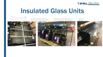
Kim et al. Biomaterials Research(2021) 25:39Page 5 of 11Fig. 2 Representative FE-SEM images of the dentin-MTA interface: 2500 magnification of the interface of A MTA, B 1% BG/MTA, C 2% BG/MTA, D5% BG/MTA, and E 10% BG/MTA. The arrows indicate the precipitates in the dentinal tubules. 2% BG/MTA group showed denser precipitates inthe dentinal tubules
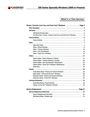
Kim et al. Biomaterials Research(2021) 25:39Page 6 of 11Fig. 3 A Results of the compressive strength. Results of experimental groups in SBF storage solution for 7 days. Same superscript means nostatistical significance (p 0.05). B The results of experimental groups in comparison of DW and SBF as storage solutionsFE-SEM analysisFigure 2 shows the FE-SEM analysis results after thepush-out bond strength measurements of the groups. Inall groups, precipitates were observed in the dentinal tubules. Among all groups, the 2% BG/MTA groupshowed a denser precipitate infiltration (Fig. 2(C)).and SBF that were storage solutions, the 2 and 5% BG/MTA groups showed a tendency to increase the compressive strength in SBF compared to DW (Fig. 3(B)).No significant interaction was observed between the materials and storage solution by two-way ANOVA (p 0.05).Compressive strengthThe compressive strength results are illustrated in Fig. 3.Among the specimens stored in SBF for 7 days, the 2%BG/MTA group showed the highest compressivestrength (Fig. 3(A)). The control group and the 1 and 2%BG/MTA groups showed statistically higher intensitiesthan that of the other BG/MTA groups (p 0.05). Comparing the compressive strength results between DWSetting timeTable 2 showed the setting time results for the groups.Compared to the MTA group, the setting time of all experimental groups decreased. The reduction of settingtime from the MTA group (428.3 min) were the 1, 2%BG/MTA groups (398.3 and 401.0 min) respectively.
Kim et al. Biomaterials Research(2021) 25:39Page 7 of 11Table 2 Setting time (min) of the groupsGroupsSetting time (min)MTA428.33 (11.93)1% BG/MTA398.33 (18.82)2% BG/MTA401.00 (26.89)5% BG/MTA406.02 (25.16)10% BG/MTA416.67 (42.52)Values are written as means (standard deviation)SolubilityThe solubility results are presented in Table 3. The solubility of the BG/MTA groups was slightly higher thanthat of the control group (p 0.05).XRD analysisThe XRD analysis results are presented in Fig. 4. In theMTA group and 1% BG/MTA group, there was no difference between the analysis immediately after the setting and after the additional 7 days of SBF maturation inthe graph. In the 2, 5, and 10% BG/MTA groups, theapatite peaks ranging from 40 to 50 in the graph wereincreased in each SBF 7-day maturation specimen, compared to the control group analysis immediately after thesetting (Fig. 4(B)).DiscussionThe biocompatibility and bioactivity of MTA have beendemonstrated in a variety of endodontic treatment indications by laboratory and clinical studies. Though it hasbeen widely used for 30 years, the disadvantage of decrease of physical properties, such as compressivestrength, dimensional stability, and anti-washout property when in contact with periodontal tissue, blood, etc.is still one of the critical considerations [4, 7]. To overcome these characteristics, we examined BG, as an additive into MTA powder, which has been reported as abiomaterial with bone-like carbonated apatite-formingability by the interaction between calcium ion and phosphate from tissue fluid. We evaluated the effect of BGon the physical properties of MTA, and demonstratedthat the BG-containing MTA improved the physicalproperties in SBF condition or dentin bonding. We alsoTable 3 Solubility (%) of the groupsGroupsSolubility (%)MTA1.17 (0.19) c1% BG/MTA1.70 (0.36) ab2% BG/MTA1.10 (0.04) bc5% BG/MTA1.25 (0.36) ab10% BG/MTA1.34 (0.12) abcValues are written as means (standard deviation)Different superscript letters indicate significant differences between thematerials (p 0.05)investigated the optimal concentration of BG supplementation into MTA, which has an increasing effect orwithout hampering effect on the mechanical propertiesof MTA itself. Recently, several studies have been demonstrated the advantages of BG’s high biocompatibilityand remineralization ability of dentin [19–23, 32].Within our knowledge, a single previous study [11] hadbeen reported that the addition of BG showed the shortening effect of the setting time of MTA Angelus and experimental Portland cement. They examined the effectof supplementation of various concentrations of BG andcalcium metasilicate (wollastonite) ranged from 10 to 30wt%. The study reported that BG addition over 10 wt%reduced the compressive strength of MTA, on the otherhand, it has the shortening effect on the setting timewithout hampering the biological response. In our study,a significant important difference from the previousstudy [11] was found that supplementation of lower concentrations of BG which are 2 and/or 5 wt%, has the effect to increase both the push-out bond strength and thecompressive strength in SBF storage condition.The push-out bond strength test is an effectivemethod for evaluating the ability to resist detachmentfrom restored sites [33]; we attempted to find the evidence of the strength by analyzing the SEM results. Allexperimental groups showed increased push-out bondstrength after 7 days of SBF storage, and the value of the2% BG/MTA group was the highest (Fig. 2). FE-SEM(Fig. 3) following the push-out bond strength was notanalyzed statistically; however, it consistently showed results that could predict uniform and dense precipitationin the 2% BG/MTA group. It is speculated that theimmediate precipitation by the formation of an amorphous calcium-phosphate layer on the BG inducesmineralization of the tag-like layer to the dentinal tubular interface during mineralization; the extrusion bondstrength appears to increase [34]. BG as an additive improved the handling properties because of an appropriate amount of silica self-agglomeration [22] during thehydration process with DW, and thus, the deposition inthe tubules increased. To investigate this, an additionalenergy dispersive X-ray spectroscopy (EDS) analysis isrequired to analyze the composition of the precipitate inthe tubule. In our preliminary study, we evaluated several mechanical and biological properties of various concentration of BG into MTA ranged from 1 wt% to 20wt%. We found that the supplementation of lower concentrations under 10 wt% of BG has the effect to increase both push-out bond strength to dentin and thecompressive strength in SBF storage condition, especiallyin the 2 wt% BG/MTA showed the high push-out bondstrength. In this study, we prepared the MTA or BG/MTA groups were prepared using the MTA and 63S BGat four different concentrations: 1, 2, 5, and 10 wt%. The
Kim et al. Biomaterials Research(2021) 25:39Page 8 of 11Fig. 4 A XRD analysis graphs shows the difference between the analysis results of immediate after setting and 7-day storage in the SBF for eachgroup. B In 2, 5, and 10% BG/MTA groups, the apatite peak was increased in each SBF 7-day storage specimen, compared to the control groupimmediately after setting (blue arrow)L/P ratio of DW to the powder was 0.3 throughout theexperiments, which was set according to the manufacturer’s recommendations. We supplemented the BG intoMTA powder so that the L/P ratio of BG/MTA composite was 0.3, however, the pure MTA L/P ratio might bedifferent. It is speculated that the BG might influencethe MTA hydration and setting reaction, due to theirion exchange between BG and phosphate from SBF solution. In addition, the different shapes and size of particles between BG and MTA powder might lead to thedifferent setting reactions of MTA in the SBF solution,followed by the different mechanical properties. Consequently, our results suggest that the critical optimal concentration of addition of BG, which could take biologicaladvantages in the SBF condition and increasing theamount of hydroxyapatite by BG without hamperingeffect on the mechanical properties, might be consideredranged from 2 to 5 wt%.Compressive strength is considered necessary in theevaluation of restorative MTA [35]. A comparison experiment was conducted to confirm the effectiveness ofBG in SBF by dividing it into two storage solutions ofDW and SBF. In the DW groups, the compressivestrength decreased as the BG content increased, and theMTA group and 1 and 2% BG/MTA groups showed thehighest values. The MTA and control groups only presented a significant decrease in SBF storage compared tothe DW storage groups. Nekoofar et al. [9] reported thatthe compressive strength of MTA tends to decrease inSBF because of the decrease in the formation of acicularcrystals. Therefore, it is speculated that this was thecause of the decrease in compressive strength in the
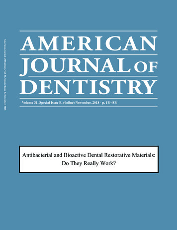
Kim et al. Biomaterials Research(2021) 25:39SBF. In the SBF storage groups, the 2% BG/MTA groupshowed the highest value, and the 2 and 5% BG/MTAgroups showed an increased compressive strength valuecompared to DW storage. The mineralization effect ofBG compensated for the compressive strength of MTA,which was reduced in SBF. Skallevold et al. [18] reportedthat the glass composition of BG contributes to poormechanical properties because of its fragility. Our resultsshowed that the compressive strength of the 10% BG/MTA group, which did not increase further even underSBF conditions, may be responsible for the amount offragile glass content rather than the increase in theamount of hydroxyapatite. The 27 mM HCO3- Tris SBFwas used as the SBF storage solution in this study. Thecomposition of 27 mM HCO3- Tris SBF is a componentcloser to plasma (27 mM HCO3- and 103 mM Cl-) thanthe original SBF (4.2 mM HCO3 and 148 mM Cl )[30], and it had an enhanced ability to induce apatitelike calcium phosphate formation [23]. According toprevious studies, there is a possibility of the autogenousprecipitation of calcium phosphate when stored at 37 Cfor several weeks [21, 36]; thus, SBF was exchanged onceevery 2 days in this study.Setting time is a property for the stable maintenanceof endodontic restorative materials. In this study, all BG/MTA groups showed a decrease in the setting time(Table 2). The initial hydration process of MTA occurson the outer surface of the particles, and the rate of thisprocess is affected by the particle size and the powderto-liquid ratio that is determined from the setting timedata. The particle sizes of MTA and BG used in thisstudy ranged from to 1–30 μm [37] and 20 μm [22], respectively. The effect of increased surface area and reactivity caused by the small size of BG [11, 38–40] couldlead to a reduction in the setting time. The known setting time of the MTA is 4–6 h; however, the setting timeof the experimental MTA was found to be 6–7 h (398–428 min), which is slightly longer. There are two types ofspecifications used for the method of measuring the setting time suggested in MTA-related studies [27]: ISO6876, dental root canal sealing materials, and ISO 9917,and dentistry water-based cements. The ISO 6876method is similar to the initial setting time because ofthe difference in the mold containing the specimen, andthe ISO 9917 method is similar to the final setting time.We demonstrated along setting time by referring to theISO 9917-1 method; however, it does not deviate significantly from the general range.The solubility test is one of the most important requisites for endodontic restorative materials because it directly contacts the periodontal tissue fluids [41] andcopes with the sealing of the wound. The solubility of allBG/MTA groups was slightly higher than that of theMTA group (Table 3); however, all groups showedPage 9 of 11values less than 3%, which is within the acceptable ISO6876 range [31]. As the conditions for measuring thesolubility of ISO were DW, calcium or phosphate ionscould not be utilized in the surrounding solution [18,42]. Therefore, it is assumed that a lower solubility valuewill be obtained if it is re-measured in a physiologicalsolution environment where the action of BG canappear.XRD was used to investigate the existence of hydroxyapatite formation in the experimental groups after 7 daysof storage in SBF (Fig. 4); the 29 peak [34, 43] increasedin the MTA, 1, 2, and 5% BG/MTA groups, and 47 and50 peaks [34, 43] increased in the 2, 5, and 10% BG/MTA groups. Although there is a limitation in that XRDonly analyzes surface components, it showed that theBG/MTA group can additionally form hydroxyapatite.However, the 2% BG/MTA group, which showed thehighest compressive strength and push-out bondstrength in SBF, did not exhibit the highest hydroxyapatite formation. From a clinical point of view, these results suggest that 2% BG/MTA improved the handlingproperties properly during the hydration process beforesetting; however, after setting, it contributed to themaintenance of physical properties by maintaining theappropriate amount of MTA along with an increase inthe appropriate amount of hydroxyapatite by BG [18].The possibility of reducing physical properties withBG at a high concentration of 10% or more can be inferred from previous studies dea
time; it was an alternative to nearly inert implant mate-rials [17]. It is a highly bioactive material with silicon di-oxide as its main component. When it encounters water from BF or simulated body fluid (SBF), BG immediately undergoes ionic dissolution and glass degradation [17]. It releases silica ions to form a silica-rich layer on the surface.