Transcription
Nordin et al. BMC Complementary and Alternative 2019) 19:290RESEARCH ARTICLEOpen AccessEffect of Nigella sativa and its bioactivecompound on type 2 epithelial tomesenchymal transition: a systematicreviewAbid Nordin1,2, Haziq Kamal1, Muhammad Dain Yazid3, Aminuddin Saim4 and Ruszymah Idrus1*AbstractBackground: Nigella sativa or commonly known as black seed or black cumin is one of the most ubiquitouscomplementary medicine. Epithelial to mesenchymal transition (EMT) of type 2 is defined by the balance betweenwound healing and tissue fibrosis, which is dependent to the state of inflammation. This systematic review isconducted to provide an overview regarding the reported effect of Nigella sativa and its bioactive compound onthe type 2 EMT.Methods: A search was done in EBSCOHOST, OVID and SCOPUS database to obtain potentially relevant articles thatwere published between 1823 and August 2019. This review includes studies that focus on the effect of Nigellasativa and its bioactive compound on the events related to type 2 EMT.Results: A total of 1393 research articles were found to be potentially related to the effect of Nigella sativa and itsbioactive compound, thymoquinone on Type 2 EMT. After screening was done, 22 research articles met inclusioncriteria and were included in this review. Majority of the studies, reported better wound healing rate or significantprevention of tissue inflammation and organ fibrosis following Nigella sativa or thymoquinone treatments. In termsof wound healing, studies included reported progression of EMT related pathological changes after treatment withNigella sativa or thymoquinone. Alternatively, in terms of fibrosis and inflammation, studies included reportedreversal of pathological changes related to EMT after treatment with Nigella sativa or thymoquinone.Conclusion: Through this review, Nigella sativa and thymoquinone have been associated with events in Type 2EMT. They have been shown to promote wound healing, attenuate tissue inflammation, and prevent organ fibrosisvia regulation of the EMT process.Keywords: Nigella sativa, Thymoquinone, Epithelial, Mesenchymal, Wound healing, FibrosisBackgroundEpithelial to mesenchymal transitionThe term epithelial-to-mesenchymal transition (EMT)was first coined by Elizabeth Hay to describe the phenotypical transition of epithelial cells into mesenchymalcells during the primitive streak formation in chick embryo [1]. This transition is a ubiquitous physiological* Correspondence: ruszyidrus@gmail.com1Department of Physiology, Faculty of Medicine, Universiti KebangsaanMalaysia, 56000 Cheras, Kuala Lumpur, MalaysiaFull list of author information is available at the end of the articleprocess that plays a major role in healing process anddisease progression [2].EMT resulted in the release of the immotile epithelialcell that was bounded to their basal membrane, into anon-polarized cell that is freely mobile [3]. The transition is further reinforced by increased production ofextracellular matrix (ECM) components [4]. At the molecular level, the EMT process is regulated by severalmolecular events such as transcription factors activation,cytoskeletal reorganization, and specific cell-surface proteins or microRNAs regulation [5]. The Author(s). 2019 Open Access This article is distributed under the terms of the Creative Commons Attribution 4.0International License (http://creativecommons.org/licenses/by/4.0/), which permits unrestricted use, distribution, andreproduction in any medium, provided you give appropriate credit to the original author(s) and the source, provide a link tothe Creative Commons license, and indicate if changes were made. The Creative Commons Public Domain Dedication o/1.0/) applies to the data made available in this article, unless otherwise stated.
Nordin et al. BMC Complementary and Alternative Medicine(2019) 19:290Page 2 of 12Classification of EMTNigella sativa and its bioactive compound in type 2 EMTEMT classification has been made according to theirfunctional consequences; namely type 1, type 2, and type3 [4]. Type 1 EMT is assigned to the embryologicaldevelopment, implantation and organ development. It isinvolved primarily during gastrulation and neurulation,whereby the cell transition between the two differentstate of epithelial and mesenchymal as needed [6]. Thesecells might be re-induced further as secondary epithelialcells that were involved in organogenesis and development [7]. Type 2 EMT is involved primarily in tissueregeneration [8] and fibrosis [9]. This happens wheninflammation initiate the production of ECM by thefibroblast, to help rebuild the damaged tissue site [10].However, persistent inflammation can lead to tissue ororgan fibrosis following incessant wound healing process[11]. Hence, the balance between wound healing andfibrosis via the inflammation process is what defines thetype 2 EMT. On the other hand, type 3 EMT, has beenspecifically assigned for neoplastic cells in regards totheir invasive capability seen in their metastasis to secondary site from their primary oncogenic origin [12].Nigella sativa or commonly known as black seed or blackcumin is one of the most ubiquitous complementarymedicine. It is widely grown in the Middle East, Africaand Asia [20]. In Islam, it was mentioned by the prophetMuhammad (PBUH) for its healing properties [21] andcurrently widely used in the Islamic practice of propheticmedicine [22]. Pre-clinical and clinical studies have shownNigella sativa to have multiple health benefits such asanti-inflammatory [23], anti-oxidant [24], anti-bacterial[25], anti-angiogenic [26], and organ-protective [27].On the other hand, a study on Nigella sativa’s chemicalcomposition by supercritical carbon dioxide revealed fewbioactive compounds; namely thymoquinone, dithymoquinone, and dihydrothymoquinone. Among these compounds,thymoquinone is the major active compound in Nigellasativa [28]. Thymoquinone by itself exhibit strong antiinflammatory [23], anti-bacterial [25], anti-diabetic [26] andanti-oxidant properties [29].Type 2 EMT plays a role in wound healing and tissuefibrosis, which is dependent to the state of inflammation.Inflammation is a complex mechanism that are influencedby bacterial activity [25], inflammatory response [30] andoxidative stress [29]. Balance between the attenuation andsustenance of these factors determine the fate of the tissue.On one hand, wound healing progresses with attenuationof inflammation. Alternatively, tissue fibrosis progresseswith the sustenance of inflammation [31]. In this review, asystematic search of the electronic databases was conductedto identify studies done on different events of EMT type 2,including wound healing, tissue inflammation and organfibrosis treated with Nigella sativa or thymoquinone.Markers for EMTIn 2009, a review was made by Michael Zeisberg andEric G. Neilson regarding biomarkers of EMT [13]. Theloss of the epithelial marker, E-cadherin, and the increase expressions of mesenchymal marker, N-cadherin,together known as the cadherin switch, has been nominated as the hallmark of EMT process [14]. The transition into mesenchymal phenotype was also indicated bythe elevation of other mesenchymal markers such asfibroblast specific protein-1 (FSP1), discoidin domainreceptor tyrosine kinase 2 (DDR2) or fibronectin [15].Activation of EMT also triggers the production of matrixmetalloproteinase [16]. This results in breaking down ofthe cell-to-cell adhesion that lead to the increased migratory capability of the resulting mesenchymal cell [13].Following the advancement of molecular biology, a review on the molecular markers of EMT was made bySamy Lamouille, Jian Xu and Rik Derynck in 2014 [5].They described regulators of EMT at the transcriptional,translational and post-translational level. In terms of transcriptional factors, some important characters are SNAIL,TWIST and ZEB family of transcription factors [5]. Twovariants of SNAIL, SNAI1 (Snail) and SNAI2 (Slug) reduce the expression of E-cadherin by repressing its promoter [17]. In cancer cell, activity of TWIST1 has beenshown to have dual transcriptional activities, namely therepression of E-cadherin and activation of N-cadherin[18]. Finally, like SNAIL and TWIST, ZEB transcriptionfactors also bind to E-cadherin promoter and function astranscriptional repressors of E-cadherin protein [19].MethodsLiterature search/search strategyA systematic review of the literature was conducted to identify relevant reports on the effects of Nigella sativa and itsbioactive compound on the EMT, particularly in type 2EMT. A comprehensive search of biomedical science journals in Ebscohost (published between 1823 to August 2019),Ovid (published between 1946 to August 2019) and Scopus(published between 1973 to August 2019) databases wasdone. The search strategy was adapted from the previouspublication looking at the effect of honey in type 2 EMT[32] involving a combination of the following two setsof keywords; (1) thymoquinone OR Nigella sativa ORdithymoquinone OR thymohydroquinone AND (2)epitheli* OR mesenchym* OR transition OR vimentinOR cadherin OR tgf*.Inclusion criteriaOnly original research articles (in vitro, in vivo or clinicalstudy) that discuss changes in epithelial to mesenchymaltransition parameters (down regulation of epithelial markers
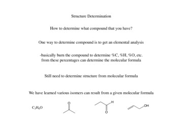
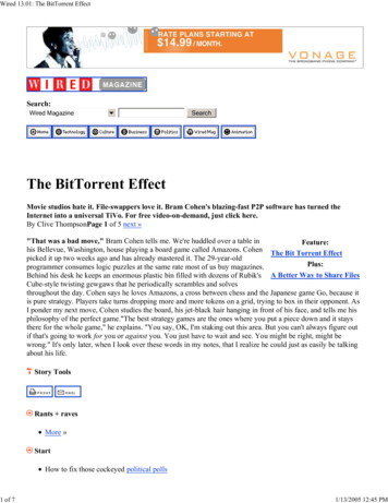
Nordin et al. BMC Complementary and Alternative Medicine(2019) 19:290and up regulation of mesenchymal marker) of type 2(wound healing, inflammation or tissue fibrosis) influenced by Nigella sativa or thymoquinone, written inEnglish language and have abstract available were included in this review. Studies included must measurethe changes of at least one of the epithelial or mesenchymal markers such as 1) cadherin switch; 2) elevationof FSP1, DDR2, or fibronectin; or 3) increased activityor expression of the metalloproteinase.Exclusion criteriaAll secondary literatures and any original articles thatwere not written in English, do not have abstract availableand do not fulfil the inclusion criteria mentioned were excluded. Studies focusing on other types of EMT such astype 1 (embryonic development) and type 3 (cancer metastasis) were excluded from the review.Data extraction and managementArticles underwent screening process prior to their inclusion in this review. Titles and abstracts were screenedfirst to ensure inclusion and exclusion criteria were adhered. Then, the full text of what remaining were readthoroughly by five independent reviewers to exclude anyarticle that did not meet inclusion criteria. All reviewersmust agree on the inclusion of selected articles for thereview before the data extraction phase begins. Any differences in opinions were resolved through discussionbetween the reviewers.Data extraction was performed independently with theuse of a data extraction form. The following data wererecorded from the studies: (1) type of experimentalmodel used; (2) treatment groups; (3) summary of theoutcomes measured in the study; (4) summary of thestudy results; and (5) conclusion of the study.ResultsSearch resultsThe literature search identified 1393 potentially relevantarticles. Four reviewers independently assessed all articles for inclusion and exclusion based on title and abstract. One thousand three hundred thirty-five of thesearticles were excluded because they were not related toNigella sativa or its bioactive compound or not relatedto EMT in wound healing, inflammation, or tissue fibrosis. From the remaining 58 articles, 36 articles wererejected based on exclusion criteria after reading theirfull text. A total of 22 articles were retrieved for furtherassessment and data extraction to be included in thisreview. A flow chart of the selection process, includingreasons for exclusion was shown in Fig. 1.Page 3 of 12Study characteristicsThere are 22 articles that were included in the review.All articles were published between the year 2010 and2018. The studies were categorized according to the typeof pathophysiology that they model which were woundhealing, inflammation and organ fibrosis.Nigella sativa was used as oil ointment in five studies[23, 24, 27, 33, 34], and as cream in one study [35]. Thymoquinone was used in 14 studies [25, 26, 29, 36–46].Nigella sativa oil and thymoquinone was compared inone study [47]. Since honey made by bee contains thebioactive compound of its nectar origin, a study thatinvestigated the effect of Nigella sativa honey and itspropolis was also included in the review [48].Wound healing of two types of tissue were modelledamong the studies included, cutaneous tissue and corneal tissue. In terms of cutaneous tissue, three types ofwound model were used, namely incisional wound, burnwound, and diabetic wound. The two studies that investigated the incisional wound physiology uses mouse [25]and rat [35]. For burn wound, three studies using ratwere included [29, 34, 48]. Only one study investigatedthe cutaneous diabetic wound [26]. In terms of cornealwound healing, one study using rat was included [27].Studies reporting effect of Nigella sativa or thymoquinone on wound healing were listed in Table 1.In terms of tissue fibrosis, 5 studies that model the tissuefibrosis of different organs were included. For cardiac fibrosis, one study using doxorubicin-induced rats was included[38]. For lung fibrosis, 2 studies with bleomycin-inducedrats [23] and paraquat-induced rats [42] were included. Forliver fibrosis, one study using carbon tetrachloride-inducedrats was included [40]. For renal fibrosis, one study usingnitrite-induced rats was included [24]. Studies reportingeffect of Nigella sativa or thymoquinone on tissue fibrosiswere listed in Table 2.Progression of wound healing and tissue fibrosis rely onthe inflammation state of the tissue [31]. As a result, type 2EMT researchers focus on the inflammatory properties ofthe diseased tissue. In terms of inflammation, 11 studiesthat model inflammation in different organs were included.In terms of the nose inflammation, rhinosinusitis, one studyusing rats induced with intranasal platelet activation factorwas included [46]. Ear inflammation, otitis, is modelled inone study using bacterial infection-induced model [36]. Interms of inflammation following lung injury, 3 studies wereincluded using cardiac ischemia-induced rats [39], cadmium chloride-induced rats [33], and ovalbumin-inducedrats [43]. In terms of inflammation in the kidney, 2 studiesusing diabetic-induced nephropathy model in rats [45, 47]and one study using reperfusion therapy-induced nephropathy in rats [41] were included. One study focussing onliver inflammation using ethanol-induced liver injury modelin mice [44] was included. Injury to testicle was modelled
Nordin et al. BMC Complementary and Alternative Medicine(2019) 19:290Page 4 of 12Fig. 1 Flowchart of the selection processin rats in one study [37]. Studies reporting effect of Nigellasativa or thymoquinone on inflammation were listed inTable 3.Potential of Nigella sativa and thymoquinone in woundhealingCutaneous normal wound healingThe literature search returns some evidence on the positiveeffect of Nigella sativa and its constituent on cutaneouswound healing. The similarities of molecular events surrounding epithelial to mesenchymal transition and woundhealing has been well described in the context of cutaneouswound [8]. Phenotypic changes following EMT in cutaneous tissue enable the progression of reepithelialisation ofthe epidermal layer in normal wound healing [49].In incisional wound, the positive effect of topical application of Nigella sativa cream (NSC) on full thicknesswound healing in rats was observed. The NSC-treatedgroups demonstrated the smallest wound size by the endof experiment (99.69% wound area reduction) comparedto the control groups (81.35% wound area reduction). Histologically, NSC also reduces inflammatory cell infiltration, and lessen cell oedema. Highest antioxidant levelswere also observed in NSC-treated rat’s tissues suggestingthe correlation between Nigella sativa antioxidant effectand its wound healing benefits [35].Delivery of thymoquinone, via the thymoquinoneloaded composite nanofiber, revealed a positive outcometo incisional wound healing in mice model. Macroscopically, application of thymoquinone-loaded dressing onmice wound produced the best wound closure. Histologically, the treatment group shows higher granulation tissue thickness in a shorter time compared to the controlgroup. It also shows highest epithelial migration after 14days. Thymoquinone delivery also resulted in the highestcollagen deposition in the healing tissue via Masson’sTrichrome staining. This study concludes that thymoquinone promotes better wound healing via its antiinflammatory capabilities [25].Taken together, both Nigella sativa and its bioactivecompound, thymoquinone, proves to be efficacious inenhancing the progression of incisional wound healing.Cutaneous burn wound healingBurn wounds can be caused by exposure to flames, liquids,chemicals, hot surfaces and radiations [48]. Burn introduced direct damage of the mechanical barrier as well asthe immune cells in the wound area. The impaired immuneresponse following burns, led to a delay in the healingprocess and inability to fight infection [29].A comparison between Nigella sativa oil (NSO) and silver sulfadiazine (SSD), the gold standard of burn woundmanagement, was done in the rat model of burn wound
Nordin et al. BMC Complementary and Alternative Medicine(2019) 19:290Page 5 of 12Table 1 Effect of Nigella sativa and thymoquinone in wound measuresResultsConclusionCutaneous Incisional WoundGomaa et al.2017 [25]Incisional woundin Balb/C mice.3% (w/w) thymoquinone1. Wound area(TQ) loaded into nanofiber. measurement.2. Histopathologyassessment.Treatment with TQimprove wound closureand tissue healing.TQ-loaded nanofibershows potentialto be used forwound dressing.Han et al.2017 [35]Full thicknesswound inWistar rats.50% topical Nigella sativa(NS) cream.1. Wound contraction.2. Biochemicalanalysis.3. Histologicalevaluation.Treatment with NScream increases woundcontraction rate andantioxidant activity buthave no effect intissue granulation.NS heals via itsantioxidanteffect in fullthickness wound.Cutaneous Burn WoundSulaiman et al.2014 [48]Burn woundmodel inalbino ratsTopical micro-emulsionof 5% NS honey and5% of its propolis.1. Wound contraction. Treatment of NS honeyand its propolis increaseswound contraction rate.Microemulsion of NShoney and propoliscontribute to fasterburn wound healing.Selcuk et al.2013 [29]Burn woundmodel inSpragueDawley rats2 mg/kg/day oral TQor 0.5% topical TQ.1. Histologicalassessment.2. Total antioxidantstate.3. Total oxidativestress.4. BacterialassessmentTopical treatment ofTQ was superior comparedto oral TQ in improvingwound histology, enhancingantioxidant activity, andreducing bacterial growth.Topical TQ is superiorto oral TQ inimprovingwound healing.Yaman et al.2010 [34]Burn woundmodel inmale, Wistaralbino rats.50% topical NS oil (NSO).1. Gross morphologyof the wound.2. Histologicalevaluation.Treatment with NSOreduces inflammationand demonstrated bettertissue granulation in wound.NSO has been shownto promote fasterburnwound healing.10% topical TQ.1. Wound contraction. Treatment with TQ2. Histologicalincreased healing andevaluation.reduced inflammatorycells and fibroblast atday 3. However, woundimprovement declineson day 7 and day 14.Cutaneous Diabetic WoundYusmin & Ahmad Chronic delayed2017 [26]wound inalloxan-induceddiabetic rats.TQ heals faster ininflammatory phasebut slower duringthe proliferativephase due to itsantiangiogenicpropertiesCorneal WoundSalem et al.2016[27]Formaldehyde-induced 40 mg/kg/day oral NSOcorneal toxicity inalbino rats.healing. NSO-treated group shows faster scab formationwith better epithelial appearances in comparison to theSSD group and the control group. NSO-treated group alsodemonstrated better tissue granulation compared to othergroups [34].In terms of thymoquinone, Selçuk et al. (2013) compared the effect of topical and systemic delivery of thymoquinone in the treatment of burn wound. The resultsof the study suggested better outcome with topical delivery as compared to systemic delivery of thymoquinone.This is supported by better vascularization and granulation tissue formation, as well as lower inflammatory cellinfiltration in the histological assessment of the tissue ofthe topical treatment groups. However, in combination,1. Histologicalevaluation.Treatment with NSO reverses NSO shown toformaldehyde-inducedresolved cornealpathological changes.injury induced byformaldehyde toxicitytopical and systemic treatment of thymoquinone wasmore superior to all other treatment groups in thestudy [29].Positive effect of Nigella sativa on wound healing wasalso apparent in the honey and propolis produced with itsnectar. Honey produced by bees has been reported to positively influence EMT of wound healing, and their effect isdependent on the source of nectar [32]. Nigella sativahoney, together with ethanolic extract of its propolis demonstrated antibacterial activity and significant promotion ofburn wound healing in comparison to SSD cream [48].The results from these studies supported the superiority of Nigella sativa and thymoquinone comparedto SSD in the management of burn wound healing.
Nordin et al. BMC Complementary and Alternative Medicine(2019) 19:290Page 6 of 12Table 2 Effect of Nigella sativa and thymoquinone on organ fibrosisReferencesExperimental modelTreatmentOutcome measuresDoxorubicin (Dox)-inducedheart failure inSprague-Dawley rats.50 mg/kg/day oral TQ. 1. Left ventricular functions.2. Atheroscleroticlesion.3. Fibrosis markers.4. Apoptosis markers.ResultsConclusionTreatment ofTQ reversesDox-inducedpathologicalchanges in theheart via inhibitionof fibrosisand apoptosis.TQ mitigatesDox-inducedcardiac damage andfibrosis.Treatment withNSO reversebleomycin-inducedpathological changesvia inductionof TGF-β1.NSO haveshown toresolve BLMinduced PFdue to itsantiinflammatoryand antifibroticpropertiesMyocardial FibrosisPei et al.2018 [38]Pulmonary FibrosisAbidi et al.2017 [23]Bleomycin-induced pulmonary 1 mg/kg/dayfibrosis in Wistar rats.oral NSO.1. Physical measurements.2. Histological evaluation.3. Liver metabolites.4. Urine metabolites.5. Expression of TGF-β1.Pourgholamhosseinet al. 2016[42]Paraquat-induced lungfibrosis in NMRI mice.20 mg/kg/day and1. Histological evaluation.40 mg/kg/day oral TQ. 2. Oxidative stress analysis.3. Hydroxyproline content.4. Gene expression.Treatment withTQ reversesparaquat-inducedlung fibrosisinhibition of fibrosisand antioxidant activity.TQ is able toreducepulmonaryfibrosis via itsanti-fibroticproperty.Carbon tetrachloride(CCl4)-induced renalfibrosis in Wistar rats.15 mg/ml oral TQ with 1. Liver functionor without 1000 IU/ml parameters.of Vitamin D3.2. Renal functionparameters.3. Histological assessment.4. Cytokines level.Treatment of TQreversesCCl4-induced renalfibrosis via inhibitionof inflammation.TQ showsanti-fibroticproperties incarbontetrachlorideinduced renalfibrosis.Sodium nitrite (NaNO2)induced renal fibrosis inSprague-Dawley rats.2.5 ml/kgoral NSO.Treatment with NSOreverses sodiumnitrite-induced renalfibrosis via antioxidative,anti-inflammatory, andanti-apoptotic properties.NSO havebeen shownto resolveNaNO2inducednephrotoxicity.Liver FibrosisAbdelghany et al.2016 [40]Renal FibrosisAl-Gayyar et al.2016 [24]Cutaneous diabetic wound healingOne of the complications of diabetes mellitus is delayed ornon-healing wound. This is primarily associated with theseries of macrovascular and microvascular alteration thatresulted in complications such as poor circulation, weakened immunity, and disrupted cellular metabolism [50].In diabetic wound, thymoquinone was shown to causeslower overall wound contraction compared to the control.However, upon histological examination at different timepoint, it was found that thymoquinone induced faster healing at early time point (Day 3) before the healing ratedecreases gradually. Observation of lesser inflammatory cellinfiltration at Day 3 suggested that thymoquinone enhancedwound healing during the inflammatory phase but started toslow down as the healing shift to proliferative phase. Theknown anti-angiogenic properties of thymoquinone was1. Renal functionparameters.2. Fibrotic markers.3. Cytokine levels.4. Protein kinase activity.5. Apoptosis markers.believed to be the reason why the healing was slower at theproliferative phase [26].Corneal wound healingCompared to the cutaneous wound healing, little isknown about the association of corneal wound healingwith the type 2 EMT. Exposure of corneal epithelial cellsto formaldehyde (FA) is known to induce ocular injury[51]. In terms of corneal wound healing, study conducted with FA-exposed corneal keratocyte demonstrated NSO ability to ameliorate inflammatory responseproduced by FA exposure and restored disrupted cornealarchitecture. NSO also ensure continuity of limbal stemcells to ensure corneal reepithelialisation [27].In terms of wound healing, all studies included in thereview demonstrated a strong evidence of positive effect
Nordin et al. BMC Complementary and Alternative Medicine(2019) 19:290Page 7 of 12Table 3 Effect of Nigella sativa and thymoquinone on tcome measuresResultsConclusionIntranasal plateletactivating factor-inducedrhinosinusitis model inSprague-Dawley rats.80 mg/kgoral TQ.1. Degree of vascularcongestion2. Intensity ofinflammatorycell infiltration3. Degree ofepithelial injuryTreatment with TQreverses the intranasalplatelet activatingfacto-inducedhistological changes.TQ have been shownto be beneficial forthe resolutionof rhinosinusitis.1. Histopathologicalassessment.2. Bacterial assessment.Treatment of TQ reversesinfection-inducedhistopathologicalchanges and reducesbacterial growth inthe ear canal.TQ shows bacteriaeradication andanti-inflammatorypropertiesRhinosinusitisCingi et al.2011 [46]OtitisDemirel et al. Bacterial infection-induced 0.1 and 0.4%2018 [36]otitis model intopical TQ.Wistar rats.Lung InflammationSezen et al.2018 [39]Cardiac ischemia-inducedlung injury inWistar rats25 mg/kg TQ.1. Apoptotic markers inTreatment of TQ reversesthe lung.cardiac ischemia-induced2. Histological assessment. histopathologychanges via suppressionof apoptosis.TQ protects against lunginjury via inhibitionof apoptosis.El-Ebiary et al. Cadmium chloride2016 [33](CdCl2)-induced lungdamage in Wistar rats1 ml/kgoral NSO.1. Histopathologicalassessment.2. Scanning electronmicroscopy.Treatment of NSOreverses histopathologicalchanges inducedby CdCl2 with normalpneumocytes morphologyand intra-alveolarseptum thickness.Treatment with NSOameliorated pathologicalchanges in CdCl2poisoned rats.Su et al.2016 [43]Ovalbumin-inducedasthmain Balb/C mice.3 mg/kgoral TQ.1. Cytokines level.2. Fibrotic markers.3. Histopathologicalassessment.4. Angiogenic factors.5. HUVEC tubeformation.6. Proteinkinase activity.Treatment of TQ reverseshistopathology changesof asthma induced byovalbumin via suppressionof inflammationand angiogenesis.TQ have anti-inflammatoryand anti-angiogenesisproperties inovalbumin-inducedasthmatic mice.Al-Trad et al.2016 [47]Streptozotocin(STZ)-inducednephropathy indiabetic rats.50 mg/kg oral 1. Renal pathologyTQ and 2 ml/kg parameters.oral NSO.2. Expression of Podocin.3. Fibrotic markers.4. Angiogenic marker.Both TQ and NSO treatmentdemonstrated comparablereversal of diabetic-inducedrenal pathology via expressionof podocin and inhibition offibrosis and angiogenesis.TQ & NSO improvespathologicalchanges indiabetic-inducednephropathy.Omran2013 [45]Nephropathy inSTZ-induceddiabetic rats.50 mg/kgoral TQ.1. Renal pathologyparameters.2. Histopathologicalassessment.3. Epithelial markers.4. Mesenchymal markers.Treatment of TQ reverses thediabetic-induced renalhistopathological changesvia inhibition of the epithelialto mesenchymal transition.TQ improves renalfunctions via inhibitionof epithelial tomesenchymal transition indiabetic nephropathy.Hammad &Lubbad2016 [41]Reperfusiontherapy-inducednephropathy inmale Wistar rats.10 mg/kgoral TQ.1. Renal pathologyparameters.2. Cytokine levels.Treatment of TQ resultedreversal of reperfusiontherapy-inducedhistopathological changesvia the inhibitionof inflammation.TQ improves renal functionsvia inhibition ofinflammationfollowing reperfusiontherapy-inducednephropathy.20 mg/kg or40 mg/kgoral TQ.1. Liver pathologyparameters.Treatment of TQ reversesthe EtOH-induced liverpathological changes viaTQ regulates LKB1 andAMPK signalling that isassociated withNephropathyLiver InflammationYang et al.2016 [44]Ethanol (EtOH)-inducedliver injury inC57/BL6 mice.
Nordin et al. BMC Complementary and Alternative Medicine(2019) 19:290Page 8 of 12Table 3 Effect of Nigella sativa and thymoquinone on inflammation come measuresResultsConclusion2. Histopathologicalassessment.3. Expression level ofSIRT1, LKB1 and AMPK.upregulation of SIRT1,LKB1, and AMPK.inflammation inethanol-inducedliver injury.1. Testicular pathologyparameters.2. Histopathologicalassessment.Treatment of TQ reverses thePb-induced testicularpathological.TQ have protectiveeffect against Pb-inducedtesticular damage.Testicular DamageMabrouk2018 [37]Lead (Pb)-inducedtesticular damagein Wistar rats.5 mg/kg/dayoral TQ.of Nigella sativa and thymoquinone. Treatment of bothNigella sativa and thymoquinone promotes EMT-relatedpathological changes in the wounded tissue.Potential of Nigella sativa and its constituent in organfibrosisMyocardial fibrosisMyocardial fibrosis contributes substantially to thepathogenesis of heart failure [52]. Prevention of myocardial fibrosis could be beneficial in the management ofheart failure. Doxorubicin, a chemother
conducted to provide an overview regarding the reported effect of Nigella sativa and its bioactive compound on the type 2 EMT. Methods: A search was done in EBSCOHOST, OVID and SCOPUS database to obtain potentially relevant articles that were published between 1823 and August 2019. This review includes studies that focus on the effect of Nigella