Transcription
Am J Cardiovasc Dis 2022;12(2):56-66www.AJCD.us /ISSN:2160-200X/AJCD0141146Original ArticleIschemic cardiomyopathy versus non-ischemiccardiomyopathy in diabetic patients: clinicalcharacteristics, management, and long-term outcomesHanan B Al Backr1*, Turki B Albacker1*, Fayez Elshaer1,2, Nur Asfina1, Fahad A AlSubaie3, Anhar Ullah4,Ahmad Hayajneh1, Osama Almogbel1, Fakhr AlAyoubi1, Waleed Al Habeeb1Department of Cardiac Sciences, College of Medicine, King Fahad Cardiac Center, King Saud University MedicalCity, King Saud University, Riyadh, Saudi Arabia; 2Cardiology Department, National Heart Institute, Cairo 11435,Egypt; 3Cardiology Department, Security Force Hospital, Riyadh, Saudi Arabia; 4National Heart and Lung Institute,Imperial College London, London, United Kingdom. *Equal contributors.1Received December 15, 2021; Accepted February 27, 2022; Epub April 15, 2022; Published April 30, 2022Abstract: Background: Diabetes mellitus causes ischemic heart disease (IHD) through macrovascular or microvascular involvement. Diabetes-associated hypertension, dyslipidemia, and obesity further increase coronary arterydisease risk and can cause left ventricular hypertrophy leading to heart failure with preserved ejection fraction independent of IHD. This study was undertaken to evaluate the differences in demographics, clinical characteristics,Echocardiographic parameters, management, and outcomes between non-ischemic cardiomyopathy (NICM) andischemic cardiomyopathy (ICM) patients in cohort of diabetes patients. Methods: This retrospective study includeddiabetes patients with reduced ejection fraction ( 40) who were hospitalized with heart failure between January2014 and February 2020. Patients were divided into two groups: group 1; ICM and group 2; NICM. Data obtainedon above mentioned features including mortality and heart failure readmissions were compared between the twogroups. Results: A total of 612 diabetes patients admitted with acute heart failure were screened of which 442 wereincluded. Group 1 (ICM) had 361 patients (81.7%) and group 2 (NICM) had 81 patients (18.3%). Patients in group1 were older, predominantly males and with higher prevalence of hypertension, smoking and insulin dependentDiabetes while group 2 patients had higher BMI and higher prevalence of cardiac rhythm problems. No significantdifference was detected in 5-year-mortality between the two groups (P 0.165). However, heart failure associatedhospitalizations were higher in group 2 though it was not statistically significant (P 0.062). Conclusion: There wasno difference in 5-years mortality between ICM and NICM in diabetes patients. However, NICM patients had higherprevalence of obesity and rhythm problems.Keywords: Heart failure, diabetes mellitus, ischemic cardiomyopathy, non-ischemic cardiomyopathyIntroductionDiabetes mellitus (DM) can affect the cardiovascular system in different aspects. It cancause ischemic heart disease (IHD) throughmacrovascular or microvascular involvement. Itis also associated with other cardiac risk factors like hypertension (HTN), dyslipidemia andobesity, which increase the risk for coronaryartery disease (CAD). Diabetes can also causeleft ventricular hypertrophy, which leads toheart failure with preserved ejection fraction(HFpEF) independent of IHD [1]. Presence ofDM in heart failure (HF) patients has prognostic significance that may be influenced by theunderlying etiology. Patients with ischemic cardiomyopathy (ICM) have worse survival ratesthan those with non-ischemic cardiomyopathy(NICM) [1, 2]. However, the impact of DM onNICM is not clearly defined. While some studiesshowed that there was no association betweenDM and mortality risk in patients with NICM [3],other studies found it resulting in increasedmortality rate only in this subgroup of patients[4-6]. Additionally, some studies supported thehypothesis of a possible direct detrimentaleffect of DM on the myocardium [7, 8]. On theother hand, a study in Denmark showed thatDM has similar adverse impact on long termprognosis of both ICM and NICM [9].
Ischemic cardiomyopathy versus non-ischemic cardiomyopathy in diabetes patientsSeveral mechanisms have been described inpathogenesis of NICM in diabetics. Myocardialinflammation is a possible pathophysiologicprocess contributing to cardiac hypertrophy,fibrosis, and dysfunction in this subgroup ofheart failure (HF) patients [10-12].Therefore, the objective of this study was toevaluate the differences in clinical characteristics, echocardiographic parameters, management, and outcomes of NICM and ICM in diabetes patients who are admitted to the hospitalwith acute heart failure (AHF).MethodsDefinition and classification of cardiomyopathyPatients with a history of coronary artery disease (CAD) documented by coronary angiography, myocardial infarction (MI), or coronaryrevascularization were classified as ischemiccardiomyopathy (ICM). For NICM, we have usedthe 2008 European Society of Cardiology (ESC)working group classification of cardiomyopathy:“A myocardial structural or functional disorderin the absence of common causes like coronaryartery disease, hypertension, valve heart disease and congenital heart disease” [13].AHF and HF with reduced EF were determinedbased on the 2021 European Society of Cardiology (ESC) HF guidelines [14]. Patientsadmitted with AHF were considered to haveacute onset of symptoms (dyspnea at restor on exercise, fatigue, tiredness, ankle swelling) and signs (tachycardia, tachypnea, elevated jugular venous pressure, pulmonary rales,pleural effusion, hepatomegaly, peripheraledema) secondary to abnormal cardiac function (with objective evidence of structural orfunctional abnormality of the heart at restsuch as third heart sound, murmurs, cardiomegaly, abnormal echocardiogram, raisednatriuretic peptide concentration).Inclusion criteriaThe study included patients (1) aged olderthan 18 years, (2) diagnosed with diabetes andon anti-diabetic medications, (3) had EF 40%by visual echocardiographic assessment, and(4) were admitted with AHF (whether acute denovo or acute on chronic HF).57Exclusion criteriaPatients with any of the following conditionswere excluded: valvular or rheumatic heart disease, congenital heart disease, alcohol cardiomyopathy, peripartum cardiomyopathy, endocrine induced cardiomyopathy, iron overloadrelated cardiomyopathy, hypertrophic cardiomyopathy, pulmonary heart disease, and/orpericardial diseases.Study design and study populationIn this retrospective, observational, single-center study, 612 consecutive patients with diabetes who were hospitalized due to AHF from January 2014 to February 2020 were screened. Ofthem, 160 patients were excluded due to HFwith preserved ejection fraction, and 10 othersbecause they met one of the exclusion criteria.Hence, a total of 442 patients were included inthe study, out of whom 361 patients had ICMand 81 patients had NICM.Data collectionBaseline data at presentation including patients’ demographics, comorbidities, clinicalhistory, physical examination findings and medications were collected from electronic medicalrecords. In this study, we included only diabetes patients with confirmed diagnosis of DMand on anti-diabetic medications. Chronic kidney disease (CKD) was defined as individualswho had an eGFR below 60 mL/min per 1.73m2 for three months or more, irrespective of thecause [15, 16]. Obesity was defined accordingto Center of Disease Control and Prevention(CDC) as body mass index (BMI) of 30 kg/m2 orhigher.Laboratory investigations and diagnostics reports including N-terminal pro-B type natriuretic peptide (NT-pro BNP) were retrieved from thehospital central lab data base. Electrocardiograms (ECG) and Echocardiography data werecollected from the cardiac non-invasive laboratory. Also, data on cardiac procedures like percutaneous interventions (PCI), Coronary ArteryBypass Grafting (CABG), implantable cardioverter defibrillator (ICD) and cardiac resynchronization therapy (CRT) were collected from thecatheterization laboratory and operating roomrecords.Am J Cardiovasc Dis 2022;12(2):56-66
Ischemic cardiomyopathy versus non-ischemic cardiomyopathy in diabetes patientsFollow-up endpointsFollow up data were collected on recurrent JFhospitalizations and/or emergency visits aswell as mortality over 5 years. Hospitalizationwas considered as HF hospitalization if thedefinition of AHF in the European Society ofCardiology guidelines (no new ischemic changes and negative troponin) was met [14].There were some patients who could not beclassified and hence, are not reported in thepresent paper. These patients included thosein whom a specific etiology could not either beascertained, even after thorough screening ofpatient files, or in whom the etiology was a mixbetween different etiology groups.The echocardiographic methodsEchocardiographic studies of AHF patientswere done and analyzed on admission in thenon-invasive core laboratory of the hospital.The echocardiograms were obtained withpatients in the left lateral decubitus positionwith the head of the examining table elevatedto about 30 , using the available echocardiographic equipment-(Philips iE33, Probe-S5-1).Standard images were obtained from multipletomographic planes. Imaging and Dopplerechocardiograms were performed. Studieswere performed using a standardized protocol and phased-array echocardiographs withM-mode, 2-dimensional, pulsed, continuouswave, and color-flow Doppler capabilities. Recordings were sent and stored in the non-invasive laboratory data base.Using the criteria from the American Society ofEchocardiography, February, 2009 [18], diastolic dysfunction was divided into: grade I diastolic dysfunction with impaired relaxation butnormal LV filling pressures at rest, grade IA LVdiastolic dysfunction with impaired relaxationand likely elevated LV end-diastolic pressure,grade II LV diastolic dysfunction with increasedLV filling pressures (pseudo normal pattern),and grade III LV diastolic dysfunction with highLV filling pressures (restrictive pattern).Quantification of Mitral regurgitation and Tricuspid regurgitation severity were based mainly on color Doppler, pulse and continuousDoppler signals from multiple windows of 2-Decho. RVSP was calculated from the tricuspidregurgitation velocity and estimated RA pressure. RA pressure was estimated from the inferior vena cava size and collapsibility. Right ventricle systolic function was assessed mainly bytricuspid annular plane systolic excursion (TAPSE; normal 1.7 cm) and visual estimation.Statistical analysisCategorized data were summarized as percentages whereas continuous data were summarized as means and standard Deviations (SD)or Medians and Inter-quartile ranges (IQR).Comparisons between different groups wereperformed using Chi-square test or Fisher’sexact test for categorical variables, whereStudent t-test or Mann-Whitney u test for continuous data. All statistical analysis were performed using SAS version 9.2 (SAS Institute,Inc. Cary, NC) and (R foundation for StatisticalComputing, Vienna, Austria).Echocardiographic measurementsResultsThe main echo data comprised of ejection fraction (EF), Grade of left ventricular hypertrophy(LVH), left ventricular (LV) size, left atrial (LA)size, right ventricular systolic pressure (RVSP),Grade of LV diastolic dysfunction and right ventricle (RV) systolic function.A total of 442 patients were included in thestudy and were divided into two groups: theICM group (361 patients, 81.7%) and NICMgroup (81 patients, 18.3%).LV internal dimension and interventricular septal and posterior wall thicknesses were measured at end diastole and end systole as recommended by the American Society of Echocardiography (ASE) [17]. When optimal orientation of the M-mode line could not be obtained,correctly oriented leading-edge linear dimension measurements were made from 2-dimensional images as per ASE recommendations.58Patients’ demographics and clinical characteristicsThe overall cohort had a mean age of 67.47 11.3 years in which 74.6% were male. Half ofthis cohort population had history of CKD withmedian GFR of 50.44 30.87, 24.2% had anemia, mean HbA1C level was 8.5 2.06 andmean NT pro BNP level was 7149 8210 withno differences in these demographics betweenthe two groups (Table 1).Am J Cardiovasc Dis 2022;12(2):56-66
Ischemic cardiomyopathy versus non-ischemic cardiomyopathy in diabetes patientsTable 1. Baseline clinical demographics and laboratory investigation of ICM vs. NICMAge (mean SD)Male, n (%)NationalitySaudi, n (%)Non-Saudi, n (%)BMI kg/m2, mean (SD)SBP mmHg median (IQR)DBP mmHg median (IQR)Resting HR B/min median (IQR)JVPNormal n (%)Raised n (%)NYHA CLASSI n (%)II n (%)III n (%)IV n (%)EF% mean (SD)HTN n (%)Dyslipidaemia n (%)Smoking n (%)Atrial fibrillation history n (%)H/O MIH/O PCIH/O CABGICD/CRTD n (%)Anaemia n (%)CKD n (%)eGFR ml/minmedian (IQR)Obesity n (%)Thyroid disease n (%)ECGNone from those 4 findings n (%)LVH n (%)LBBB n (%)Paced n (%)AF in ECG n (%)HgB gm/dl, median (IQR)Serum Cr μmol /L median (IQR)K, mmol/L median (IQR)Iron, µmol/L median (IQR)Ferritin, mcg/L median (IQR)HbA1C% median (IQR)NT pro BNP pg/mL, median (IQR)ICMN 361 (81.67%)68.23 10.97278 (77.01%)NICMN 81 (18.33%)64.09 12.2252 (64.20%)TotalN 44267.47 11.31330 (74.66%)270 (75.00%)90 (25.00%)28.19 5.90119.9 21.5167.80 12.3878.13 14.1270 (86.42%)11 (13.58%)30.83 7.28122.1 22.3569.23 13.5977.56 16.12340 (77.10%)101 (22.90%)28.68 6.26120.4 21.6668.07 12.6178.03 14.50179 (66.30%)91 (33.70%)27 (52.94%)24 (47.06%)206 (64.17%)115 (35.83%)0.06892 (31.51%)134 (45.89%)50 (17.12%)16 (5.48%)29.19 8.68308 (93.05%)93 (31.85%)90 (27.86%)66 (19.02%)228 (63%)120 (33%)100 (28.6%)77 (22.32%)84 (23.27%)190 (52.63%)49.98 30.61199 (56.53%)37 (10.69%)14 (24.14%)29 (50.00%)13 (22.41%)2 (3.45%)28.00 7.9170 (86.42%)19 (23.75%)7 (8.75%)25 (31.25%)0 (0%)0 (0%)0 (0%)28 (34.57%)23 (28.40%)37 (45.68%)52.50 32.1553 (65.43%)10 (12.35%)106 (30.29%)163 (46.57%)63 (18.00%)18 (5.14%)28.98 8.55378 (91.75%)112 (30.11%)97 (24.07%)91 (21.31%)228 (63%)120 (33%)100 (28.6%)105 (24.65%)107 (24.21%)227 (51.36%)50.44 30.87252 (58.20%)47 (11.01%)0.535171 (58.97%)22 (7.59%)31 (10.69%)29 (10.00%)37 (12.76%)119.5 20.56168.5 106.84.41 0.619.28 5.38202.4 218.58.58 2.057214 823819 (26.03%)8 (10.96%)14 (19.18%)14 (19.18%)18 (24.66%)117.1 20.15170.1 129.44.26 0.639.97 5.76259.6 659.88.11 2.136850 8128190 (52.34%)30 (8.26%)45 (12.40%)43 (11.85%)55 (15.15%)119.1 20.48168.8 111.24.38 0.629.40 5.44212.4 338.08.50 2.067149 8210P-value0.0030.0170.027 0.0010.4150.3560.7510.2590.0520.162 0.669 0.0010.3240.9040.0540.4060.2780.0940.728Values are the number of patients (%), unless indicated otherwise. BMI: Body mass index; HTN: hypertension; HR: Heart Rate;SBP: systolic blood pressure; NT-proBNP: N-terminal Pro B-type natriuretic peptide; EF: ejection fraction; ICD: implantable cardioverter defibrillator; CRT: cardiac resynchronization therapy; NYHA: New York heart association; SBP: systolic blood pressure;DBP: diastolic blood pressure; HR: heart rate; bpm: beats per minute; LV: left ventricular; IQR: interquartile range; AF: atrialfibrillation; GFR: glomerular filtration rate.59Am J Cardiovasc Dis 2022;12(2):56-66
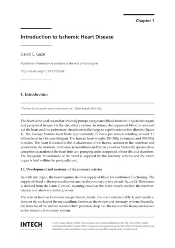
Ischemic cardiomyopathy versus non-ischemic cardiomyopathy in diabetes patientsTable 2. Echocardiographic parameters of ICM vs. NICMICMN 361 (81.7%)NICMN 81 (18.3%)TotalN 442P-value343 (95.01%)76 (93.82%)419 (94.8%)0.41718 (4.99%)5 (6.17%)23 (5.2%)LVH gradeNone to mildModerate to severeLV sizeNone to mild271 (75.06%)56 (69.13%)327 (74%)Moderate to severe90 (24.93%)25 (30.86%)115 (26%)None to mild202 (55.95%)36 (44.44%)238 (53.8%)Moderate to severe159 (44.04%)45 (55.55%)204 (46.2%)59 (25.43%)12 (37.50%)71 (26.89%)0.168LA size0.040Grade of diastolic dysfunctionImpaired relaxation (Grade I) n (%)Impaired relaxation with high filling pressure (Grade IA) n (%)4 (1.72%)0 (0.00%)4 (1.52%)Pseudo normal (Grade II) n (%)69 (29.74%)7 (21.88%)76 (28.79%)Restrictive (Grade III) n (%)100 (43.10%)13 (40.63%)113 (42.80%)0.437RVSPNone to mild260 (72.02%)56 (69.13%)316 (71.5%)Moderate to severe101 (27.97%)25 (30.86%)126 (28.5%)0.347RV functionNone to mildModerate to severe263 (72.85%)55 (67.90%)318 (71.9%)98 (27.14%)26 (32.09%)124 (28.1%)266 (73.68%)55 (67.90%)321 (72.6%)95 (26.31%)26 (32.09%)121 (27.4%)289 (80.05%)61 (75.30%)350 (79.2%)72 (19.94%)20 (24.69%)92 (20.8%)0.222Mitral regurgitationNone to mildModerate to severe0.179Tricuspid regurgitationNone to mildModerate to severe0.210LVH, left ventricle hypertrophy; LV, left ventricle; LA, left atrium; RVSP, Right ventricle systolic pressure; RV, right ventricle.The ICM group included older patients(68.23 10.97 vs. 64.09 12.22, P 0.003),more males (77% vs. 64.2%, P 0.017) andhigher prevalence of hypertension (93% vs.86.42%, P 0.05), smoking (27.86% vs. 8.75%,P 0.001), and insulin dependent Diabetes(58.12% vs. 40%, P 0.004) than the NICMgroup. Additionally, patients in this group hadthe following medical history: 63% myocardialinfarction (63%), PCI (one-third), and CABG(one-third).The NICM patients had higher BMI (30.83 7.28 vs. 28.19 5.90, P 0.001) and higherprevalence of rhythm problems (LBBB, pacedrhythm and atrial fibrillation) (63% vs. 34.87%,P 0.001) compared to ICM patients. Additionally, more patients in the NICM groupreceived ICD/CRTD therapy (34.6% vs. 22.3%,P 0.021). Other demographic and clinicalCharacteristics are described in Table 1.60Echocardiographic dataMean EF was 28.98 8.55. 26% had moderateto severe LV dilatation, 28.5% had moderate tosevere pulmonary hypertension and 28% hadmoderate to severe RV dysfunction with no significant differences between both groups. Also,we have looked at the grade of left ventriculardiastolic dysfunction (LVDD) in these twogroups and found that 42.8% of patients hadGrade III LVDD (Restrictive pattern), 29% hadGrade II LVDD (Pseudo normal pattern) and27% have grade I LVDD (impaired LV relaxation) with no significant differences betweenboth groups. Echocardiographic characteristicsare shown in Table 2.In-hospital managementIn terms of HF medications, it was found thatLasix was used in 93.3%, B-blockers in 96.4%,ACEI in 39%, ARB in 17.3% and ARNI in 12% ofAm J Cardiovasc Dis 2022;12(2):56-66
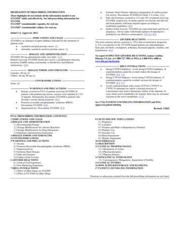
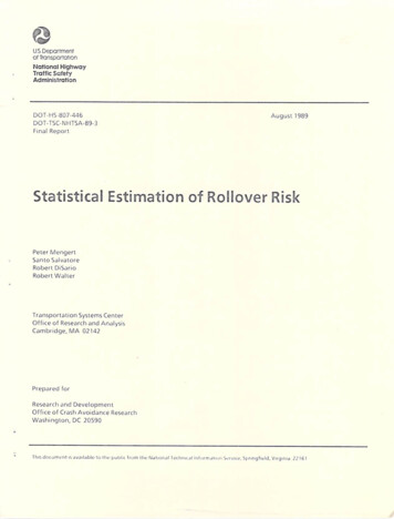
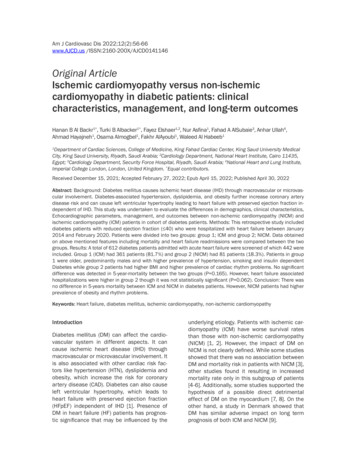
Ischemic cardiomyopathy versus non-ischemic cardiomyopathy in diabetes patientstion was provided to 88% ofthe study population abouttheir disease, side effects oftheir medications, compliance with diet and other medications. No significant differences were found betweenthe two groups (Figure 1).Figure 1. In-hospital heart failure medications, vaccination and health education in ICM vs. NICM. B blockers, beta blockers; ACEI, Angiotensin-converting-enzyme inhibitors; ARB, Angiotensin receptor blockers; ARNi, Angiotensinreceptor-neprilysin Inhibitors; MRA, mineralocorticoid receptor antagonist;ASA, acetylsalicylic acid (aspirin); ISDN, isosorbide dinitrate.A total of 254 patients(57.5%) were on oral hypoglycemic agents only while 240patients (54.5%) were oninsulin. The patients in theICM group used more insulinthan those in the NICM group(58% vs. 40%, P 0.004).One-third of the patientswere on DDP4 (26%) and onlyfew (5%) patients were onSGLT2 inhibitors (Figure 2).Long term mortality andheart failure hospitalizationFigure 2. Diabetic medications prescribed in ICM vs. NICM. OHA, oral hypoglycemic agents; DDP4, Dipeptidyl peptidase-4 inhibitor; SGLT2 inhibitors,Sodium-glucose cotransporter-2 Inhibitors.patients in the whole cohort with no differences between the two groups.The use of Aspirin (ASA), Isosorbide Dinitrateand statins was more in ICM compared toNICM (78.33% vs. 33.8%, 42% vs. 22.5% and93% vs. 74% respectively, P 0.001). The use ofanticoagulation and Spironolactone was morein NICM compared to ICM (35.4% vs. 26%,P 0.091) and (46% VS. 30%, P 0.011) respectively (Figure 1).About two thirds of the cohort patients(63.75%) received yearly Influenza and Pneumococcal vaccine with no significant difference between the two groups. Health educa61There was no significant difference in 5-year-mortalitybetween ICM and NICM(39.5% vs. 34.1%, P 0.165).However, compared to ICMgroup, there was a higherrate of HF hospitalizations inNICM group though it wasjust shy of statistical significance (49.38% vs. 36.11%,P 0.062) (Table 3).DiscussionIn the current study we aimed to analyze thedifferences in clinical characteristics, Echocardiographic parameters, management andoutcomes between ICM and NICM in diabetespatients to study the differential impact of diabetes on mortality and recurrent HF hospitalization in the absence of ischemic burden.Diabetes patients with reduced EF who wereadmitted with acute decompensated HF wereincluded and the cohort was divided into twogroups-ICM and NICM. Patients in the ICMwere older, with more males and had higherprevalence of other cardiac risk factors. TheNICM patients had higher BMI with higherprevalence of rhythm problems (LBBB, pacedrhythm and atrial fibrillation).Am J Cardiovasc Dis 2022;12(2):56-66
Ischemic cardiomyopathy versus non-ischemic cardiomyopathy in diabetes patientsTable 3. Long term outcomes mortality and Hf talization for HFNoYesUnknownDiabetic ICMPDiabetic NICMPTotalP-value128 (35.56%)123 (34.17%)109 (30.28%)33 (40.74%)32 (39.51%)16 (19.75%)161 (36.51%)155 (35.15%)125 (28.34%)0.165129 (35.83%)130 (36.11%)101 (28.06%)26 (32.10%)40 (49.38%)15 (18.52%)155 (35.15%)170 (38.55%)116 (26.30%)0.062On analysis, no difference was found in 5-yearmortality between the ICM and NICM patients.However, a trend of increased hospitalizationdue to recurrent HF was noted in the NICMgroup compared to ICM though was not statistically significant. The available data on theimpact of DM on long term outcomes of HFpatients without ischemic heart disease is conflicting [3-9]. Anderson et al [9] demonstratedthat HF patients with concomitant diabeteshad an approximately 40% increased risk ofdeath, compared to HF patients without diabetes and this increased mortality was similaramong patients with ischemic and non-ischemic HF, which corroborates with findings. Thefact that the present study found the prognosisto be similar in both groups, and the fact thatsome of the previous studies have reported nointeraction between ischemic HF etiology anddiabetes suggest that the mechanism behindthe increased mortality risk associated withdiabetes is multi-factorial and not an effect ofserious ischemic heart disease alone.A retrospective analysis of 6,797 participantsof the Studies of Left Ventricular Dysfunction(SOLVD) Prevention and Treatment trials foundthat diabetes imparted an increased risk for allcause mortality in patients with ischemic cardiomyopathy (relative risk [RR] 1.37, p 0.0001),but not in those with nonischemic cardiomyopathy (RR 0.98, p 0.98) [3]. On the contrary, a12-year prospective survival analysis [4] doneon patients hospitalized with AHF, half of whomhad a history of ischemic cardiomyopathy,found that DM increases mortality among HFpatients without ischemic cardiomyopathywhether they have preserved or reduced LVSF.Obesity is common in our population as shownin this cohort and it is one of the commoncomorbidities in female patients with diabetes62that also include hypertension, atrial fibrillationand AHF with preserved or reduced ejectionfraction. The trend of increased hospitalizationin NICM patients compared to ICM points outthe difficulty in managing HF in this group ofpatients that may require special attention totheir risk factors including obesity and dysrhythmias.Although the primary etiology of HF in NICM isnon-ischemic, patients with diabetes might stillhave some ischemic features, such as diffusevascular or microvascular disease, comparedto patients without diabetes. Additionally, diabetes induced cellular and biochemical changes like, altered myocyte metabolism, impairedglucose utilization, increased lipolysis andmyocardial fibrosis [19], may contribute to theadverse outcomes in this group.It is to be noted that HF patients in our population present at a relatively younger age, have amuch higher incidence of DM, and predominantly have LV systolic dysfunction, which ismainly ischemic in origin, compared with patients in other ethnic groups [20]. More than 30years ago, the Framingham Heart Study presented data showing an increased mortalityrate of HF in patients with diabetes, comparedto those without [21]. However, studies on thedifferential effect of DM on the outcome of HFpatients with presence or absence of ischemiashowed conflicting evidence [5-9].We have reported several findings in thiscohort. The average age of our patients was60-70 years in both ICM and NICM groups.This was 10 years more than what was reported before in our national registries SPACE andHEARTS [22, 23], where the average age of HFpatients was 57-60 years in both registries.This may imply improvement in the manageAm J Cardiovasc Dis 2022;12(2):56-66
Ischemic cardiomyopathy versus non-ischemic cardiomyopathy in diabetes patientsment of HF over the last five to seven yearswhich also reduced mortality rate of thesepatients. In SPACE HF [22] the prevalence ofHF complicating acute coronary syndrome(ACS) increased considerably with age, whichmatches our findings in this study. It was interesting to note in our findings that, compared toICM, patients who presented with HF due toNICM were more likely to be women, obese,have higher EF and have atrial fibrillation. Thisis expected because HF in the presence of LVHis the most common subtype of HF in women,with higher prevalence compared to men andassociated with several comorbidities, such asolder age, obesity, diabetes mellitus, hypertension, and hyperlipidemia, which define phenotypic profiles associated with HFpEF [24].History of AF was found in 21% of patients whopresented to the hospital with HF. This wascomparable to the findings in the atrial fibrillation HF sub study of HEARTS, where out of2593 patients admitted with HF, 449 (17.8%)had AF at presentation [25]. In our cohort wealso noted that AF is more common in NICMcompared to ICM patients.In the present study, 50% of the patients inthis cohort had impaired kidney function andthis may explain the lower use of ACEI and ARBin both groups. The use of beta-blockers, ASAand isosorbide dinitrate (ISDN) was higher inthe ICM group compared to NICM which isexpected in patients with coronary artery disease. The use of anticoagulants was muchhigher in NICM, most likely reason being a higher rate of AF compared to ICM.On the other hand, devices like ICDs and CRTswere under prescribed. This could be attributed to lack of application of knowledge to clinical practice in our population. It could also berelated to improve EF after revascularization inthe ICM group which obviates the need forthese devices, which we had earlier stated inHEARTS-chronic sub study [26]. Verifying theindication for an ICD-/CRT-D implantation isimportant as patients with DM and HF are atincreased risk of malignant ventricular arrhythmias and sudden cardiac death. This was shown in the CHARM trial that revealed a higherrate of sudden cardiac death in patients withDM compared to non-diabetics irrespective ofHF phenotype [27].63Besides the advancement in HF treatmentthere are new anti-diabetic medications thathave shown a significant outcome improvement in terms of both mortality and recurrenthospitalization needs in HF patients. Two largerandomized controlled trials investigating thecardiovascular safety of sodium-glucose cotransporter type 2 (SGLT2) inhibitors, such asempagliflozin and canagliflozin, have shown asignificant reduction in hospitalization need inHF patients with both drugs [28, 29]. Recentlythe DAPA-HF trial showed a significant reduction in deterioration or death from cardiovascular causes among patients with HF andreduced EF by dapagliflozin compared to placebo. Interestingly, this effect was independent of the presence of diabetes [30]. In ourstudy, very few patients were on SGLT2 inhibitors as part of anti-diabetic regimen that mightbe attributed to non-availability of the drug inour hospital at the time of patients’ recruitment. Additionally, the poor glycemic control, inthis cohort (HbA1C level 8.5 2.06), may beresponsible for the worse outcomes in our patients independent of the underlying HF etiology. Lawson reported that patients grouped bylevels of HbA1c showed a U-shaped relationship with outcomes, with the lowest and highest HbA1c groups associated with the highestrisk of first hospitalization and all-cause mortality [31]. Evidence on HbA1c impact on outcomes in HF has been inconsistent. This is anarea of potential future investigation in diabetes patients with HF.Despite the fact that amongst diabetics NICMhad similar mortality and HF re-hospitalizationrates to ICM patients, we think that NICM indiabetics may have different precipitating riskfactors for HF re-hospitalization like dysrhythmias, obesity and poor glycemic control asshown in our study. This should be investigatedfurther in future studies and should be thefocus of future efforts in managing thesepatients.Study limitationsThe study was limited by its relatively smallsample size. Additionally, it was limited to diabetes patients admitted with acute decompensated HF, and hence it does not represent theuniverse of CHF. However, the aim of this studywas to describe, for the first time, the clinicalAm J Cardiovasc Dis 2022;12(2):56-66
Ischemic cardiomyopathy versus non-ischemic cardiomyopathy in diabetes patientscharacteristics and outcomes of NICM compared to ICM specifically in diabetes patients.Higher numbers of patients with more geographical representation is needed to confirmthe findings of our study.[4]ConclusionThere was no difference in 5-year-mortalitybetween ischemic and non-ischemic cardiomyopathy in diabetes patients. However, diabetespatients with no
Ischemic cardiomyopathy versus non-ischemic cardiomyopathy in diabetes patients 57 Am J Cardiovasc Dis 2022;12(2):56-66 Several mechanisms have been described in pathogenesis of NICM in diabetics. Myocardial inflammation is a possible pathophysiologic process contributing to cardiac hypertrophy, fibrosis, and dysfunction in this subgroup of