Transcription
Neth Heart J (2015) 23:232–240DOI 10.1007/s12471-015-0673-xOriginal Article - Design Study ArticleRight ventricular function in dilated cardiomyopathy andischemic heart disease: assessment with non-invasive imagingS. Schalla · C. Jaarsma · S.C. Bekkers · J. Waltenberger · R. Dennert ·H.J. Crijns · J. Wildberger · S. Heymans · H-P. Brunner-La RoccaPublished online: 6 March 2015 The Author(s) 2015. This article is published with open access at Springerlink.comAbstractBackground Dilated cardiomyopathy and ischaemic heartdisease can both lead to right ventricular (RV) dysfunction.Direct comparisons of the two entities regarding RV sizeand function using state-of-the-art imaging techniques havenot yet been performed. We aimed to determine RV function and volume in dilated cardiomyopathy and ischaemicheart disease in relation to left ventricular (LV) systolic anddiastolic function and systolic pulmonary artery pressure.Methods and results A well-characterised group (cardiacmagnetic resonance imaging, echocardiography, coronaryangiography and endomyocardial biopsy) of 46 patients withdilated cardiomyopathy was compared with LV ejectionfraction (EF)-matched patients (n 23) with ischaemic heartdisease. Volumes and EF were determined with magneticresonance imaging, diastolic LV function and pulmonaryartery pressure with echocardiography.After multivariable linear regression, four factors independently influenced RVEF (R2 0.51, p 0.001): LVEF(r 0.54, p 0.001), ratio of peak early and peak atrialS. Schalla ( ) · C. Jaarsma · S.C. Bekkers · R. Dennert ·H.J. Crijns · S. Heymans · H.-P. Brunner-La RoccaDepartment of Cardiology, Maastricht University Medical Center,P. Debyelaan 25,P.O. Box 5800, 6202 AZ Maastricht, The Netherlandse-mail: s.schalla@mumc.nlJ. WaltenbergerDepartment of Cardiology, University Hospital Muenster,Muenster, GermanyJ. WildbergerDepartment of Radiology, Maastricht University Medical Center,Maastricht, The Netherlands13transmitral Doppler flow velocity as measure of LV fillingpressure (r 0.52, p 0.001) and tricuspid regurgitation flow velocity as measure of pulmonary artery pressure (r 0.38, p 0.001). RVEF was significantly worsein patients with dilated cardiomyopathy compared withischaemic heart disease: median 48 % (interquartile range(IQR) 37–55 %) versus 56 % (IQR 48–63 %), p 0.05.Conclusions In patients with dilated cardiomyopathy andischaemic heart disease, RV function is determined byLV systolic and diastolic function, the underlying causeof LV dysfunction, and pulmonary artery pressure. It wasdemonstrated that RV function is more impaired in dilatedcardiomyopathy.IntroductionRight ventricular (RV) dysfunction and dilatation are correlated to limited exercise capacity and poor outcome [1–4],but often neglected in the clinical setting [5, 6]. Dilatedcardiomyopathy (DCM) and ischaemic heart disease (IHD)can both lead to RV dysfunction. Direct comparisons ofthe two entities with respect to RV size and function usingstate-of-the-art imaging techniques have not yet been performed. Such a comparison may help to better understandthe underlying pathophysiology of RV dysfunction. Therefore, we determined RV function and volume in relationto left ventricular (LV) systolic and diastolic function, andpulmonary artery pressure in patients with DCM to assessthe main mechanisms of RV dysfunction. In addition, aftermatching for LV ejection fraction (LVEF), patients withIHD due to infarction of the left coronary artery were alsoexamined.
Neth Heart J (2015) 23:232–240MethodsStudy populationTwo groups of patients were included in the study: patientswith DCM from Maastricht University Medical Center andpatients with IHD from our infarct database matched forLVEF.Study subjects in the DCM group comprised 46 consecutive patients with DCM referred to our heart failure program.All underwent a standard diagnostic evaluation includingelectrocardiogram (ECG), coronary angiography, endomyocardial biopsy, echocardiography and cardiac magnetic resonance imaging (CMR). LV and RV volumes and systolicfunction were determined by CMR. LV diastolic functionand RV systolic pressure were assessed with echocardiography as described later in the text. Patients with a historyof myocardial infarction, history of cardiotoxic agents, significant coronary artery stenosis on coronary angiography,valvular heart disease on echocardiography as well as otherknown causes of impaired systolic function such as inflammation or infiltrative disorders on endomyocardial biopsywere excluded from the study. Patients with permanentpacemakers, rhythm other than sinus and significant chronicrenal failure ( stage 3 kidney disease) were also excluded.Study subjects in the IHD group were included from ourinfarct database matched for LVEF as measured by CMR.Thus, 23 patients with impaired systolic LV function due tochronic non-inferior myocardial infarction (infarction in leftanterior descending (LAD) or left circumflex (LCX) coronaryartery territory to avoid inclusion of patients with RV infarction) and without significant valvular disease were included.The study was approved by the local ethics committee.Magnetic resonance imagingPatients were examined with a 1.5-T scanner (GyroscanIntera,Philips Medical Systems, Best, the Netherlands).ECG-gatedcine images were acquired for functional analysis using a steady-state free precession sequence (slice thickness 6 mm, gap 4 mm, repetition time/echo time 3.8/1.9 ms,flip angle 50 , field of view 350 mm, matrix 256 256,22–25 phases per cardiac cycle). A breath-hold multisliceT1-weighted three-dimensional inversion-recovery gradient-echo sequence (acquired/reconstructed slice thickness12/6 mm, average repetition time/echo time 3.9/2.4 ms,multi-shot [50 profiles/shot] segmented partial echo readout, flip angle 15 , field of view 400 mm, matrix 256 256)to evaluate the presence of myocardial infarction of the leftand right ventricle was used to acquire images 10 min afterintravenous administration of 0.2 mmol/kg body weightgadolinium diethylenetriaminepentaacetic acid (Magnevist,233Bayer, Germany). Inversion times were adjusted to null normal myocardium (200–280 ms).CMR images were analysed by an investigator blindedto clinical and echocardiographic data. Endocardial andepicardial LV and endocardial RV contours were manuallytraced in end-diastolic and end-systolic phases on short-axisslices to determine LV and RV end-diastolic volume (EDV),end-systolic volume (ESV), ejection fraction (EF) and LVend-diastolic mass. Areas of late enhancement were visuallyassessed to confirm the presence and extent of infarction inthe LAD or LCX territory as the reason for impaired LVEFin the IHD group.EchocardiographyEchocardiography was used to determine LV diastolic function, LA volume and peak tricuspid regurgitation (TI) Doppler velocity. Transthoracic echocardiograms were performedusing dedicated systems (Sonos 5500 with S3 or IE33 withS5-1 transducers, Philips Medical Systems, Andover, MA,USA). Echocardiographic investigations were performed inthe standard views, according to the recommendations of theAmerican Society of Echocardiography [7]. LV diastolic function was assessed by measuring mitral (ratio of peak early andpeak atrial transmitral Doppler flow velocity, E/A) and pulmonary vein flow velocities (ratio of peak systolic and diastolicpulmonary vein Doppler flow velocity, S/D), tissue Dopplerflow velocities from the basal septal and lateral wall to calculate the ratio of early transmitral inflow and myocardialtissue velocity (E/e’) and left atrial (LA) volumes. Since wedid not include patients with normal LV function, we used theE/A ratio as an easily obtainable measure of LV filling pressure. Peak tricuspid regurgitation (TI) Doppler flow velocitywas used as a measure of pulmonary artery systolic pressure(PAP). Images were digitally stored and analysed off-line byan investigator blinded to CMR results and clinical data.Statistical analysisVariables are expressed as percentage, mean standarddeviation or median (interquartile range (IQR)) as appropriate. The Kolmogorov–Smirnov test was applied to testwhether continuous variables were normally distributed.Group comparisons were performed by using the Pearsonχ2 test, Student’s t-test or Mann–Whitney U test, as appropriate. For correlations, Pearson’s r was used. Finally, independent variables influencing RVEF were sought by usingthe multivariable linear regression model with a backwardprocedure (inclusion p 0.05, exclusion p 0.1).13
234Neth Heart J (2015) 23:232–240Table 1 Clinical, cardiac magnetic resonance imaging and echocardiographic characteristics of the patients with dilated cardiomyopathy (DCM)and ischaemic heart disease (IHD)Characteristicp ValueDCM (n 46)IHD (n 23)Age, years49 1459 16 0.010.18Male, n (%)27 (59 %)18 (78 %)Body surface area, m20.281.92 0.251.97 0.19Dyspnoea0.48NYHA 1/2, n (%)38 (83 %)21 (91 %)NYHA 3/4, n (%)8 (17 %)2 (9 %)Duration HF, months4 (1–18)3 (0–19)0.98Diabetes mellitus0.62Type 1, n (%)1 (2 %)0 (0 %)Type 2, n (%)4 (9 %)1 (4 %)0.71COPD, n (%)6 (13 %)2 (9 %)0.24Hypertension, n (%)9 (18 %)8 (35 %)Systolic BP0.80125 20123 19Diastolic BP0.7575 1274 10Heart rate, beats/min0.3277 1473 13PR duration, ms0.66150 57157 60QRS duration, ms0.10111 3192 430.17LBBB, n (%)16 (35 %)4 (17 %)0.11RBBB, n (%)0 (0 %)2 (9 %)Creatinine, µmol/l0.7492 2795 45Beta-blocker0.3436 (78 %)18 (78 %)ACE-inhibitor or AT-II-receptor blocker0.2040 (87 %)21 (91 %)Diuretic0.0332 (70 %)9 (39 %)Aldosterone antagonist0.2812 (26 %)4 (17 %)Calcium channel blocker0.130 (0 %)1 (4 %)RV EDV, ml/m278 (65–92)71 (63–78)0.03RV ESV, ml/m241 (35–51)32 (24–39)0.03RV SV, ml/m235 (28–43)38 (29–43)0.98RVEF, %48 (37–55)56 (48–63)0.05LV EDV, ml/m2120 (96–158)131 (101–165)0.79LV ESV, ml/m282 (64–117)85 (66–128)0.63LV SV, ml/m239 (29–46)38 (34–43)0.75LVEF, %31 (22–40)34 (18–39)0.77LV mass, g/m275 (62–84)68 (62–86)0.86RA volume, ml/m220 (16–32)22 (17–26)0.43LA volume, ml/m232 (26–53)37 (28–46)0.86E max velocity, cm/s71 (55–82)68 (53–98)0.67E/A1.00 (0.70–1.40)0.96 (0.63–1.78)0.99dt E-top ms170 (130–205)160 (130–220)0.68S/D1.04 (0.81–1.33)1.00 (0.62–1.45)0.79E/e’ IVS10.0 (8.0–13.0)6.5 (4.9–7.5) 0.001E/e’7.7 (5.4–9.7)8.4 (6.6–11.5)0.31TI peak velocity, m/s2.13 (1.86–2.44)2.42 (2.02–3.03)0.04Values represent mean standard deviation, median (interquartile range) or n, numbers of patients (%)DCM dilated cardiomyopathy, IHD ischaemic heart disease, NYHA New York Heart Association class, COPD chronic obstructive pulmonarydisease, HF heart failure, BP blood pressure, LBBB left bundle branch block, RBBB right bundle branch block, ACE angiotensin convertingenzyme, AT II angiotensin II, RV right ventricular, LV left ventricular, EDV end-diastolic volume, ESV end-systolic volume, SV stroke volume,EF ejection fraction, PASP pulmonary artery systolic pressure, RA right atrial, LA left atrial, E peak early transmitral Doppler flow velocity, E/Aratio of peak early and peak atrial transmitral Doppler flow velocity, dt E-top deceleration time of peak early transmitral Doppler flow signal,S/D ratio of peak systolic and diastolic pulmonary vein Doppler flow velocity, E/e’ IVS ratio of peak early transmitral Doppler flow velocityand peak early diastolic tissue Doppler flow velocity from the basal septal left ventricular wall, E/e’ ratio of peak early transmitral Dopplerflow velocity and peak early diastolic tissue Doppler flow velocity from the basal lateral left ventricular wall, TI peak tricuspid regurgitationDoppler flow velocity13
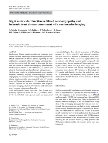
Neth Heart J (2015) 23:232–240ResultsClinical characteristics of patients with DCM and IHDThe clinical characteristics of the DCM and IHD patientsare summarised in Table 1. Patients in the IHD group wereolder and less often treated with diuretics. All other parameters were not significantly different between the groups.CMR image analysis was possible in all patients. In threepatients, however, image quality on echocardiography wasnot sufficient to obtain reliable pulmonary vein flow Doppler signals. In two of them, image quality was also not sufficient to obtain tissue Doppler signals, but the E/A ratiocould be determined in all patients.A total of 21 patients with IHD had infarcts in theLAD territory and 2 patients in the LAD and LCX territory (Fig. 1). Infarction of the RV free wall was recognisedin only one patient of the IHD group: a small area of lateenhancement of the RV apex was visible on CMR images.This patient had a transmural infarction in the LAD territoryshowing late enhancement from the anterior wall to the apexof the LV continuing to the RV apex (Fig. 2). None of the235patients with DCM had focal fibrosis of the RV free wallmyocardium on CMR late enhancement images.RV volume and functionDifferences between patients with DCM and IHDThe RVEF was more impaired and RV size larger inpatients with DCM in comparison with patients with IHD(Figs. 3 and 4) despite similar LV systolic and diastolicdysfunction. There was an isolated increase in E/e’ of theseptum in DCM patients, but an opposite trend laterallyand no further differences were seen in diastolic parametersbetween the groups, suggesting no difference in diastolicLV function. LV EDV and ESV were not different betweenthe groups. All CMR and echocardiography parameters aresummarised in Table 1.Parameters influencing RV functionAs depicted in Table 2, the RVEF was significantly correlated with LVEF and the volumes of all four cardiacchambers. Furthermore, parameters of LV diastolic func-Fig. 1 Late gadolinium enhancement (a, b) and cine (four-chamber view in end-diastole (c) andend-systole (d)) magnetic resonance images of a patient with alarge myocardial infarction (leftventricular ejection fraction 15 %)of the left anterior descendingcoronary artery (right ventricularejection fraction 56 %)13
236Neth Heart J (2015) 23:232–240Fig. 2 Late gadolinium enhancement (four-chamber view (a), 2chamber view (b)) and cine (fourchamber view in end-diastole (c)and end-systole (d)) magneticresonance images of a patientwith a large myocardial infarction(left ventricular ejection fraction22 %) of the left anterior descending coronary artery, continuingfrom the left ventricular apex tothe right ventricle (right ventricular ejection fraction 57 %) (whitearrow) A corresponding akineticregion of the right ventricularapex can be depicted on theend-systolic cine image (D, blackarrow)tion negatively correlated with RV function. Also, the TIvelocity as measure of PAP was negatively correlated withRVEF. Finally, age and heart rate showed significant correlations with RVEF. RVEF was higher in women comparedwith men (55 10 versus 44 13 %, p 0.001). Other clinical parameters had no influence on RVEF (data not shown).Overall, there were no significant differences in these correlations between patients with DCM and IHD (data notshown).Multivariable analysis revealed that the RV function (i.e.RVEF) was correlated with LVEF, DCM compared withIHD, E/A ratio as measure of LV filling pressure, and TIvelocity as measure of PAP as depicted in Table 3. Althoughthe relationship with TI velocity was of borderline significance, it was in addition to a measure of LV filling pressure,e.g. a possible active process in addition to passive elevationof PAP due to elevated LV filling pressures.DiscussionIn the current study, the non-invasive imaging techniquesCMR and echocardiography were applied in patients withDCM and IHD without RV free wall infarction, to evaluate13RV function and volumes. RV systolic function was influenced by different factors including the underlying diseaseprocess, i.e. the presence of DCM, systolic and diastolicfunction of the LV and elevation of the PAP.In ST-segment elevation myocardial infarction (STEMI)patients with cardiogenic shock, RV dysfunction had beenidentified as a prognostic parameter [8]. Between patientswith and without RV dysfunction, no significant differencesin infarct-related artery and infarct location were observed.RV dysfunction in patients in whom the right coronary artery(RCA) was not the infarct-related artery was also present.This suggests that not only direct RV infarction causes RVdysfunction but also mechanisms such as reduced bloodsupply of the septum, low RV preload due to low outputand diminished contribution of LV contraction to RV systole[8]. RV function may depend on LV septal contractile contribution transmitted through systolic ventricular interaction[9–11] and the septum itself may contribute to the systolicfunction of both ventricles [12]. RV function was experimentally as much impaired with septal ligation as withRCA ligation [13]. Our results, however, suggest that otherfactors such as elevated LV filling pressure and PAP are ofadditional importance to explain reduced RV function.
Neth Heart J (2015) 23:232–240237Fig. 3 Late gadolinium enhancement (a, b) and cine (fourchamber view in end-diastole (c)and end-systole (d)) magneticresonance images of a patientwith idiopathic dilated cardiomyopathy (left ventricular ejectionfraction 24 %, right ventricularejection fraction 45 %) The leftventricular systolic function ofthis patient is slightly better thanthat of the patients with ischaemicheart disease from Figs. 1 and 2while the right ventricular function is more impaired. Areas oflate enhancement are not presentRV infarction detected by late enhancement CMR andRV systolic function were outcome predictors in a generalpopulation of STEMI patients [14]. In inferior STEMI, RVsystolic function was related to the presence and extent ofRV infarction, while in anterior STEMI it was related to LVsystolic dysfunction. When imaging more early after infarction, RV infarction was not related to prognosis [15]. Thisdiscrepancy could be explained by the fact that RV functionmay recover to a greater extent than LV function, depending on recovery of septal function [9]. Thus, global RV performance recovered within days after infarction, regardlessof artery patency [16]. Moreover, global RV performanceimproved greatly even in chronic RCA occlusion, despitepersistent severe RV free wall dysfunction [17]. To excludethe influence of direct RV free wall infarct on RV function,we included only patients with infarcts of the left coronaryartery territory (LAD and LCX infarct). To our knowledge,this has not been applied in previous comparative studies ofpatients with DCM and IHD. We observed a small extension of infarct from the left to the RV myocardium in onepatient only, which is in some contrast to a recent CMRstudy in 20 patients with reperfused proximal LAD occlusion where a small RV infarct was observed in 40 % of thepatients [18]. They found a relatively large area of the RV atrisk for necrosis, although the resulting final RV infarctionsize was small. A possible explanation for the discrepancybetween this and our findings is that only a small portion ofour patients had transmural infarcts of the whole septum.The underlying process also determines RV function.Patients with DCM experienced larger RV volumes and moresevere impairment of RV function. In contrast to previoussuggestions [19, 20], assessing systolic RV function alone isnot sufficient to distinguish between DCM and IHD in individual patients. Few studies have directly compared the RVfunction in patients with DCM and IHD. Our findings aresupported by some earlier studies where patients with DCMshowed more severe RV dysfunction than patients withIHD using radionuclide angiography, thermodilution andinvasive RV angiography [19–21]. However, these findingshave not been uniform. By applying tissue Doppler imaging, the RV dysfunction was more pronounced in patientswith IHD than DCM in an echocardiographic study [22].Since only two patients had inferior wall infarction basedon ECG criteria, the authors concluded that this worse RVfunction had not been due to more infarcted RV myocardium. However, patients were not sufficiently matched withrespect to other factors potentially influencing RV function.Thus, patients with IHD exhibited more severe LV diastolic13
238Neth Heart J (2015) 23:232–240Fig. 4 Late gadolinium enhancement (a, b) and cine (fourchamber view in end-diastole (c)and end-systole (d)) magneticresonance images of a patientwith idiopathic dilated cardiomyopathy with more pronounceddilation of the right ventricle (leftventricular ejection fraction 36 %,end-diastolic volume 211 ml;right ventricular ejection fraction 23 %, end-diastolic volume269 ml) Although elevated leftventricular filling pressure and TIvelocity were present, the degreeof right ventricular dilation is outof proportion, possibly suggestingan active unknown process affecting the right ventricle more thanthe left ventricleTable 2 Correlations of clinical, cardiac magnetic resonance imaging(CMR) and echocardiographic characteristics with right ventricularejection fraction as measured with CMRVariableCorrelationp ValueAge0.290.02Systolic BP0.130.28HR 0.46 0.001LVEF0.54 0.001LV EDV0.02 0.29RV EDV 0.50 0.001LA volume 0.41 0.001RA volume 0.37 0.001E/A ratio 0.52 0.001S/D ratio0.48 0.001TI velocity0.001 0.38BP blood pressure, HR heart rate, LV left ventricular, RV rightventricular, EF ejection fraction, EDV end-diastolic volume, LAleft atrial, RA right atrial, E/A ratio of peak early and peak atrialtransmitral Doppler flow velocity, S/D ratio of peak systolic anddiastolic pulmonary vein Doppler flow velocity, TI peak tricuspidregurgitation Doppler flow velocitydysfunction and higher PAP. Such a difference in PAP anddiastolic function was not present in our patient groups, butPAP and diastolic function were independently related to13Table 3 Regression analysis testing the association between right ventricular ejection fraction and various potential predictors in the entirestudy population (dilated cardiomyopathy and ischaemic heart disease)VariableRegression coefficient p ValueLVEF (per %)0.50 0.0001DCM versus IHD9.410.0005E/A ratio (per unit)0.02 3.60TI velocity (per m/s)0.06 4.84(R 2 0.51)DCM dilated cardiomyopathy, IHD ischaemic heart disease,LVEF left ventricular ejection fraction, E/A ratio of peak early andpeak atrial transmitral Doppler flow velocity, TI peak tricuspidregurgitation Doppler flow velocityRV dysfunction. Moreover, assessment of RV dimensionsand systolic function by echocardiography has importantlimitations. In the present study, we therefore used the current reference standard CMR to assess RV volumes and EF[6, 23, 24].Elevated LV filling pressure, which leads to passive elevation of PAP, was one of the factors related to RV dysfunction in the current study. Since the prognosis of patients withimpaired RV function is worse in comparison with patientswith impaired systolic LV function only [25, 26], assessment of RV function and possibly more aggressive treatment
Neth Heart J (2015) 23:232–240of elevated LV filling pressure as suggested by Stevensonet al. [27] might be important, irrespective of the underlying cause. An additional active component of elevated pulmonary artery pressure, i.e. out-of-proportion pulmonaryhypertension leading to fixed pulmonary hypertension, mayalso have contributed to worsening of RV function sinceelevated PAP was related to RV dysfunction independentlyof LV filling pressure.The current study has some limitations. Patients in theIHD group were older and less often treated with diuretics.Older age had a negative influence on RV function. However, although the patients in the IHD group were older, theyshowed a less impaired RV function than the patients fromthe DCM group. Thus, not matching for age did not resultin a relevant bias. Treatment with diuretics can lower LVfilling pressure and PAP and might have had an influence onthe lower TI peak velocity measured in patients with DCM[28]. In contrast to IHD, DCM usually comprises a heterogeneous group of diseases in many studies. However, witha comprehensive diagnostic routine including endomyocardial biopsy, imaging and blood tests, certain diseases suchas acute myocarditis, infiltrative and storage diseases wereexcluded from our study to make the DCM study population as uniform as possible. Regional RV wall motion asmeasured by, e.g. speckle tracking was not assessed. Earlystages of dyssynchronicity between the right and left ventricle were therefore not detected. Other potential mechanismswere also not addressed, e.g. interventricular interaction andeffects of changes of geometry. Finally, this was a diagnostic study. However, the study revealed different potentialmechanisms of RV dysfunction that may be therapeuticallyinfluenced. It is important to evaluate RV function, especially in DCM, and it may be necessary to treat elevatedfilling pressure and PAP more aggressively or with newtherapeutic strategies such as phosphodiesterase inhibitorsto protect the right ventricle.In conclusion, RV systolic function was influenced bydifferent factors including the underlying disease process,i.e. the presence of DCM, systolic and diastolic functionof the LV and elevation of PAP. A better understanding ofthese mechanisms may help to define therapeutic targets forfuture studies in these patients with RV dysfunction knownto have a poor outcome.Funding None.Compliance with ethical guidelinesConflict of interest None declared.This study was approved by the Medical Ethics Committee of theMaastricht University Medical Center, Maastricht, the Netherlands.239Open Access This article is distributed under the terms of the CreativeCommons Attribution License which permits any use, distribution, andreproduction in any medium, provided the original author(s) and thesource are credited.References1. Baker BJ, Wilen MM, Boyd CM, et al. Relation of right ventricular ejection fraction to exercise capacity in chronic left ventricularfailure. Am J Cardiol. 1984;54:596–9.2. Di Salvo TG, Mathier M, Semigran MJ, et al. Preserved right ventricular ejection fraction predicts exercise capacity and survival inadvanced heart failure. J Am Coll Cardiol. 1995;25:1143–53.3. Gavazzi A, Berzuini C, Campana C, et al. Value of right ventricular ejection fraction in predicting short-term prognosis of patients with severe chronic heart failure. J Heart Lung Transplant.1997;16:774–85.4. de Groote P, Millaire A, Foucher-Hossein C, et al. Right ventricularejection fraction is an independent predictor of survival in patientswith moderate heart failure. J Am Coll Cardiol. 1998;32:948–54.5. Jefferies JL, Towbin JA. Dilated cardiomyopathy. Lancet2010;375:752–62.6. Mertens LL, Friedberg MK. Imaging the right ventricle-currentstate of the art. Nat Rev Cardiol. 2010;7:551–63.7. Gottdiener JS, Bednarz J, Devereux R, et al. American Society ofEchocardiography recommendations for use of echocardiographyin clinical trials. J Am Soc Echocardiogr. 2004;17:1086–119.8. Engstrom AE, Vis MM, Bouma BJ, et al. Right ventricular dysfunction is an independent predictor for mortality in ST-elevationmyocardial infarction patients presenting with cardiogenic shockon admission. Eur J Heart Fail. 2010;12:276–82.9. Popescu BA, Antonini-Canterin F, Temporelli PL, et al. Rightventricular functional recovery after acute myocardial infarction:relation with left ventricular function and interventricular septummotion. GISSI-3 echo substudy. Heart. 2005;91:484–8.10. Goldstein JA, Tweddell JS, Barzilai B, et al. Importance of leftventricular function and systolic ventricular interaction to rightventricular performance during acute right heart ischemia. J AmColl Cardiol. 1992;19:704–11.11. Sharkey SW, Shelley W, Carlyle PF, et al. M-mode and two-dimensional echocardiographic analysis of the septum in experimental right ventricular infarction: correlation with hemodynamicalterations. Am Heart J. 1985;110:1210–8.12. Banka VS, Agarwal JB, Bodenheimer MM, et al. Interventricularseptal motion: biventricular angiographic assessment of its relativecontribution to left and right ventricular contraction. Circulation.1981;64:992–6.13. Fixler DE, Monroe GA, Wheeler JM. Hemodynamic alterationsduring septal or right ventricular ischemia in dogs. Am Heart J.1977;93:210–5.14. Miszalski-Jamka T, Klimeczek P, Tomala M, et al. Extent of RVdysfunction and myocardial infarction assessed by CMR are independent outcome predictors early after STEMI treated with primary angioplasty. JACC Cardiovasc Imaging. 2010;3:1237–46.15. Hombach V, Grebe O, Merkle N, et al. Sequelae of acute myocardial infarction regarding cardiac structure and function and theirprognostic significance as assessed by magnetic resonance imaging. Eur Heart J. 2005;26:549–57.16. Steele P, Kirch D, Ellis J, et al. Prompt return to normal of depressed right ventricular ejection fraction in acute inferior infarction. Br Heart J. 1977;39:1319–23.17. Laster SB, Shelton TJ, Barzilai B, et al. Determinants of the recovery of right ventricular performance following experimental chronic right coronary artery occlusion. Circulation. 1993;88:696–708.13
24018. Bodi V, Sanchis J, Mainar L, et al. Right ventricular involvementin anterior myocardial infarction: a translational approach. Cardiovasc Res. 2010;87:601–8.19. Iskandrian AS, Helfeld H, Lemlek J, et al. Differentiation between primary dilated cardiomyopathy and ischemic cardiomyopathy based on right ventricular performance. Am Heart J.1992;123:768–73.20. La Vecchia L, Zanolla L, Varotto L, et al. Reduced right ventricularejection fraction as a marker for idiopathic dilated cardiomyopathycompared with ischemic left ventricular dysfunction. Am Heart J.2001;142:181–9.21. Juilliere Y, Buffet P, Marie PY, et al. Comparison of right ventricular systolic function in idiopathic dilated cardiomyopathy andhealed anterior wall myocardial infarction associated with atherosclerotic coronary artery disease. Am J Cardiol. 1994;73:588–90.22. Parcharidou DG, Giannakoulas G, Efthimiadis GK, et al. Rightven
234 Neth Heart J (2015) 23:232-240 Table 1 Clinical, cardiac magnetic resonance imaging and echocardiographic characteristics of the patients with dilated cardiomyopathy (DCM) and ischaemic heart disease (IHD) Characteristic DCM (n 46) IHD ( 23) p ValueAge, years 49 14 59 16 0.01 Male, n (%) 27 (59 %)18 (78 0.18 Body surface area, m2 1.92 0.25 1.97 0.19 0.28