Transcription
J. of Animal and Poultry Production, Mansoura Univ., Vol 12 (9):305 - 314, 2021Journal of Animal and Poultry ProductionJournal homepage: www.japp.mans.edu.egAvailable online at: www.jappmu.journals.ekb.egCharacteristics, Yield, Flow Cytometer, DNA Fragmentation, and CometAssay Parameters of Goat Spermatozoa in Semen of Zaraibi and BaladiBucks at Young and Old AgesZaghloul, H. K. 1*; M. M. El-Maghraby2; A. A. El badawy2 and A. I. A. Yousif2Cross Mark1HighInstitute For Agricultural Co-Operation, Shoubra, Egypt.2Animal Production Research Institute, Agricultural Research Center, Egypt.ABSTRACTThe aim of this study was to determine goat breed (Zaraibi and Baladi) or buck age (young andold) effect on characteristics, production, flow cytometer, DNA fragmentation and comet assay parametersof spermatozoa. Twelve Baladi (BG) and Zaraibi (ZG) bucks, 3 old (2-4 years) and 3 young (8-12 months)from BG or ZG used for semen collection for 24 weeks. Volume (EV), sperm progressive motility (PM),livability (LS), abnormality (SA), and concentration (SCC) were evaluated, sperm outputs (SO) werecalculated. Sperm apoptosis, DNA fragmentation and comet assay were analyzed. Results revealed thatPM, LS, SA, SCC, total, motile, live, normal, and functional SO per ejaculate were significantly higher inZG than BG and in old than young bucks in each breed. Both EV and LS were significantly higher, whileSCC was significantly lower in old than young ZG. Each of EV, PM, and SCC was significantly higher,while SA was significantly lower in old than young BG. Viable sperm, haploid and cell cycle percent wassignificantly higher, while early apoptotic, apoptotic, and necrotic spermatozoa percent was significantlylower in old than young bucks of each breed. Spermatid percent in ZG and diploid percent in BG weresignificantly higher in old than young bucks. Tailed sperm, tail length, tail DNA percentage, and tailmoment were significantly lower in ZG than BG, and in old than young bucks of each breed. It can bestated that Zaraibi bucks has a good potential for semen production than Baladi breed.Keywords: Semen, goats, breed differences, sperm function, apoptosis, DNA damage.INTRODUCTIONFor centuries, goat was not only an economic animalfor people living in arid regions, but was also valued as agood source of meat and milk. In developing countries, goatprovides is an important source of milk and meat. Goatreproductive, genetic and environmental factors are factorsaffecting the productive efficiency (Hossain et al., 2004;Song et al., 2006). Reproductive performance male goatbefore mating is important to achieve breeding success(Ford et al., 2009). Libido and semen characteristics arelimited factors for male reproduction, being different inrelation to breed (Karagiannidis et al., 2000) and age (Mahalet al. 2013).In goats, proper selection of breeding buck for thenatural mating or artificial insemination (AI) is needing forgoat breeding, especially for AI, which require in to increaseand disseminate genotypes of quality breeding stock andimprove a herd (Ngoma et al. 2016). The breeding buckexamination is important to produce good fertility of a flock.This process includes semen evaluation as managerialpractice in AI breeding program of goats (Gore et al., 2020).Genetic, hormonal, and spermatogenesisdisorders, poor semen quality, and damage of DNA tospermatozoa are attributed to decreased male fertility(Esteves et al., 2012). The sperm DNA fragmentationroutes and cell death were reported to be not fully caspasedependent (Cande et al., 2004). Variety of assays have*Corresponding author.E-mail address: helmyzaghlool@yahoo.comDOI: 10.21608/jappmu.2021.95942.1021been developed to detect DNA fragmentation levelsincluding terminal deoxynucleotidyl transferase dUTPnick-end labeling (TUNEL) assay (Mitchell et al., 2011),comet assay (Lewis et al., 2008), DNA breakagedetection fluorescence in situ hybridization (DBD-FISH)(Fernández et al., 2000), potential of oxidative reduction(Agarwal et al., 2017) assays DNA damage andchromatin structure of spermatozoa (Fernández et al.,2005).Both Egyptian Baladi (BG) and Zaraibi (ZG)goats are mostly found in Nile valley and delta (Galal etal., 2005). It is well known that ZG is related to theEgyptian Nubian breed, in the same time it considers asthe most important native goat breed evaluated on thebasis of milk and meat production (Marai et al., 2002;Galal et al., 2005). In Egypt, the Zaraibi goat, as a nativebreed, has a high genetic potential for production of milk(Dwidar et al., 2018). In developing countries, programsof genetic selection in dairy goats are still uncommon, butthey have been great successful for improvement ofbreeding strategies for production of milk and meat fromgoat (Shrestha and Fahmy, 2007; Vakka et al., 2018).The current study aimed to evaluate the effect ofgoat breed (Zaraibi and Baladi) or age of buck (youngand old) on characteristics, production, flow cytometer,DNA fragmentation, and comet assay parameters ofspermatozoa.
Zaghloul, H. K. et al.MATERIALS AND METHODSSite and location of the study:The experimental work was conducted at AnimalProduction Research Station, Sakha, KafrelsheikhGovernorate, located in the northern part of Nile Delta(latitude 31o 15’N and longitude 31o 45’E), belonging toAnimal Production Research Institute (APRI), AgriculturalResearch Center, Ministry of Agriculture, Egypt, during theperiod from January, 2021 until June, 2021.Animals and feeding system:Twelve bucks of Egyptian local goat breed Baladi(BG) and Zaraibi (ZG) goat bucks were used in this study.Baladi goats (n 6) included 3 old bucks (2-4 years of ageand 50.66 1.36 kg LBW) and 3 young bucks (8-12 monthsof age and 23.66 1.36 kg LBW), while ZG included 3 oldbucks (2-4 years of age and 53.33 1.36 kg LBW) and 3young bucks (8-12 months of age and 26.33 1.36 kg LBW).All bucks were used for semen collection by artificialvagina.Requirements of feeding were determined after tothe recommendations of APRI, Ministry of Agriculture,Egypt. Each buck was fed Concentrated Feed Mix,including crude protein (14%), total digestible nutrients (%),plus four kg of fresh Egyptian alfalfa (Trifoliumalexandrinum) during the period from December toFebruary or one kg of alfalfa hay through March-June, withfree access to mineral salt blocks and drinking water all thetime.Semen collection and evaluationFor semen collection period of 24 weeks, semen wascollected once/week before feeding (7-8 a.m.) by aconventional artificial vagina. Semen ejaculates weretransferred immediately after collection to the laboratory,and then placed in water bath at 37oC.Ejaculate volume was determined and semen wasassessed for percentage of progressive sperm motility ineach semen sample (10 µL diluted semen) using a phasecontrast microscope supplied with a hot stage adjusted to 37 C. Smears were prepared using nigrosin/eosin staining fordetermining the percentages of live and abnormalspermatozoa. The concentration of spermatozoa wasdetermined with a Neubauer /ejaculate), in terms of total (TSO), motile (MSO), live(LSO), normal (NSO) and functional (FSO) as thefollowing:TSO ejaculate volume (ml) x sperm concentration (x109/ml)MSO progressive sperm motility (%) x TSOLSO live sperm (%) x TSONSO (sperm abnormality (%) – 100) x TSOFSO TSO x sperm motility (%) x sperm livability (%) xnormal sperm (%)Sperm apoptosis:Phosphatidylserine (PS), a protein ordinarily foundin the inner leaflet of the plasma membrane of healthy cells,travels to the outer layer of the plasma membrane during theearly stages of apoptosis, where it is exposed to the cell'souter surface. Anexin-V binds to PS with a high affinity, andannexin staining with fluorochrome V is utilised to detectapoptosis (Plesca et al., 2008).For acquisition and analysis, flow cytometry wasperformed on an Accuri C6 cytometer (BD Biosciences, SanJose, CA) with Accuri C6 software (Becton Dickinson)(Masters and Harrison, 2014). The sperm subpopulationswere classified as percentage of viable sperm (A-/PI-), earlysperm (A /PI-), apoptotic sperm (A /PI ), and apoptoticsperm (A-/PI-) based on flow cytometry results.Spermatozoa that have died (A-/PI ) (Chaveiro et al.,2007).Cellular morphology and DNA fragmentation (CellCycle method):The capacity of flow cytometry to determine cellularDNA concentration is reliant on the fluorometry of dyes thatbind to DNA in a stoichiometric manner. Ploidy DNA is ameasure of the DNA content of the cells under inquiry as apercentage of the normal (diploid) control. Due to the factthat DNA content repeats before cell division, mathematicalmodels have been developed to predict the percentage ofcells in various phases of the cell cycle (G0/1, S-phase,G2/M), diploid and aneuploid cycles, CV, and death ratio.Apoptotic cells, DI, diploid percentage, and anueploidpercentage are all terms used to describe cells that havereached the end of their life cycle (Spyratos, 1993).The sub-G1 approach for diagnosing cell death isbased on the idea that during extended fixation, fragmentedlow molecular weight DNA is liberated from cells afterDNA internal cleavage. This would result in a population ofcells that bind the quantitative DNA pigment, PI, to a lesserlevel than G1 cells; G1 is the longest phase of the cell cycle,and as a result, the majority of cells are in G1. As a result, acluster of cells to the left of the G1 apex will form.One cc of ice-cold 100% alcohol per tube was usedto fix lymphocytes or tissue cells, which were then stored at 4 C until analysis. The material was centrifuged again atleast 12 hours after fixing to eliminate the excess ethanol(Vindelov, 1977). In a 15-ml (2095) Falcon Compoundtube, the cell suspension (200 l) was put in citrate solution.During preparation, storage, and staining, the propidiumiodide solution was shielded from light with foil. In another5 ml tube, the solution was combined and the sample wasfiltered through a 30 mm pore diameter nylon mesh filter toremove nuclear masses (12 x 75 mm, cat. no. 2058, FalconCompounds). Within 1 hour of adding propidium iodide,samples were run using a flow cytometer.MODFIT DNA analysis programme (veritysoftware house, Inc. Po Box 247, Topsham, ME 04086USA, version: 2.0 power Mac with 131072 KB Registrationnumber: 42000960827-16193213) was used to analyse thedata. Manufacturing date: September 16, 1996). Thissoftware calculated the coefficient of variation (CV) aroundthe G0/G1 peak, the percentage of apoptosis, the DNAindex (DI), and the percentage of cells in each Phase(G0/G1, S, and G2/M) of the DNA cell cycle for eachsample, as well as DNA ploidy (diploid cycle and aneuploidcycle percent).The comet assay analysis:Comet assay based on the method of Badawy et al.(2018) was conducted. A fluorescent microscope with a 40xmagnification and a 420-490 nm excitation filter was usedto examine the migration patterns of DNA fragments from100 cells/sample. Kinetic Imaging Ltd.'s Komet 5 imageanalysis software was used. (Liverpoo1, UK) used a CCDcamera to measure the length of DNA migration and theproportion of migrating DNA to determine the quantitative306
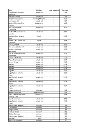
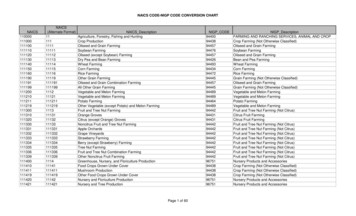
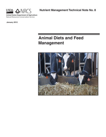
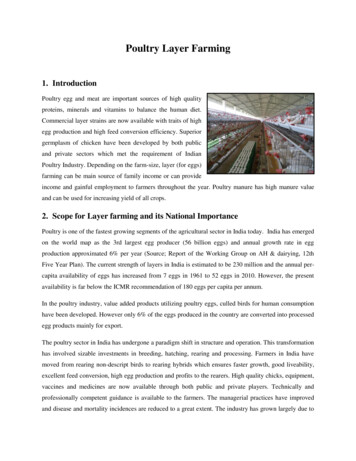
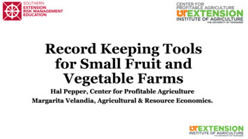
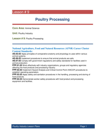
J. of Animal and Poultry Production, Mansoura Univ., Vol 12 (9), September, 2021and qualitative amount of DNA damage in cells. The tails ofthe comets were measured from the middle of the nucleus tothe tail's termination.Fertility trial:Totally, 126 healthy mature goats (71 Zaraibi and 55Baladi) were naturally mated during September breedingseason (from 1st to end of September). Conception rate(Number of lambed goats/number of inseminated goats)x100 and Litter size (LS)/ goat (Number of bornlambs/number of lambed goats) according to Zonturlu et al.(2011) were considered as an index of fertility andreproductive performance.Statistical analysis:Data were statistically analyzed by T-test to comparebetween the two breeds, two ages, and two ages within eachbreed using computer program SAS (2004).RESULTS AND DISCUSSIONResultsPhysical semen characteristics:Physical semen characteristics of BG and ZG ofyoung and old bucks are presented in Table 1. Resultsrevealed that semen parameters including the percentage ofprogressive motility (PM), livability (LS), abnormality(SA), and sperm cell concentration (SCC) were better(P 0.0001) in ZG than in BG, but ejaculate volume (EV)was not affected (P 0.05) by goat breed. As affected by age,EV (P 0.0001) and LS (P 0.05) were higher, while SCCwas lower (P 0.05) in old than in young of ZG, but PM andSA were not affected by age. However, EV (P 0.001), PM,and SCC (P 0.01) were higher, while SA was lower(P 0.05) in old than in young of BG, but LS was not affectedby age.Table 1. Effect of breed (Zaraibi and Baladi) and age (Young and old) on physical semen characteristics of goat bucks.SpermvariableEV (ml)PM (%)LS (%)SA (%)SCC (x109/ml)BreedZaraibiBaladi0.82 0.050.70 0.0580.33 0.75 67.83 0.7479.23 0.85 69.83 1.1611.16 0.63 20.36 0.822.48 0.681.85 0.80ZaraibiPValueOldYoung0.111.03 0.05 0.62 0.040.0001 81.67 1.05 79.00 1.000.0001 80.93 1.03 77.53 1.240.0001 10.80 0.86 11.53 0.930.0001 2.61 0.09 2.34 0.080Sperm outputs per ejaculate:Results in Table 2 show that sperm outputs interms of total, motile, live, normal, and functional ung0.91 0.050.50 0.0470.00 0.84 65.67 0.9571.06 1.52 68.60 1.7618.53 1.21 22.20 0.922.07 0.111.63 0.08PValue0.00010.00210.29970.02280.0047ejaculate was higher (P 0.001) in ZG than in BG, and in oldthan in young bucks in each breed.Table 2. Effect of breed (Zaraibi and Baladi) and age (Young and old) on sperm outputs in ejaculate of goat eedZaraibiBaladi1.98 0.981.35 0.131.59 0.850.93 0.101.57 0.860.96 0.101.76 0.911.09 0.111.13 0.720.54 Young2.40 0.081.61 0.121.92 0.081.27 0.091.90 0.081.25 0.092.09 0.081.43 0.111.40 0.080.87 0.06Sperm flow cytometry parameters:Sperm flow cytometry parameters of ZG and BG inyoung and old bucks are shown in Table 3 and illustrated inFig. 1. Results revealed insignificant differences in thepercentage of viable, early apoptotic, apoptotic, and OldYoung1.93 0.170.78 0.061.36 0.120.51 0.041.38 0.130.54 0.051.58 0.140.61 0.050.80 0.090.28 a between the two goat breeds. However,percentage of viable spermatozoa was higher (P 0.0001),while the percentage of early apoptotic, apoptotic, andnecrotic spermatozoa was significantly lower in old than inyoung bucks of each breed.Table 3. Effect of breed (Zaraibi and Baladi) and age (Young and old) on sperm flow cytometer percentages of goat bucks.ItemViableEarly apoptosisApoptosisNecrosisBreedZaraibiBaladi81.95 1.44 79.70 1.474.15 0.38 4.26 0.406.29 0.37 6.20 0.457.59 0.97 9.83 0.84PValue0.280.840.870.09ZaraibiOldYoung87.74 0.39 76.16 0.532.85 0.235.45 0.395.50 0.367.08 0.543.90 0.4511.28 0.65Sperm DNA fragmentation:Sperm DNA parameters of ZG and BG in young andold bucks are shown in Table 4 and illustrated in Fig. 2.Results showed insignificant differences in the percentage ofsub G1, haploid, spermatid, diploid, and cell cycle betweenthe two goat breeds. However, percentage of sub G1 waslower (P 0.0001), while the percentage of haploid and cellcycle was higher in old than in young bucks of each breed.Only, spermatid percentage in ZG, and diploidpercentage in BG were significantly higher in old than inyoung 85.48 0.55 73.91 0.773.10 0.315.43 0.494.70 0.337.70 0.466.71 0.4112.95 0.64PValue0.00010.00110.00010.0001Comet assay parameters:Comet assay parameters of sperm cells of ZG and BGin young and old bucks are shown in Table 5 and illustratedin Fig. 3. Results showed that percentage of untailed spermcells was higher (P 0.001), while percentage of tailedspermatozoa, tail length, tail DNA percentage, and tailmoment were lower (P 0.05) in ZG than in BG, and lower(P 0.001) in old than in young bucks of each goat breed.307
Zaghloul, H. K. et al.Zaraibi oldBaladi oldZaraibi youngBaladi youngFig. 1. Flow cytometry contour plots in old and young Zaraibi and Baladi bucks spermatozoa labelled with annexin Vfluorescein isothiocyanate (FITC) fluorescence vs. propidium iodide (PI) fluorescence. The lower left quadrant(LL) contains viable spermatozoa which are negative for Annexin-V and exclude PI staining (A-/PI-). The lowerright quadrant (LR) shows early apoptotic spermatozoa which bind Annexin-V but exclude PI (A /PI-). The upperright quadrant (UR) represents apoptotic spermatozoa binding Annexin-V and PI (A /PI ). The upper leftquadrant (UL) represents necrotic cells excluding Annexin-V and binding PI (A-/PI ).Table 4. Effect of breed (Zaraibi and Baladi) and age (Young and old) on sperm DNA fragmentation of goat bucks.Parameter(%)Sub G1HaploidSpermatidDiploidCell cycleBreedZaraibiPValueZaraibiBaladiOldYoung12.15 1.58 16.12 2.44 0.186.48 0.93 17.82 1.3079.10 1.03 77.45 1.61 0.39 81.74 1.01 76.45 1.344.88 0.804.17 0.76 0.527.15 0.932.61 0.783.86 0.782.25 0.58 0.104.61 0.663.11 1.4287.84 1.58 83.87 2.44 0.18 93.51 0.93 82.17 oung6.53 0.91 25.72 1.2483.66 0.52 71.24 1.065.41 0.942.93 1.094.40 0.540.10 0.0293.47 0.90 74.27 1.24Zaraibi oldBaladi oldZaraibi youngBaladi youngPValue0.00010.00010.10660.00010.0001Fig. 2. Flow cytometry contour plots in old and young Zaraibi and Baladi bucks spermatozoa labelled with propidiumiodide (PI) fluorescence. Analysis of cell number % of each cell cycle phase relative to total phases, Sub-G1:represents the percentage of DNA fragmentation according to the staining of apoptotic bodies or fragments ofnecrotic cells in the sample. Haploid%: represents the percentage of (IN) haploid sperm (DNA content).Spermatid%: represents the round spermatid and very closed in the content of DNA (IN). Diploid%: representsthree types of cells: diploid sperm (2N) (DNA content), diploid cells lymphocyte (2N) and two sticky sperm (N)represents as one sperm.308
J. of Animal and Poultry Production, Mansoura Univ., Vol 12 (9), September, 2021Table 5. Effect of breed (Zaraibi and Baladi) and age (Young and old) on sperm comet assay parameters of goat bucks.ItemsTailed (%)Untailed (%)Tail length (µm)Tail DNA (%)Tail momentBreedZaraibiBaladi15.34 1.56 30.41 0.8884.65 1.60 69.58 0.905.46 0.469.64 0.324.80 0.398.28 0.2329.32 4.45 80.60 4.24ZaraibiPValueOldYoung0.0001 9.02 0.40 21.66 0.520.0001 90.97 0.40 78.33 0.520.0001 3.79 0.337.13 0.330.0001 3.40 0.296.21 0.290.0001 13.53 2.29 45.12 oung27.04 0.56 33.77 0.4272.95 0.56 66.22 0.428.69 0.3510.60 0.327.83 0.298.74 0.3168.53 4.82 92.67 4.10Zaraibi oldBaladi oldZaraibi youngBaladi youngPValue0.00010.00010.00110.05140.0015Fig. 3. Typical images of spermatozoa obtained from comet assay in old and young Zaraibi and Baladi bucks semen.(A) Spermatozoa with intact nuclei, undamaged comet (without DNA fragmentation). (B) Spermatozoa withlow damaged comet (low DNA fragmentation). (C) Spermatozoa with high damaged comet (high DNAfragmentation). Arrow ( ) pointed to a degree of tail length (Ethidium bromide stain, x 400).(2012) indicated that breed of ram had no effect (P 0.05) onConception rate and litter size:Results in Table 6 showed that conception rate and the semen volume. However, Hassanin et al. (2013) reportedlitter size of goat does mated with old bucks in the same significant (P 0.05) breed effect, for Najdi and Harri breeds,breed were higher in ZG than in BG.on EV (1.02 0.01 and 0.96 0.01ml, respectively). Althoughthe present study indicated significant effect of goat age onTable 6. Conception rate and litter size of goats matedEV, being higher with old than young bucks, some authorswith old Zaraibi and Baladi bucks.found that age did not influence semen volume of bucksGoatBuck Mated Pregnant Conception No. of Litter(Gore et al., 2020) or rams (Tabbaa et al., 2006). Age is aBreedNo. goat does goat doesRateLambs size1232191.30462.19phenomenon, being in paralleling in association with differentZaraibi2231982.61382reproductive risks such as decreased testis volume, changes in3252184.00422Total716185.9126 2.07testicular tissue, lower B/FSH ratio compatible with reduced1171588.24312.06Sertoli cell mass (Mahmoud et al., 2003). Semen qualityBaladi2201575.00291.93including PM and SCC are important variables for tion, serving as an indirect measure of metabolicactivity and sperm vitality (Marcus-Braun et al., 2004).DiscussionSemen quality, in terms of semen volume, sperm cell Several studies showed that very low sperm motility was aconcentration and sperm motility, are among the most good predictor of poor fertilization in IVF or ICSI (Freemanimportant variables estimated for male fertility centers et al., 2001). The differences observed in sperm motility(García-Ferreyra, 2015). In our study, there were insignificant parameters between ZG and BG were also reported betweendifferences in EV between ZG and BG EV. In old BG bucks, goat breeds (Nelson, et al. 1987), sheep breeds (KubovičováEV was 0.91 0.05 ml, being nearly similar to the ejaculate et al., 2011). Also, Ayoub et al. (2013) found that a significantvolume of Baladi bucks (0.81 0.01 ml) as reported by El- difference in SCC between Awassi (imported from Syria) andRuby et al. (2010). This value is close to those reported by local Awassi in Egypt. However, mass motility and SCCFurstoss et al. (2009) and Barkawi et al. (2006). Also, EV of were not significantly affected by breed for Najdi and HarriZG old bucks is within a range from 0.92 to 1.27 ml in breeds, respectively (Hassanin et al., 2013). In agreementdifferent goat breeds were recorded by Al-Ghalban et al. with the present results, buck age affect PM and SCC, bucks(2004). In accordance with the present results, Aller et al. aging 1-2 years showed lower PM and SCC than bucks aging3-6 years (Gore et al., 2020). Moreover, SCC elevated with in309
Zaghloul, H. K. et al.old bucks (Al-Ghalban et al. 2004; Mahal et al. 2013) and inold rams (Benia et al., 2018). However, PM and SCC werenot affected by age. Generally, age, breed, nutrition andproduction system were found to affect semen quality (Kridliet al., 2005; Tabbaa et al., 2006).In accordance with the breed differences between ZGand BG, Abdel-Rahman et al. (2000) found significant effectof breed on LS percentage in Najdi and Naemi breeds, beinghigher in than in Merino, Somalian and Sudanese breeds.Despite the significant effect of age on LS of BG bucks andSA in ZG bucks, some authors reported insignificant effect ofage on live sperm cells and normal morphology in goats(Gore et al., 2020) and rams (Tabbaa et al., 2006). Totalsperm output per ejaculate is very important factor affectingfertility and depends on both EV and SCC. Factors affectingthe EV can also affect the sperm concentration and in turntotal sperm output per ejaculate (El-Maghraby, 2007). Also,Kridli et al. (2005) reported negative correlation betweenSCC and EV. These relationships are pronounced in eachbreed and with advancing age of goat bucks. It is of interest tonote that the results of sperm flow cytometry parameters werenot affected by goat breed, bur are in parallel with physicalsemen characteristic in old and young bucks. Viable spermpercentage increased, while sperm early apoptosis andapoptosis, and necrosis percentages were lower in old than inyoung bucks. Both apoptosis and necrosis are two cell deathtypes. Necrosis of injury cells causes swelling and membranerupture of the cells, but apoptosis affects single cells withoutany accompanying inflammation in the surrounding tissues(Wyllie et al., 1980).Various divisions (mitotic and meiotic), maturehaploid and polarized sperm cells from a diploidspermatogonium occurred during spermatogenesis asspermatocytogenesis and spermiogenesis (Kumaresan et al.,2020). During the latter part of spermatogenesis, the Ourresults indicated slight differences in spermatogenesis withinthe seminiferous tubules between both goat breeds. However,there are more development processes may be occurred byage advancing between old and young bucks in both breeds,being more pronounced in ZG than in BG bucks.Results of comet assay parameters indicated highersperm DNA fragmentation in BG than in ZG, and in youngthan in old bucks, in terms of decreasing percent untailedsperm and increasing percent tailed sperm, tail length, tailDNA, and tail moment. The sperm DNA integrity is centralto the transmission of genetic information duringreproduction and DNA damage can a result in paternalfertility problems (Evenson et al., 2002; Agarwal and Said,2003). The tail DNA percentage is a marker of the intensityof the relative fluorescent in head and tail of sperm cells(Collins, 2004), while tail moment is a measurement of thegenetic material migration, the relative amount of DNA in thetail, and a product of the tail length and tail DNA percentage(Lovell and Omori, 2008). Tail length and comet length werepositively correlated with the tail moment (Gliozzi et al.,2011). Generally, Hourcade et al., (2010), suggested thatdefects in DNA fragmentation are likely to inhibit spermmotility, as a result of changes in the overall spermmorphology, and eventually impede fertilization orsubsequent embryo development (Fatehi et al., 2006).It has also been mentioned that DNA integrity ofspermatozoa may be a more marked marker of spermfunction in contrast to standard semen analysis (Saleh et al.,2003; Avdatek et al., 2010), and may be more reliable forpredicting potential fertility. Semen fertility using acombination of laboratory tests to predict the characteristicsof different sperm. It has been documented that loss ofchromatin integrity is associated with sperm morphologicalabnormalities (Tavalaee et al., 2009), loss of viability andprogressive motility (Ozmen et al., 2007), and decreasedconcentration and maturation of sperm (Virro et al., 2004).Furthermore, there is a strong relationship betweenchromatin integrity loss and implantation or spontaneousabortion (Carrell and Liu, 2001). Therefore, sperm cells willlater support the embryonic development pregnancymaintenance (Kumaresan et al., 2020). DNA fragmentation isthe hallmark of apoptosis (Nagata et al., 2003), providing abasis for the development of two types of flow cytometricassays to identify apoptotic cells (Huang et al., 2005). Thisfinding was observed in each breed and by advancing age,whereas increasing DNA fragmentation was associated withelevating percentage of apoptotic spermatozoa. Since DNAfragments are lost from apoptotic nuclei and DNA content canbe easily measured by flow cytometry, after staining DNAwith specific fluorochromes (Nunez, 2001), methods havebeen developed to perform a quantitative assessment ofapoptotic nuclei. At a late stage of apoptosis, DNA may belost due to elimination of apoptotic bodies containingchromatin fragments (Halicka et al., 2000).Simultaneous measurement of DNA and RNA in thecells makes it possible to distinguish whether apoptosis ispreferable to G1 or G0 cells (Halicka et al., 2002). However,sub-G1 DNA content cannot be used as a single marker ofapoptotic cells because DNA fragmentation into micro- ormononuclear fragments does not always occur duringapoptosis (Darzynkiewicz et al., 2001). Consistent with theresults obtained by García-Ferreyra et al. (2012), the DNAfragmentation, progressive motility, and morphology ofsperm cells are associated with advanced paternal age. Theeffect of DNA integrity of spermatozoa on the fertility andsemen quality has been studied in stallion (Neuhauser et al.,2019), pig (Khezri et al., 2019), and ram (Falchi et al., 2018;Peris-Frau et al., 2019). In this way, Belloc et al. (2009) foundsignificant increase in DNA damage of spermatozoa withmale age progress. A stronger inverse correlation wasreported between DNA damage and SCC (Irvine et al., 2000)or sperm motility (Saleh et al., 2003). A higher fragmentationof DNA was found in younger than old animals. Fortes et al.(2012) found that Nellore bulls aging 1.8–2 y and 8–14.3 ywere more susceptible to DNA integrity than bulls aging 3.5–7 y. The young bulls exhibited more defective protaminationthan old bulls and old bulls had more nuclear oxidativedamage (Carreira et al., 2017). Sperm DNA integrity had animportant role in male fertility, which is in association withpresence of undeveloped sperm cells, in terms of proximalcytoplasmic droplets and change in shape of sperm head(Boe-Hansen et al., 2018). These findings revealedsuperiority of ZG bucks in sperm characteristics and clearedan indication that these semen characteristics improve withincreasing age of bucks in each goat breed (Gore et al., 2020).The difference may be attributed to breed variation (Nelson,et al. 1987) and advancing age (Al-Ghalban et al., 2004) interms of testicular volume, testosterone level, andspermatogenesis. The superiority of Zaraibi bucks in physical310
J. of Animal and Poultry Production, Mansoura Univ., Vol 12 (9), September, 2021characteristics of semen may be related indirectly to the roleof testosterone on spermatogenesis (Massoud et al. (1991).Rate of sperm production was found to be affected mainly bytesticular size. These effects may be attributed to alterations inthe seminiferous tubules size and spermatogenesis efficiency(Abdel-Khalek et al., 2000). Effects on sperm function areaccompanied by changes in the endocrine testicular function(testosterone or inhibin). Improving some physiologicalcharacteristics of semen may be related indirectly to the roleof testosterone on spermatogenesis (Massoud et al. (1991).Litter size is an important marker for the fertility andreproduction performances in sheep and goat (Cao et al. 2011;Tang et al. 2012; Dinçel et al.
For acquisition and analysis, flow cytometry was performed on an Accuri C6 cytometer (BD Biosciences, San Jose, CA) with Accuri C6 software (Becton Dickinson) (Masters and Harrison, 2014). The sperm subpopulations were classified as percentage of viable sperm (A-/PI-), early sperm (A /PI-), apoptotic sperm (A /PI ), and apoptotic