Transcription
(2021) 26:11Bartoszewska et al. Cell Mol Biol ular & MolecularBiology LettersOpen AccessRESEARCHTriazoloacridone C‑1305 impairs XBP1splicing by acting as a potential IRE1αendoribonuclease inhibitorSylwia Bartoszewska1*, Jarosław Króliczewski2, David K. Crossman3, Aneta Pogorzelska4, Maciej Bagiński5,James F. Collawn6 and Rafal .pl;rafalbar@gumed.edu.pl1Department of InorganicChemistry, Medical Universityof Gdansk, Hallera 107,80‑416 Gdańsk, Poland2Department of Biologyand Pharmaceutical Botany,Medical University of Gdansk,Hallera 107, 80‑416 Gdańsk,PolandFull list of author informationis available at the end of thearticleAbstractInositol requiring enzyme 1 alpha (IRE1α) is one of three signaling sensors in theunfolding protein response (UPR) that alleviates endoplasmic reticulum (ER) stress incells and functions to promote cell survival. During conditions of irrevocable stress,proapoptotic gene expression is induced to promote cell death. One of the three signaling stressors, IRE1α is an serine/threonine-protein kinase/endoribonuclease (RNase)that promotes nonconventional splicing of XBP1 mRNA that is translated to splicedXBP1 (XBP1s), an active prosurvival transcription factor. Interestingly, elevated IRE1αand XBP1s are both associated with poor cancer survival and drug resistance. In thisstudy, we used next-generation sequencing analyses to demonstrate that triazoloacridone C-1305, a microtubule stabilizing agent that also has topoisomerase II inhibitoryactivity, dramatically decreases XBP1s mRNA levels and protein production during ERstress conditions, suggesting that C-1305 does this by decreasing IRE1α’s endonucleaseactivity.Keywords: XBP1s, UPR, ER stress, IRE1αIntroductionDuring tumor development and progression, transformed cells adapt to increaseddemands on protein and lipid production that are needed for rapid proliferation [1] byenhancing endoplasmic reticulum (ER) function and expansion. To accomplish this,cancer cells take advantage of the adaptive multifunctional signaling pathway called theunfolded protein response (UPR) [1]. The normal function of this pathway is to protectcells against the accumulation of unfolded or misfolded proteins in ER. The UPR doesthis by activating three ER transmembrane sensors: inositol-requiring protein 1 alpha(IRE1α encoded by ERN1), protein kinase RNA-like ER kinase (PERK) and activatingtranscription factor 6 (ATF6) [2]. The function of the UPR is to promote cell survivaland restore proper ER function, or alternatively during irrevocable stress, to trigger celldeath when cellular homeostasis cannot be restored [3]. Interestingly, cancer cells avoid The Author(s) 2021. Open Access This article is licensed under a Creative Commons Attribution 4.0 International License, which permitsuse, sharing, adaptation, distribution and reproduction in any medium or format, as long as you give appropriate credit to the originalauthor(s) and the source, provide a link to the Creative Commons licence, and indicate if changes were made. The images or other thirdparty material in this article are included in the article’s Creative Commons licence, unless indicated otherwise in a credit line to the material. If material is not included in the article’s Creative Commons licence and your intended use is not permitted by statutory regulation orexceeds the permitted use, you will need to obtain permission directly from the copyright holder. To view a copy of this licence, visit http://creat iveco mmons .org/licen ses/by/4.0/.
Bartoszewska et al. Cell Mol Biol Lett(2021) 26:11this UPR transition from prosurvival to apoptosis, and therefore strategies that inhibitthe prosurvival pathways have become an attractive target for novel anticancer therapies.Although the all three UPR sensors provide appealing therapeutic candidates, IRE1αactivity has been a major focus because it promotes a protumoral phenotype in several cancers and furthermore, elevated levels of IRE1α are associated with poor cancerprognosis (reviewed in [4, 5]). IRE1α is ubiquitously expressed and displays both serine/threonine kinase and endoribonuclease (RNase) activities. Activation of IRE1α’s RNAseactivity requires trans-auto phosphorylation and subsequent oligomerization of IRE1α[6]. Once activated, IRE1α’s RNAse is utilized to degrade ER bound RNAs, termed asregulated IRE1α-dependent decay (RIDD). A second important function is to remove a26-base intron from X-box binding protein 1 (XBP1) mRNA in an unconventional splicing reaction that produces a highly active spliced XBP1 (XBP1s) transcription factor[7–12].XBP1s’s function is to enhance the expression of ER-resident chaperones and genesinvolved in ER associated protein degradation (ERAD) [13] as well as ER expansion [11].Given that dysregulation of IREα and XBP1s levels are associated with poor cancerpatient survival and drug resistance [14–19] and IRE1’s kinase and endoribonucleaseactivities are required for generating XBP1s, IRE1α has become an attractive therapeutictargets for novel anticancer therapies. Although numerous small molecules that inhibitIRE1α activity has been developed, their use in cancer therapy remains limited [4, 5],and therefore the search for new drug candidates that inhibit IRE1α activity is clearlyneeded. In this study, we demonstrate that triazoloacridone C-1305, a microtubule stabilizing agent that also has topoisomerase II inhibitory activity [20, 21], efficiently preventsXBP1 splicing, therefore suggesting that C-1305 inhibits IRE1’s endoribonuclease activity. Given C-1305’s multiple activities, it could be considered as a potential lead compound to include in the development of future cancer therapeutic strategies.Materials and methodsCell lines and culture conditionsHuman bronchial epithelial 16HBE14o-cells were obtained as previously described(Sigma-Aldrich, Catalog no. #SCC150) [22]. HeLa S3 cells (ATCC CCL‐2.2) and humannon-small lung carcinoma A549 cells (ATCC CCL-185) were obtained from AmericanType Culture Collection (ATCC, Manassas, VA, USA). 16HBE14o-cells were culturedin Minimum Essential Modified Eagle’s Medium (Invitrogen) with 10% fetal bovineserum (FBS), while HeLa S3 cells were cultured in Minimum Essential Modified Eagle’sMedium (Thermo Fisher Scientific, Waltham, MA, USA) with 2 mM l‐glutamine (MilliporeSigma, Burlington, MA, USA), antibiotics (100 U/ml of penicillin and 100 µg/mlof streptomycin (MilliporeSigma) with 10% FBS. A549 cells were cultured in RPMI 1640media supplemented with 10% FBS in a humidified incubator at 37 C in 5% CO2. Allexperiments were conducted at a final confluence of one (C18H19N5O2, MW 337.38 g/mol) (compound C-1305) was synthesized by the Faculty of Chemistry, University of Gdansk, Poland, and verified for purity and identity withPage 2 of 17
Bartoszewska et al. Cell Mol Biol Lett(2021) 26:11RP-HPLC, elemental analysis, 1H NMR spectroscopy and HR MS spectrometry as previously described in [21, 23, 24]. The C-1305 chemical formula is provided in Fig. 1a. Twoindependent batches of C-1305 were tested. C-1305 was stored in the dark at 4 C. Priorto the experiments, the compound was always freshly dissolved in DMSO as a 3 mMstock solution. DMSO (Catalog no. S-002-D) was purchased from Sigma-Aldrich. TheIRE1 activity inhibitor, 4µ8C, was purchased from Sigma-Aldrich (SML0949) and dissolved in DMSO prior to use.Induction of ER stress and activation of the UPRPharmacological induction of ER stress and activation of the UPR were performed as wepreviously described [25]. Briefly, cells were treated with the compounds for the timeperiods specified: tunicamycin (0.5 μg/mL; Sigma, T7765) and thapsigargin (Tg, 25 nMSigma, T9033). Control (CTRL) cells were treated with vehicle CTRL, DMSO ( 0.5%v/v; Sigma, D2650).Isolation of RNATotal RNA (containing both mRNA and miRNA) was isolated using miRNeasy kit (Qiagen). RNA concentrations were calculated based on the absorbance at 260 nm. RNAsamples were stored at 70 C until use.Next‐generation RNA sequencing analysesCells treated with C-1305 or DMSO vehicle (as specified in “Results” section) were usedfor the RNA isolation and analyses. Following rRNA depletion, the remaining RNA fraction was used for library construction and subjected to 75-bp single-end sequencingon an Illumina NextSeq 500 instrument (San Diego, CA, USA). Sequencing reads werealigned to the Gencode human reference genome assembly (GRCh38 p7 Release 25)using STAR version 2.5.3b [26]. Transcript assembly and estimation of the relative abundance and tests for differential expression were carried out with HTSeq-count version0.9.1 and DESeq2 for those samples with biological replicates, and with Cufflinks andCuffdiff for those samples without biological replicates [26–28]. The resulting data werevalidated with quantitative real-time PCR. The heat map generation and hierarchicalclustering were performed with the Morpheus Web server (Morpheus, https ://softw are.broad insti tute.org/morph eus). The Enrichr Web server (https ://amp.pharm .mssm.edu/Enric hr/) [29] was applied to assign the NGS results into the ‘Gene Ontology BiologicalProcess’ categories with the selection based on a p‐value below p 0.05. Furthermore,the analyses were limited to experimentally verified interactions and no extended geneenrichment set analyses were performed.Measurement of mRNA quantitative Real Time PCR (qRT‑PCR)We used TaqMan One-Step RT-PCR Master Mix Reagents (Applied Biosystems)as described previously [30] using the manufacturer’s protocol (retrotranscription:15 min, 48 C). The relative expressions were calculated using the 2 ΔΔCt method[31] with the Glyceraldehyde 3-phosphate dehydrogenase (GAPDH—Hs02786624g1) and (TBP Hs00427620 m1) genes as reference genes for the mRNA. TaqManprobes ids used: HSPA5—Hs00607129 gH, BBC3 (PUMA)—Hs00985031 g1, DDIT3Page 3 of 17
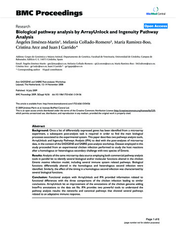
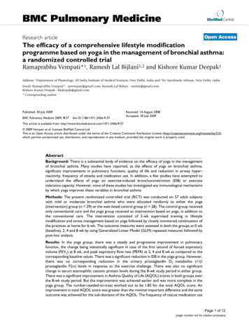
Bartoszewska et al. Cell Mol Biol Lett(2021) 26:11Fig. 1 The NGS and pathway analyses of early gene transcripts dysregulated by C-1305 treatment in A549cells. a The 2D chemical structure of C-1305 1,2,3]triazolo[4,5,1-de]acridin-6-one; C18H19N5O2). b The unique early gene transcripts dysregulated after 8 hof treatment of A549 with 3 µM C-1305 were selected from the NGS experiments. Well-established genetranscripts with greater than 10 RPKMs per sample and with significance (p 0.05) greater for change inexpression between C-1305-treated and control groups (no treatment and 24-h treatment) were used inpathway analyses. The Gene Ontology clusters are depicted for selected genes; clusters are listed followedby the p values and enrichment scores calculated by Enricher, which is used to determine the percentage ofthe chart. The longer bar the lower p-value, while the darker the color, the more enriched the cluster. Onlyclusters with p 0.001 were considered. c The heat map representing C-1305 exposure related expressionchanges in genes related to UPR and ER stress as observed in NGS experiments in A549 cells exposed to 3 µMand 10 µM C-1305 for 8 h. Heat maps were generated with the Morpheus software (Morpheus, https ://softw are.broad insti tute.org/morph eus). The color scale and values depict fold change (c)Page 4 of 17
Bartoszewska et al. Cell Mol Biol Lett(2021) 26:11(CHOP)—Hs00358796 g1, ERN1 (IRE1)—Hs00980095 m1, PMAIP1 (NOXA)—Hs00560402 m1, XBP1 (isoform 1and 2)—Hs00231936 m1, XBP1s—Hs03929085 g1.Western blotsXBP1s protein detection was performed as described in [32]. Briefly, cells were lysedon ice for 15 min in RIPA buffer [150 mM NaCl, 1% NP‐40, 0.5% sodium deoxycholate, 0.1% SDS, and 50 mM Tris‐HCl (pH 8.0)] supplemented with Protease InhibitorComplete Mini (000000011836170001; Roche, Basel, Switzerland). The insoluble material was removed by centrifugation at 15,000g for 15 min. Protein concentrations weredetermined by Bio‐Rad Protein Assay (Bradford‐based method; Bio‐Rad, Hercules, CA,USA) using bovine serum albumin (BSA; MilliporeSigma) as the standard. Followingthe normalization of protein concentrations, lysates were mixed with an equal volumeof 2 Laemmli sample buffer (Bio‐Rad) and incubated for 5 min at 95 C before separation by SDS‐PAGE on stain‐free TGX gradient gels (Bio‐Rad). Following SDS‐PAGE,the proteins were transferred to PVDF membranes (300 mA for 90 min at 4 C). Themembranes were then blocked with BSA dissolved in PBS and Tween‐20 (3% BSA and0.5% Tween‐20 for 1–2 h) followed by immunoblotting with the primary antibody foreach experiment for XBP1s (mAb 12782; diluted at 1:1000; Cell Signaling Technology,Danvers, MA, USA). After the washing steps, the membranes were incubated with goatanti‐rabbit IgG (H L) horseradish peroxidase-conjugated secondary antibodies (Bio‐Rad) and detected using ECL (Amresco, Solon, OH, USA). Densitometry was performedusing Image Lab software v.4.1 (Bio‐Rad).Molecular dockingThe crystal structure of human phosphorylated IRE1α in complex with ADP-Mg (PDBID code 4YZD, RSCB Protein Data Bank) [33] was used for molecular docking performed in this study. The structure of IRE1 was prepared for docking analysis on theUCSF Chimera software package [34] where the multi-chain structure was converted toa monomeric structure (chain A), and water and ADP-Mg were deleted to enable testingof studied ligand binding. A mol2 file with all hydrogens and 3D coordinates of C-1305,4µ8C IRE1 [35], and 3,6 DMAD ramethylacridine-3,6,9-triamine) [36] IRE1 inhibitors were generated using MarvinSketch (ver. 20.18.0 ChemAxon Ltd) [36]. Energy optimizations of C-1305, 4µ8C, and3,6-DMAD with 500 steps of the steepest descent algorithm using the MMFF94 forcefield were carried out for all 2D structures using the Avogadro 1.2.0 package software[37]. The minimized structures were used as inputs for ligand binding modeling. Furthermore, all three compounds were docked into their potential binding pockets of thenative 3D structure of IRE1 proteins (PDB ID code 4YZD) with the aid of EDock thatis based on replica-exchange Monte Carlo simulations that identifies high-quality blinddocking sites [38]. Only protein fragments that are not well ordered in the crystal structure were predicted using the I-TASSER (Iterative Threading ASSEmbly Refinement), apart of EDock server. This simulation does not disturb the native IRE1 binding pocketstructure. RMSD (UCSF Chimera) between 143 pruned atom pairs is 0.000 angstroms(across all 143 pairs: 0.000). For the docking analyses, only the best scoring results wereused. The results of docking were visualized with UCSF Chimera.Page 5 of 17
Bartoszewska et al. Cell Mol Biol Lett(2021) 26:11In vitro kinase assayERN1 (IRE1α) Kinase Enzyme assays were performed using the luminescent ADPGlo assay kit (VA7146, Promega) that measures the generation of ADP by the protein kinase reaction that is measured by an increase in the luminescence signal in thepresence of luminescent ADP-Glo reagent. All reagents were prepared accordingto the manufacturer’s instructions. To determine the appropriate enzyme concentration for use in each inhibitor dose–response curve determination, a kinase titrationwas performed using 10 µM ATP. Based on these results, a concentration of 5 ng ofIRE1 enzyme per reaction was chosen to determine the kinase inhibitory activity ofC-1305 and 4µ8C (SML0949, Sigma-Aldrich). Staurosporine (S6942-200UL, SigmaAldrich) was used as a control kinase inhibitor. All reactions were measured using the GloMax -Multi Detection System (Promega). The results were expressed as remaining activity, normalized to the uninhibited control for 100% activity and to the fullyinhibited one for 0% activity. To normalize the data from the following equation wasused:′′% Remaining activity 100 (S B/U B)","S" being the signal obtained for each concentration of compound, "U" the signalproduced by the uninhibited control (enzyme alone), and "B" the background produced by the fully inhibited control (no enzyme). The data were plotted and fit to sigmoidal curves using Origin Pro ver 7.0 (OriginLab Corporation, Northampton, MA,USA) to determine the I C50.Statistical analysisResults were expressed as a mean standard deviation. Statistical significance wasdetermined using the Student’s t-test and ANOVA on ranks with p-values belowp 0.05 considered significant.ResultsWe previously utilized a genome-wide approach to identify transcriptomic signatures of C-1305 cytotoxicity in lung cell lines and demonstrated that C-1305 promotes direct microtubule stabilization as a part of its mechanism of action that leadsto apoptosis [21]. We also observed that C-1305 at concentrations up to 10 µM hadno significant effect on growth and survival of an adenocarcinoma lung cell line,A549, up to about 18 h [21]. To further evaluate the early transcriptomic signalingthat contributes to C-1305’s biological effects, we focused on the genome-wide consequences of 8 h exposure of A549 cells to 3 µM C-1305, a concentration that didnot affect cell survival during this time of exposure [21]. In this experiment, totalRNA was extracted from the A549 cells that were exposed to C-1305 for 8 h and from8 h DMSO vehicle-treated cells (controls), and then subjected to RNA sequencing.To focus only on the C-1305 early transcriptome changes, we corrected (subtractedout) the A549-C-1305-8 h treated gene set with the A549-8 h vehicle-treated geneset and also the previously obtained A549-C-1305-24 h treated gene set [21]. Ouranalysis was narrowed to the most significant expression changes (p 0.05). ThisPage 6 of 17
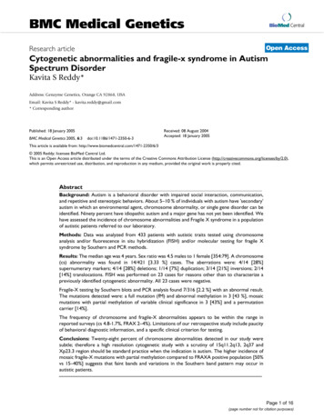
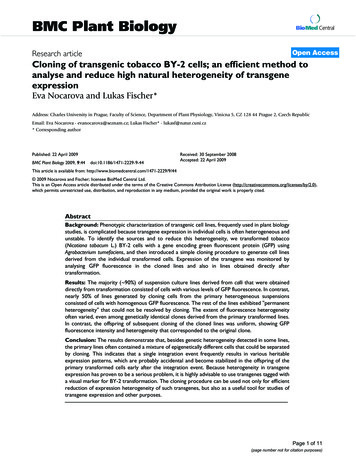
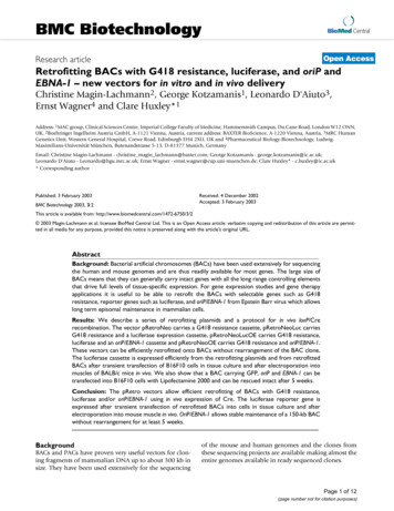
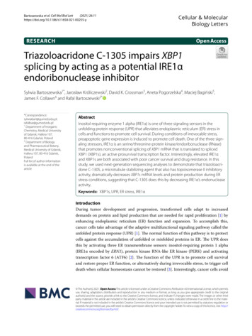
Bartoszewska et al. Cell Mol Biol Lett(2021) 26:11approach resulted in the identification of 182 genes dysregulated specifically at theC-1305 treatment 8 h time point and these genes were further subjected to geneontology analysis using Enricher [29]. As shown in Fig. 1b, the cellular response tounfolded/misfolded proteins was the most significant assignment. Within this cluster of gene transcripts, the following genes’ mRNA levels were downregulated uponC-1305 exposure: heat shock protein family A member 5 (HSPA5 aka BiP), XBP1,heat shock protein family A member 8 (HSPA8), heat shock protein family A member 1A (HSPA1A) and heat shock protein family A member 1B (HSPA1B) (Fig. 1c).Notably, the upregulated genes in this set were Bcl-2 binding component 3 (BBC3 akaPUMA), phorbol-12-myristate-13-acetate-induced protein 1 (PMAIP1 aka NOXA),activating transcription factor 3 (ATF3), DNA damage inducible alpha (GADD45A) aswell as ERN1 (aka IRE1) and DNA damage inducible transcript 3 (DDIT3 aka CHOP)(Fig. 1c). Notably, similar and even more dramatic transcriptomic changes were alsoobserved up on 8 h exposure to 10 µM concentration of C-1305 (Fig. 1c).Based on the results of the RNAseq-based approach, we speculated that C-1305 mayimpair endoplasmic reticulum (ER) protein homeostasis via downregulation of transcripts of chaperone (BiP) and proadaptive transcription factors (XBP1s) and this couldlead to activation of UPR pathways. Furthermore, the UPR proapoptotic factors likeCHOP, PUMA, NOXA and GADD45A were elevated at this time. To verify this observation, we performed qPCR analysis of these UPR related transcripts levels after 8 hC-1305 treatments at 1 µM, 3 µM and 10 µM concentrations in A549 as well as in a noncancer lung cell line 16HBE14o-, and in HeLa cells. We included HeLa cells since thiscell line is a well recognized and characterized model for studying pharmacological ERstress and UPR induction, including the IRE1/XBP1s branch [39–47].As shown in Fig. 2, the 8 h C-1305 exposure of all cell lines significantly reduced BiPexpression and resulted in significant induction of ERN1 mRNA levels. Although, thecell lines differed between each other in terms of C-1305 dose response dependence andmagnitudes of transcriptomic changes, the general compound’s effects on BiP and ERN1(IRE1) mRNA levels correlated fairly well with the C-1305 concentrations except for the10 µM concentration in the HeLa cells. In examining the effects of C-1305 on the activeisoform of XBP1, XBP1s, the results indicated that the levels of this mRNA were dramatically lower in all three cell lines (Fig. 2c) which was surprising given that the ERN1mRNA levels were increased.In terms of the proapoptotic factors, there was no significant induction of CHOPobserved in airway cells, 16HBE14o- and A549, after exposure to 1 µM and 3 µMC-1305, whereas it was in the HeLa cells at all three concentrations (Fig. 3a). Nevertheless, a moderate CHOP induction was observed in the NGS analysis of A549 exposedto 3 µM C-1305, however, this discrepancy results from the different gene expressionnormalization methods applied for NGS and qPCR data (global—DESeq2 and individual—GAPDH and TBP reference genes, respectively). In contrast, there was a significantinduction of both proapoptotic NOXA and PUMA mRNA expression at all three concentrations in all of the tested cell lines (Fig. 3b, c). The collective data in Figs. 2 and 3suggest that C-1305 treatment induced IRE1, but paradoxically, decreased XBP1s mRNAexpression, and that for the most part, the UPR prosurvival genes decreased and theproapoptotic genes increased.Page 7 of 17
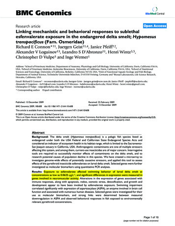
Bartoszewska et al. Cell Mol Biol Lett(2021) 26:11Fig. 2 C-1305-induced changes in a BiP, b ERN1, c XBP1s mRNA levels in A549, 16HBE14o- and HeLa cellsafter 8 h of treatment. The results from 3 independent experiments (n 9) are plotted normalized to GAPDHand TBP mRNA levels and expressed as a fold-change over the vehicle controls. Error bars represent standarddeviations. Significant changes (p-value 0.05) are marked with an asteriskBased on these results, we next tested whether the decrease in XBP1s was due to ainhibition of IRE1α endoribonuclease activity. We compared the C-1305 effectiveness in preventing IRE1α-dependent XBP1 mRNA splicing with the efficiency of commonly used IRE1 inhibitor 4µ8C [35, 48] (Fig. 4a). As an ER stress induced XBP1 mRNAsplicing model, we used HeLa cells treated for 6 h with the classical pharmacologicalstressor tunicamycin (Tm, 0.5 ug/ml), a glycosylation inhibitor. We used HeLa cells forthis experiment since they are commonly used in ER stress and UPR studies [39–43].Previous studies using this model indicated that maximal XBP1s mRNA levels areobserved after 6 h of treatment [44]. Furthermore, both treatments of HeLa cells withC-1305 for 6 h or co-treatments with Tm and Tg had a limited effect on cell survivalPage 8 of 17
Bartoszewska et al. Cell Mol Biol Lett(2021) 26:11Fig. 3 C-1305-induced changes in a CHOP, b NOXA, c PUMA mRNA levels in A549, 16HBE14o- and HeLa cellsafter 8 h of treatment. The results from 3 independent experiments (n 9) are plotted normalized to GAPDHand TBP mRNA levels and expressed as a fold-change over the vehicle controls. Error bars represent standarddeviations. Significant changes (p-value 0.05) are marked with an asterisk (IC50 37,62 µM for C-1305, I C50 34,80 µM for C-1305 and Tm; I C50 21,6 µM forC-1305 and Tg; as monitored by the MTT assay, Additional file 1: Figure S1).As shown in Fig. 4b, the use of Tm resulted in about a sevenfold induction of XBP1smRNA, whereas use of 1 µM, 10 µM and 50 µM of C-1305 resulted in dramatic reduction of XBP1s mRNA levels and appeared very similar to the Tm 4µ8C treatments.Furthermore, the XBP1s protein reduction was similar for both the C-1305 and 4µ8C(Fig. 4c, d), suggesting that C-1305 inhibited the IRE1 endoribonuclease activity andthe subsequent production of XBP1s.To characterize the C-1305 as a potential IRE1 inhibitor, we used the HeLa modelto determine C-1305’s IRE1α endoribonuclease activity I C50 values based on thePage 9 of 17
Bartoszewska et al. Cell Mol Biol Lett(2021) 26:11Fig. 4 C-1305-prevents IRE1α-dependent XBP1 mRNA splicing during ER stress. a The 2D chemical structureof 4µ8C de, C 11H8O4). b HeLa cells were treated withER stressors (Tm, 0.5 µg/ml) for 6 h in a presence of specified concentrations of C-1305 and 4µ8C. Followingthe treatments, the XBP1s mRNA levels were accessed with qPCR and expressed as a fold-change over theno stress controls. The results from 3 independent experiments (n 9) are plotted normalized to GAPDH andTBP mRNA. Error bars represent standard deviations. Significant changes (p-value 0.05) are marked with anasterisk. c The corresponding changes in XBP1s protein levels were evaluated by Western blot (d) normalizedto total protein levels and related no stress control. The data from 3 independent experiments were analyzed.*p 0.05 was considered significant. The raw data are provided in Additional file 1Page 10 of 17
Bartoszewska et al. Cell Mol Biol Lett(2021) 26:11(See figure on next page.)Fig. 5 The XBP1s mRNA-based determination of C-1305 impact on IRE1α endoribonuclease activity inER stressed HeLa cells. HeLa cells were treated with ER stressors (Tm, 0.5 µg/ml and Tg, 25 nM), for 6 h ina presence of specified concentrations of C-1305 and 4µ8C. Following the treatments, the XBP1s mRNAlevels were accessed with qPCR and expressed as a change versus ER stressed cells treated with vehicle. Theresults from 3 independent experiments (n 9) are plotted normalized to GAPDH and TBP mRNA. Error barsrepresent standard deviations. Concentration response curves and IC50 values of a C-1305 and b 4µ8C inTm treated cells were determined using Sigma Plot 1.1 software. Similar approach was used to determineConcentration response curves and IC50 values of c C-1305 and d 4µ8C in Tg treated cells. The IRE1α kinaseassays were performed in the presence of serial dilution and were measured with ADP-Glo kinase assay.Concentration response curve and IC50 values of e C-1305, f 4µ8C and g staurosporine (used as a control ofkinase activity) were determined using Origin Pro software. The results were expressed as remaining activity,normalized to the uninhibited control for 100% activity and to the fully inhibited one for 0% activity asdescribed in “Materials and methods”. Data are plotted as means SD from triplicate measurements of twoindependent measurements (n 6)measurement of XBP1s mRNA levels and used 4µ8C as our control. As shown inFig. 5a, in Tm treated HeLa cells, the I C50 for C-1305 was 1.017 µM, whereas the I C50for 4µ8C under the same conditions was 0.142 µM (Fig. 5b). Next, we tested anothercommon ER stress model thapsigargin (Tg, 25 nM), a noncompetitive inhibitor of theendoplasmic reticulum Ca2 ATPase that results in higher induction of XBP1s mRNA[23, 32]. In this case, higher concentrations of both C-1305 and 4µ8C were necessaryto reduce the XBP1s mRNA levels (IC50 16.821 µM for C-1305 and IC50 1.66 µMfor 4µ8C, Fig. 5c, d). Taken together, the results suggest that C-1305 is an efficientinhibitor of IRE1α and this compound works independently of the mechanism ofpharmacological ER stress induction.Using purified IRE1 protein, we next determined C-1305’s and 4µ8C’s effects on theIRE1α kinase activity in vitro and used staurosporine as a reference control. As shownin Fig. 5e, the C-1305 has no effect on IRE1α kinase activity, whereas 4µ8C was able toinhibit half of IRE1 phosphorylation at 31.25 µM concentration (Fig. 5f ). These resultssuggest that C-1305 mechanism of action is based on inhibiting IRE1α endoribonucleaseactivity rather than kinase activity, whereas 4µ8C can block both endoribonuclease andkinase activity of IRE1. That being said, 4µ8C IC50 value for the kinase activity is remarkably high when compared with staurosporine (1.98 nM, Fig. 5g), and significantly higherthan the concentrations required to reduce the endoribonuclease activity of IRE1α basedon the XBP1s message readout.Since our results suggested that C-1305 may inhibit IRE1α via direct interactionwith its RNAse domain, we evaluated docking of C-1305 targeting the catalytic siteof the RNase domain using molecular docking analysis (Fig. 6). The model of IRE1αin complex with C-1305 inhibitor obtained in the docking analysis suggests that thismolecule can form hydrophobic interactions with F889 and L886 residues, a hydrogen bond with Y892 and N906 (Fig. 6a). Molecular docking results clearly suggestthat triazole ring can interact with both Lysine via H-bonding and Leucine via a pialkyl interaction. These interactions could stabilize C1305 in the ribonuclease activesite of IRE1α. In contrast, the docking of 4µ8c to IRE1α shows interactions withH910, N906, K907, and E913 and was similar to the previous docking model published by [35], Hanwell, Curtis [37] indicated that Y892, F889, N906, K907, and H910residues of IRE1α interact with 4µ8c (Fig. 6b). The mode of action of 4µ8c is relatedPage 11 of 17
Bartoszewska et al. Cell Mol Biol Lett(2021) 26:11to the formation of a Schiff base with K907 residue. Despite the fact that C1307 doesnot have an aldehyde group, it is conceivable that it forms an imine bond with K907due to the presence of a carbonyl group in its structure. Since structurally C-1305is related to a known IRE1α inhibitor, acridine derivative 3,6-DMAD [36], we alsoprepared a docking model of 3,6-DMAD with IRE1α (Fig. 6c) and showed the interaction of 3,6-DMAD with H910, N906, K907, L886, Y892, F889, and E913 residuesof IRE1α that are present in the catalytic site of the RNAse domain of IRE1α. TakenPage 12 of 17
Bartoszewska et al. Cell Mol Biol Lett(2021) 26:11Fig. 6 Prediction of C-1305, 4µ8C, and 3,6-DMAD binding mode in the and in the endoribonuclease(RNase) domain of IRE1α. The binding mode of a C-1305, b 4µ8C, and c 3,6-DMAD was modeled using theEDock server (https ://zhang lab.ccmb.med.umich .edu/EDock /). The native and modeled structures of t
Bartoszewska et al. Cell Mol Biol Lett 1 11 Page 4 of 17 Fig. 1 The NGS and pathway analyses of early gene transcripts dysregulated by C-1305 treatment in A549 cells. a The 2D chemical structure of C-1305 1,2,3] iaztr olo[4,5,1-de]acridin-6-one; C 18H 19N 5O 2). b The unique early gene transcripts dysregulated after 8 h