Transcription
Eshkoor et al.Egyptian Journal of Medical Human (2022) 23:105Egyptian Journal of MedicalHuman GeneticsOpen AccessREVIEWMicroRNAs influence and longevitySima Ataollahi Eshkoor1,4*, Nooshin Ghodsian2 and Mehrnoosh Akhtari‑Zavare3AbstractBackground: MiRNAs play critical roles in the regulation of cellular function, life span, and the aging process. Theycan affect longevity positively and negatively through different aging pathways.Main text: MiRNAs are a group of short non-coding RNAs that regulate gene expressions at post-transcriptional lev‑els. The different types of alterations in miRNAs biogenesis, mRNA expressions, and activities of miRNA-protein com‑plexes can affect the regulation of normal post-transcriptional gene process, which may lead to aging, age-relateddiseases, and an earlier death. It seems that the influence of deregulation of miRNAs on senescence and age-relateddiseases occurring by targeting aging molecular pathways can be used for diagnosis and prognosis of them. There‑fore, the expression and function of miRNAs should be studied more accurately with new applicable and validatedexperimental tools. However, the current review wishes to highlight simply a connection among miRNAs, senescenceand some age-related diseases.Conclusion: Despite several research indicating the key roles of miRNAs in aging and longevity, further investiga‑tions are still needed to elucidate the essential roles of miRNAs in controlling mRNA regulation, cell proliferation,death and/or protection during stress and health problems. Besides, more research on miRNAs will help to identifynew targets for alternative strategies regarding effectively screen, treat, and prevent diseases as well as make slow theaging process.Keywords: Aging, Cancer, Cognition, Longevity, MiRNA, StressMicroRNAs (miRNAs)MicroRNAs (miRNAs) are a large class of small noncoding RNAs functioning as the important regulatorsof a wide range of cellular processes [1] that have beenidentified firstly in C. elegans in 1993 [2–4]. They act asmediators that can regulate post-transcriptional geneexpressions [5–7]; therefore, they are able to controlcellular behavior, balance in biological processes, development, and diseases [7, 8] by modulating gene expression [1]. The post-transcriptional gene effect of miRNAshappens by base-pair binding on their related mRNAstargeting untranslated regions (UTRs) of genes and multiple sites within a single UTR [9]. Each miRNA can target multiple mRNAs, and one mRNA can be regulated by*Correspondence: simaataolahi@yahoo.com1Department of Psychiatry, Regionshospitalet Randers, Randers, Denmark8930Full list of author information is available at the end of the articlemultiple miRNAs [10, 11]. MiRNAs have been found inplants, animals, bacteria and some viruses [12] by theirgene expression profiling. In animal models, miRNAscontribute into genetic networks and metabolic pathways. It seems that miRNAs play critical roles in theoccurrence of pathological conditions like neurodegeneration and cancer following the alterations in the expression of specific miRNAs in the brain and/or homeostasisin the body [6, 9].Besides, some miRNAs are related to the regulation ofsenescence in a variety of human cells [8]. As it is shownin Table 1, cancer, cognition, and senescence can be associated with miRNAs [2, 6, 13–17]. Until now, more than950 miRNAs in humans have been found [18]. It has beenindicated that some of miRNAs are tissue- or cell-specificand some of them are house-keeping molecules. However, a relatively small number of miRNAs have probablykey functions in order to regulate the human genome andaffect post-transcriptional physiological process. It seems The Author(s) 2022. Open Access This article is licensed under a Creative Commons Attribution 4.0 International License, whichpermits use, sharing, adaptation, distribution and reproduction in any medium or format, as long as you give appropriate credit to theoriginal author(s) and the source, provide a link to the Creative Commons licence, and indicate if changes were made. The images orother third party material in this article are included in the article’s Creative Commons licence, unless indicated otherwise in a credit lineto the material. If material is not included in the article’s Creative Commons licence and your intended use is not permitted by statutoryregulation or exceeds the permitted use, you will need to obtain permission directly from the copyright holder. To view a copy of thislicence, visit http:// creat iveco mmons. org/ licen ses/ by/4. 0/.
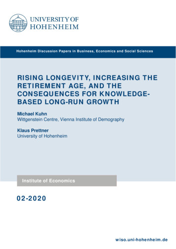
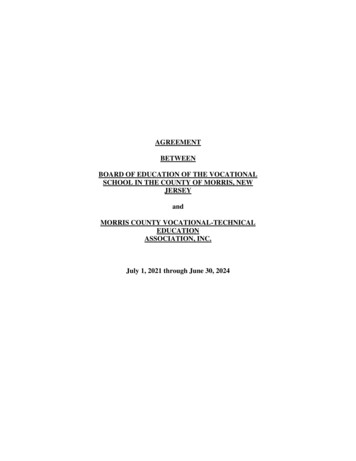
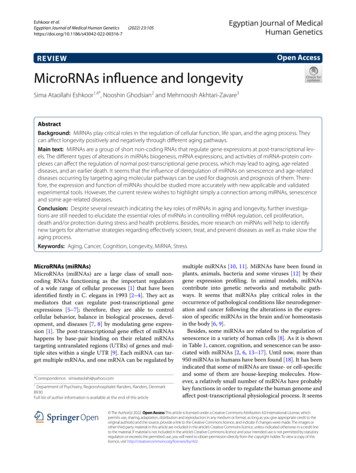
Eshkoor et al. Egyptian Journal of Medical Human Genetics(2022) 23:105Table 1 Showing the correlation of miRNAs with cancer,cognitive function and senescenceMiRNAsCancerNeurodegenerationand cognitionSenescence1miR-1 2Let-7 3Let-7b? 4MiR-9 5miR-17 6MiR-21 7MiR-26b 8MiR-29a 9MiR-29b 10miR-30a 11MiR-31 12MiR-34 13miR-34a 14miR-71? 15miR-10016MiR-107 17MiR-124 18MiR-125b 19MiR-132 ?20MiR-137 21MiR-146 22MiR-146a 23MiR-155 24miR-199a 25Mir-206 26MiR-210 27miR-211‐5p 28MiR-212 29MiR-217 ? 30MiR-221 31MiR-222 32miR-320 33MiR-340‐3p ? 34MiR-371-3 ?35MiR-374a‐5p 36MiR-376c‐3p 37miR-483 ? 38MiR-484 39MiR-567 ?41MiR-1225‐3p ? 42MiR-5095 ? presence of correlation?there are not studies obviously indicating the correlationthat they can influence differentiation and tumor suppression in cells, which may relate to some pathologicalPage 2 of 16processes such as carcinogenesis or senescence [10].Thus, miRNAs have been suggested to be used as biomarkers to evaluate many diseases and aging [9], eventhough it is difficult to estimate miRNAs quantificationdue to their small size, low copy number, interferencefrom other small RNAs, and contamination by degradation products of mRNAs or other RNA species [2].Despite the presence of miRNAs in tissue cells, theycan also be found in the body fluids and extracellularenvironments such as plasma, serum, urine, saliva, seminal fluid, ascites, pleural effusions, and cerebrospinal fluid[4, 19]. They are injected to the circulation in differentways. It can happen through a passive leakage followingapoptosis, necrosis, inflammation, or an active secretionby exosomes/microvesicles, lipoproteins, and RNA–protein complex. Circulating miRNAs following packinginto exosomes have been found specific to a tissue or adisease, which can indicate degree of tumor progressionand stage of cancer. Several studies have also shown thatthe abnormality of specific circulating miRNAs can beassociated with the manifestation, development, invasion, and metastasis of cancer [19]. However, miRNAsrelated studies will increase our understanding regardingage-related gene regulation and improve miRNA-basedbiomarker development for an advance in RNA-baseddiagnosis and therapies [9]. Thus, the aging process andage-related problems and conditions can be better monitored. In addition, further studies will help to better identify specific miRNAs and their changes related to caloricrestriction, aging and age-related diseases pathways.Biogenesis of miRNA in animalsMiRNAs are a group of small non-coding RNA [8, 14, 20,21] with approximately 21–24 nucleotide (nt) in length[3] that are widespread and probably regulate 50% ofthe human genome [22]. They are produced from precursor molecules (pri-miRNAs), which are made throughtranscription by RNA polymerase II from independentgenes or derived from introns after splicing. Pri-miRNAsare subsequently converted to pre-miRNAs by Droshaenzyme and exported to the cytoplasm. Dicer enzymecleaves them to the mature approximately 20-bp miRNA5p/3p pairs. One strand of this duplex will incorporateinto the miRNA-inducing silencing complex (miRISC) [3,23–25]. The process is summarized in Fig. 1.The transcription of DNA coding for miRNAs and therelated protein-coding genes occurs in a similar way. Themature miRNA directly interacts with a member of theArgonaute protein family, which results in the formationof the RNA-induced silencing complex (RISC). As a component of RISC, miRNAs direct the post-transcriptionalrepression. Thus, miRNAs show regulatory functions
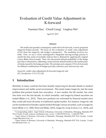
Eshkoor et al. Egyptian Journal of Medical Human Genetics(2022) 23:105Page 3 of 16Fig. 1 Gene silencing procedure by miRNAregarding gene expression and can play a central role inseveral cellular processes including cell growth, differentiation, proliferation, and apoptosis [18].MiRNAs are potent negative regulators of gene expressions [8]. They regulate their target genes through eithertranslational repression or mRNA degradation. Suchfunctions happen via binding to the complementaryregions of messenger transcripts [14] and targeting specific messenger RNAs (mRNAs) [21]. Each miRNA cantarget up to hundreds of mRNAs [8, 18, 26] and they areable to regulate the expression of more than 60% of protein-coding genes of the human genome [27]. The effectof miRNAs on the regulation of many human genes [28–30] indicates their critical roles in a variety of biologicalprocesses [8, 21].MiRNAs are important epigenetic regulators [14] andmany of them are also self-regulated epigenetically [8].For instance, epigenetically transcriptional repression bymiRNAs can be through DNA methylation and histonemodifications in which to affect subsequently mostlyCpG islands located in the promoter regions of genesand result in silencing of genes [31]. Moreover, miRNAscan also down-regulate mRNAs [26] by declining targetmRNAs or the levels of translation into proteins [32]. Thesilence of target mRNAs is based on base-pairing recognition sites [8] and the interaction of miRNAs with the3’ UTR of target mRNAs. Thus, mRNA degradation and/or translational repression result in gene silencing [4, 29,33, 34].However, miRNAs are known as major elements thatcan contribute into complex functional pathways, control cellular processes [35], and the regulation of biological functions [36] such as differentiation, proliferation,apoptosis, replicative senescence [21], development, andstress responses [35] in plants and animals (Table 2).MiRNAs are in both intracellular and extracellularregions of the body [61]. Extracellular miRNAs can beused as biomarkers in which to evaluate a variety of diseases such as liver fibrosis and hepatocellular carcinoma.Besides, miRNAs play important roles in intercellularcommunication. Such miRNAs can be delivered to targetcells, where they have hormone-like activities and mayact as autocrine, paracrine, and/or endocrine regulators.It can modulate cellular activities [4] by changing theexpression of proteins following the specific inhibition ofmRNA targets in cell-free miRNAs, which may originatefrom one cell type and acquired by another cells or othercell types [11]. In addition, miRNAs, gene expression patterns, physiology, and homeostasis in cells are related tothe effects of different factors such as stress [135].
iR-175miR-34aMiR-9412Let-7213miR-11MiRNAsCell cycle influenceInhibiting proliferation, invasion, and migration, inducing apoptosis. synapticplasticity and cognitionEffect on tumor suppressive genes, aggressiveness and prognosis of malig‑nant neoplasms, cell cycle regulation, fibrosis and neuronal regulations,tumorigenesis and cellular senescence, osteogenic differentiationCell proliferation, apoptosis, angiogenesis, invasion, metastasis, drug resist‑ance and chemotherapy resistanceInhibits the cell cycle and cell proliferation[65][2, 70, 71][67–69][2, 53, 66]Proliferation, differentiation, metabolism, apoptosis, drug resistance, tumorimmunity, diagnosis, prognosis and clinical treatment of tumors, neuroninflammationProliferation, differentiation, proliferation, apoptosis, and growth of cells aswell as immunity[53, 75][73, 74]Cell cycle influence, the progression of cell growth, migration, and invasion in [72]various cancers,Subtle, maintaining cell viability, cognitionLong-term survivalCell cycle influence, response to hypoxia stress[61–64][2, 59, 60][54–58][54, 55][53][50–52](2022) 23:105NF-kB pathway, CDKN2A pathway, p53 pathway, PI3K/Akt/mTOR pathway,ErbB2 pathway, Wnt pathway,Notch signaling pathway, miR124/AMPK/mTOR pathway, miR-124-STAT3pathway, TLR signaling pathway, JNK and p38 pathways, MiR-124-PTPN1signaling pathwayWnt/β-catenin signaling pathway,FGFR3 pathway, HIF-1α and HIF-2α dependent pathway, mTOR pathway,MAPK pathway, PI3K pathway, PKB pathwayp38 pathway, KGB-1 on DAF-16 involved pathways, PI3K pathway, UNC-31–mediated InsR/PI3K signalingFoxM1/c-Myc pathway, p53 pathway, HIF-1a pathwayMEK/ERK signaling pathway, E2F pathwayRAS/MAPK pathway, RAS/MARK pathway PI3K/AKT pathway, RB/E2F pathway, Proliferation, Tumorigenesis, migration, anti-apoptosis, improving cognitionP53 pathway, Rho/ROCK pathway, NF-κB pathway, E2F pathway, WNT path‑way,TOR pathway, Wnt pathway, Fgf signaling pathway, Wnt/β-catenin pathway,MTDH/PTEN/Akt pathway, PI3K signaling pathway, PI3K/Akt/mTOR signalingpathway, EZH2/miR-30a/KPNB1 signaling pathwayPI3K-Akt signaling pathways, PTEN/AKT/β-catenin signaling pathway, AKT/GSK-3β/β-catenin signaling pathway, AKT/mTOR pathway, PTEN/AKT/GSK3β/β-catenin signaling pathway. Wnt-β-catenin pathwayMMP2 signaling pathway, Smad4-dependent way, TGF-β signaling pathway,Wnt-β-catenin signaling pathway, PTEN/AKT/GSK3β signaling pathway,TGF-beta1/miR-29a/HDAC4 pathway, Hedgehog signaling pathway, NF-κBpathway, p42-44 MAPK pathway and Wnt-β-catenin pathwayRb1-E2F and p27(kip1) pathways, Wnt-β-catenin signaling pathway,[45, 46][2, 40–44][37–39]ReferencesAutophagy, apoptosis, growth, chemoresistance, regulated genes involved in [47–49]cell cycle control and tumor development, aging, cognitionNeurogenesis, angiogenesis, regulation of lymphatic inflammatory andlymphangiogenic pathways, cognition, cell proliferation, migration, invasionand angiogenesisSenescence, age-dependent changes in the vasculature and vascular aging,tumor suppressor and oncogene, apoptosis, migration, and invasion, poten‑tial diagnostic and prognostic marker, neurodegenerationDifferentiation of cardiac muscle, skeletal muscle differentiation followingaging, inhibit tumor growth and metastasis, cognitionFunctions involvedRasA1 signaling pathway KRAS, RAS/MEK/extracellular signal-regulated kinase Promotes cell proliferation, anti-apoptosis signaling and malignant transfor‑(ERK) signaling pathway, PI3K/AKT pathwaymation, inflammation-related miRNAs, advanced oxidation protein products(AOPP), with the presence of frailty, age-related frailtythe P53, ERB, and MAPK signaling pathwaysHIF-1α-VEGF signaling pathway, p16-Prb pathwayDAF-12 signaling pathway, insulin/IGF signaling (IIS) pathway, LOX-1-ROSp38MAPK-NF-κB signaling pathway, LOX-1-ROS-PKB-eNOS pathway, TGF‐βpathway, RAS and KIT signaling pathwaysIGF1 and HDAC4, mTORmiR1HDAC4follistatin pathway, Notch signalingpathwayPathwaysTable 2 Examples of miRNAs, targets and functions involvedEshkoor et al. Egyptian Journal of Medical Human GeneticsPage 4 of 16
-222miR-3202526272829303132References[2, 91–94][87–90]Amyloid-beta deposition, mitochondrial metabolism, angiogenesis, the DNAdamage response, cell proliferation, apoptosisTumorigenesis and progression, chemotherapy resistance, cancer cellproliferation, invasion and metastasis, stemness promotion, cell survival, therelapse of cancer cells, cognition, life span?Hemostasis, normal growth, and maintenance of cells, proliferation, apopto‑sis, transcription regulation and development, invasion, drug resistanceServe as an oncogene or tumor suppressor, oncogenesis, development andmetastasis of cancer, proliferation, invasion, Metastasis, cognition, life span[2, 49, 103–105][2, 102][76, 79, 100, 101][41, 98, 99][53, 85, 97]Neuronal differentiation, brain development, functions in neuromuscular syn‑ [37, 95, 96]apse development and maintenance, muscle cell proliferation; inhibits tumorcell proliferation, migration, and invasion, and induces apoptosisMaster regulators for cellular processes, function in immune system, potenttumor suppressor, cognition, proliferation, migration, invasion, apoptosis,autophagy and glycometabolismTumor inflammation and growth, chemotherapy resistance, regulatesimmune system including B cells and T cells. cognitionPLA2/PKCα signaling pathwaymTOR pathwayinsulin/IGF signaling (IIS) signaling pathway, AQP1-mediated proliferativesignaling pathway,[113–115][61, 112][48, 110, 111][2, 107–109](2022) 23:105Proliferation, migration, invasion, chemoresistance, prognosis, life span,cognitiontumor growth and metastasis, drug resistance, overall survivalAffecting life span, Tumoral survival, Human longevityNeurodegenerative disease, tumor suppressor, to promote tumorigenesis,life spanPTEN pathway, p27kip1 and c-kit pathway, PTEN-mediated PI3K/Akt pathway; Tumor initiation and progressions, cancer cell proliferation, invasion and[2, 48, 103, 104, 106]p53 pathwaymetastasis, stemness promotion, cell survival, chemoresistance, the relapse ofcancer cells, cognition,PTEN pathway, AKT pathway, JAK/STAT3 signaling pathway, PTEN-mediatedPI3K/Akt pathwaySirtuin signaling pathway, HIF-1α pathway, MAPK signaling pathway,Sirtuin signaling pathway, Hedgehog signaling pathway, Wnt/βcatenin path‑way, AKT signaling pathway,[82–84]Neuroinflammation, Amyloid-beta deposition, inflammation, innate immu‑[51, 53, 85, 86]nity, and cancer, as well as to regulate mitochondrial functions, inflammationaging, inhibition of proinflammatory pathwaysCell proliferation, invasion, and metastasis, act as a tumor suppressor, inflam‑mationNeuroprotective effects, neural development, neoplastic transformation, act [80, 81]as a tumor suppressor, cell proliferation, differentiation, migration and metas‑tasis, and induce cell cycle arrest, differentiation and apoptosisEzrin/Fak/Src signaling pathway, JAK/STAT3 pathway, PERK pathway, MiR-211- Aging, proliferation, prognosis in various cancers, apoptosis, cell proliferation,5p-NUAK1 Pathwayinvasion, apoptosis and immunity, cognitionIncreased ROS pathway, protein kinase B (Akt) pathway and p53-dependentpathway, mTOR pathway, HIF-1α pathway, PI3K pathway, Akt pathway, ERKpathway, NFκB pathway, p53 pathwayIGF1 pathway, MRFs pathway, BDNF pathway, Fstl1 pathway, MEF2 pathway,IL-17 pathway, HIF-1α pathway; WNT signaling pathway; mitochondrialapoptosis pathway;HIF-1α pathway, Toll-like receptor signaling pathway, insulin signalingpathway, adipocytokine signaling pathway (KEGG), PI3K/AKT/mTOR signalingpathway, Wnt/β-catenin signaling pathwayMiR-374a‐5p mTOR signaling pathwaymiR-199a24BCR pathway, Toll-like receptor (TLR) pathway, ERK pathway, c-Jun N-terminalkinase pathway (JNK), SOCS1 pathway, JAK2/STAT3 pathway, RhoA pathway,PI3K/AKT pathway35MiR-15523NF-kB pathway, TLR4/NF-κB pathway, TNFα inflammatory pathway, IL-1αsignaling pathwayMiR-340‐3pMiR-146a22RAS pathway, NOTCH pathway, NF-κB pathway;MiR-371–3MiR-14621miR-137/Src/MAPK signaling pathway, ERK1/2 pathway33MiR-13720Functions involvedHIF-1α pathway, PTEN/AKT/FOXO3 signaling pathway, AKT signaling pathway, Neuronal health, neuron morphogenesis and plasticity, suppresses the migra‑ [2, 76–79]tion and invasion of cancerPathways34MiR-13219MiRNAsTable 2 (continued)Eshkoor et al. Egyptian Journal of Medical Human GeneticsPage 5 of 16
miR-483MiR-484MiR-567MiR-1225‐3p mTOR/p70S6K pathway, Melatonin pathway, MMEJ pathway,MiR-50953738394041Wnt/b-catenin signaling pathway, melatonin pathwayPI3K/AKT/c-Myc pathway, cMras/miR-567/PTPRG regulatory pathway;mTOR signaling pathway, Hippo signaling pathway, estrogen signaling path‑way, interferon pathwayWnt/β-catenin signaling pathway, glucose/OGT/miR-483-3p signaling path‑way, MAPK/ERK pathway, TGF-β pathway,MiR-376c‐3p Wnt/β-catenin pathwayPathways36MiRNAsTable 2 (continued)References[51, 119]Life span, inhibited cell proliferation, migration and invasion of glioblastoma;esophageal tumor development and progressionLife span, role in repairing single-strand breaks during DNA replication,uncontrolled proliferation and progression of cancer,Biological processes in the neuronal cells, neuronal differentiation and braindevelopment, tumorigenesis and development, cell proliferation, migrationand invasion, induced G1/S transition, chemoresistance, tumor suppressor;[129, 132–134][129–131][123–128]Mitochondrial fission, synaptic transmission and regulation of synaptic plas‑[120–122]ticity, synaptic function, ATP generation, presynaptic calcium level, oncogene,tumor suppressor, negative prognostic biomarkerAntiaging properties, inflammation, proliferation, apoptosis, promote tumordevelopmentLife span, cell cycle progression, proliferation, migration, invasion, survival and [55, 116–118]poorly response to chemotherapy, inhibits apoptosis, cognitionFunctions involvedEshkoor et al. Egyptian Journal of Medical Human Genetics(2022) 23:105Page 6 of 16
Eshkoor et al. Egyptian Journal of Medical Human Genetics(2022) 23:105StressStress is caused by different stressors and is divided intoacute, chronic and several forms. It can play an importantrole in daily life in which to threat an individual’s homeostasis, well-being, health, and/or survival. Stress is witha systemic physiological response such as inflammatoryand cellular reactions, metabolic processes, and epigenetic regulation [18]. The occurrence of stress in cellsmay happen due to sudden or frequent changes in environmental factors. Stress can damage existing macromolecules in living cells such as proteins, mRNAs, DNA, andlipids, which in turn can increase the risk of death, metabolic imbalances [136], and chronic diseases [20] such ascognitive decline and cancer with advancing age [2].The severity and duration of stress can affect cellularhomeostasis in which to return to stable state or modifyto a new state. However, responses to stress in the bodycan occur through several mechanisms [136] including induction of molecular chaperones [137, 138], rapidclearance of damaged macromolecules [139], activationof specific gene expressions, growth arrest [140], and celldeath [20].MiRNAs play critical roles in main cellular processes.Their identifying helps to understand the cellular stressresponses and the occurrence of senescence associatedwith environmental changes [20]. Nowadays, there is abetter understanding regarding the miRNAs involving inphysiological reactions and reaction pathways as well astheir regulatory roles. For instance, the alteration of miRNAs concentration such as miR-21 has been detected insome stress-related pathological conditions and diseasessuch as psychiatric diseases [18]. MiRNAs have shownregulatory role in the control of specific mRNA translation. It leads to a gene-specific control over protein translation, which reversibly and rapidly can regulate cellularlandscape and also enables survival during periods ofextreme stress with efficient utilization of ATP turnover [12]. Responses of cells to stresses are in the form ofrestoring or reprogramming of gene expression patternsmediated by miRNA functions, the amounts of miRNAs,the amount of mRNA targets, and the activity of miRNAprotein complexes. The levels of cells reactions can determine specificity, time, and concentration of genes relatedto products expressed at each stress situation [20].Several studies indicate that miRNAs can have a majorregulatory influence over a number of cellular processesin which to play essential roles in prolonged environmental stress survival [12]. For instance, several miRNAs areinvolved in the regulation of psychological stress effectson the genesis and maintenance of many diseases [18].In addition, stress regulates both transcription and thebiogenesis of miRNAs, which results in the accumulationof pre-miRNAs, the reduction of mature miRNAs, orPage 7 of 16facilitating the processing of some miRNAs. Such regulatory effect happens by mediating of some important factors such as SMADs, p53 and breast cancer 1 (BRCA1)protein [24]. Besides, psychological stress results inoxidative stress by induction of the sympathetic-adrenal-medullary (SAM) and later the hypothalamic–pituitary–adrenal (HPA) axis, which in turn can cause proteindamages and induce specific cellular stress responsepathways [18].Such connections between several miRNAs and pathways in the body can probably explain the effect of stresson aging and age-related changes. For example, there isan association between the expression of some miRNAs (miR-217, miR-100, miR-34a, miR-199a, and miR132) and different factors of sirtuin 1 (SIRT1) protein,chemokine production, and hypoxia-inducible factor-1alpha (HIF-1α). It seems that such relation can affecthemostasis, normal growth, and maintenance of cells inthe body [2]. The effect of miRNAs happens through recognizing partly complementary sequences in their ownmRNA targets and inhibition of their expression by translational repression or degradation of the target mRNAs.As follows, it results in controlling protein synthesis incells, which in turn plays an important role in regulatingcell proliferation, development, and aging [135].AgingAging is due to the accumulation of genetically and environmentally damages [8, 141, 142] accompanied with theunregulated repair systems of DNA [8]. The accumulations of cellular and molecular damages can cause agingand the functional decline of organs, which results in theincreased risk of susceptibility to diseases and mortality[21, 143].The nine hallmarks indicating aging are genomic instability, telomere reduction, epigenetic alterations, loss ofproteostasis, deregulated nutrient sensing, mitochondrialdysfunction, cellular senescence, stem cell exhaustion,and altered intercellular communication. Besides, furtherstudies show the presence of other hallmarks such as dysregulation of the extracellular matrix in aging, for example aging lung [144].The senescent cell phenotype is under the combinationeffects of cell changes in morphology, behavior, structure,and functions. These changes occur due to alterationsprobably happening in gene expressions [145], proteinsecretions [146], and inducibility of apoptosis, which mayincrease in senescent fibroblasts [147] and decrease inendothelial cells [148].The harmful altered senescent cells functions mayaccelerate senescence and/or loss of cells within tissues,resulting in the age-associated decline of body functionand the increased rate of age-associated diseases [8].
Eshkoor et al. Egyptian Journal of Medical Human Genetics(2022) 23:105It has been indicated that tissue micro-environmentchanges happening due to the age-related accumulationof senescent cells can promote age-related phenotypes,cancer [149–151], and neurological disorders [8]. However, changes in patterns of gene expression in cellsfollowing the effects of non-coding RNAs, particularlymiRNAs can lead to different functions in senescentcells [8].As it has been noted, miRNAs play key roles in theregulation of development, apoptosis and metabolismin the body; therefore, they regulate aging and processes responsible for life span determination in vertebrates [152]. Studies on aging in animal models havesupported the major roles of miRNAs in modulatinglife span and the aging process [2]. Age‐related diseasescan also be associated with changes in the expressionof circulating miRNAs in the body fluids includingserum and plasma. Such miRNAs are released duringtissue injury or shed from the plasma membranes ofvarious cell types. They are remarkably stable as wellas resistant to heat, pH changes, long time storage, andrepeated freeze and thaw cycles. Despite these findings,exact molecular pathways underlying aging are not yetwell understood [8] and also little is known about therole of circulating miRNAs related to aging in humans[21].Some of factors and pathways involved in the agingprocess are insulin/insulin-like growth factor (IGF)-1,phosphoinositide 3-kinase (PI3K), target of rapamycin (TOR), mitogen-activated protein kinase (MAPK),AMP-dependent protein kinase (AMPK), protein kinaseC signaling pathway (PKC), nuclear factor kappa-lightchain-enhancer of activated B cells (NF-ĸb), transforming growth factor beta (TGF-β), WNT signaling pathway(wingless-related integration site), Notch signaling pathway, receptor tyrosine kinase (c-Kit), and H2A histonefamily member X (H2AX). It seems that miRNAs affectthe aging process by targeting these mentioned pathwaysincluding insulin/IGF-1 pathway [2, 153]. For example,miR-100, miR-30a, and miR-34a contribute into TORpathway (miR-100, miR-30a) and aging signaling pathway(miR-34a) as the common pathways affecting life spanand aging. It has also been found that changes in miRNAs expression with age are opposite to mRNAs expression [2]. However, miRNAs have roles in the regulatingaging and age-related specific phenotypes of tissue andcell type with up-regulation, down-regulation and targeting the genes involved in aging pathways during cellular senescence. Various miRNAs expressions involvedin aging process can be associated with specificity of tissue and the tissue-specific functions of aging signalingpathways. Despite the up-regulation of some of miRNAs,such as miR-34 and miR-71 with aging, the vast majorityPage 8 of 16of C. elegans miRNAs expression is down-regulated withaging. Such differences in their expression can be due tothe globality or specificity of those miRNAs related to atissue or differences found among plants, animals andviruses. Thus, it is important to find various factors thatcan actively affect up- or down-regulation of miRNAswith aging [153].Recent studies have shown miRNAs effects on agingand age-related diseases. For instance, miRNAs can regulate all related aspects of cutaneous biogenesis, functionality, and aging. It has been found that some miRNAs,such as le
e transcription of DNA coding for miRNAs and the related protein-coding genes occurs in a similar way. e mature miRNA directly interacts with a member of the Argonaute protein family, which results in the formation of the RNA-induced silencing complex (RISC). As a com - ponent of RISC, miRNAs direct the post-transcriptional