Transcription
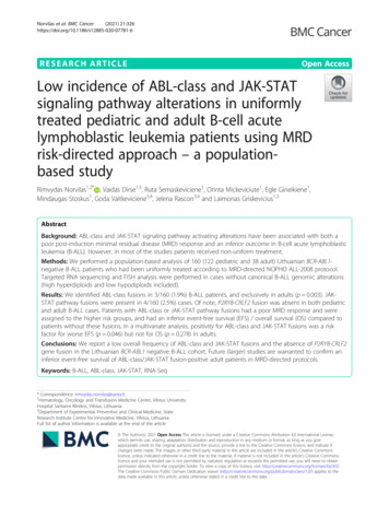
Norvilas et al. BMC Cancer(2021) SEARCH ARTICLEOpen AccessLow incidence of ABL-class and JAK-STATsignaling pathway alterations in uniformlytreated pediatric and adult B-cell acutelymphoblastic leukemia patients using MRDrisk-directed approach – a populationbased studyRimvydas Norvilas1,2* , Vaidas Dirse1,3, Ruta Semaskeviciene1, Orinta Mickeviciute1, Egle Gineikiene1,Mindaugas Stoskus1, Goda Vaitkeviciene3,4, Jelena Rascon3,4 and Laimonas Griskevicius1,3AbstractBackground: ABL-class and JAK-STAT signaling pathway activating alterations have been associated with both apoor post-induction minimal residual disease (MRD) response and an inferior outcome in B-cell acute lymphoblasticleukemia (B-ALL). However, in most of the studies patients received non-uniform treatment.Methods: We performed a population-based analysis of 160 (122 pediatric and 38 adult) Lithuanian BCR-ABL1negative B-ALL patients who had been uniformly treated according to MRD-directed NOPHO ALL-2008 protocol.Targeted RNA sequencing and FISH analysis were performed in cases without canonical B-ALL genomic alterations(high hyperdiploids and low hypodiploids included).Results: We identified ABL-class fusions in 3/160 (1.9%) B-ALL patients, and exclusively in adults (p 0.003). JAKSTAT pathway fusions were present in 4/160 (2.5%) cases. Of note, P2RY8-CRLF2 fusion was absent in both pediatricand adult B-ALL cases. Patients with ABL-class or JAK-STAT pathway fusions had a poor MRD response and wereassigned to the higher risk groups, and had an inferior event-free survival (EFS) / overall survival (OS) compared topatients without these fusions. In a multivariate analysis, positivity for ABL-class and JAK-STAT fusions was a riskfactor for worse EFS (p 0.046) but not for OS (p 0.278) in adults.Conclusions: We report a low overall frequency of ABL-class and JAK-STAT fusions and the absence of P2RY8-CRLF2gene fusion in the Lithuanian BCR-ABL1 negative B-ALL cohort. Future (larger) studies are warranted to confirm aninferior event-free survival of ABL-class/JAK-STAT fusion-positive adult patients in MRD-directed protocols.Keywords: B-ALL, ABL-class, JAK-STAT, RNA-Seq* Correspondence: rimvydas.norvilas@santa.lt1Hematology, Oncology and Transfusion Medicine Center, Vilnius UniversityHospital Santaros Klinikos, Vilnius, Lithuania2Department of Experimental, Preventive and Clinical Medicine, StateResearch Institute Centre for Innovative Medicine, Vilnius, LithuaniaFull list of author information is available at the end of the article The Author(s). 2021 Open Access This article is licensed under a Creative Commons Attribution 4.0 International License,which permits use, sharing, adaptation, distribution and reproduction in any medium or format, as long as you giveappropriate credit to the original author(s) and the source, provide a link to the Creative Commons licence, and indicate ifchanges were made. The images or other third party material in this article are included in the article's Creative Commonslicence, unless indicated otherwise in a credit line to the material. If material is not included in the article's Creative Commonslicence and your intended use is not permitted by statutory regulation or exceeds the permitted use, you will need to obtainpermission directly from the copyright holder. To view a copy of this licence, visit http://creativecommons.org/licenses/by/4.0/.The Creative Commons Public Domain Dedication waiver ) applies to thedata made available in this article, unless otherwise stated in a credit line to the data.
Norvilas et al. BMC Cancer(2021) 21:326BackgroundB-cell acute lymphoblastic leukemia (B-ALL) is an aggressive and genetically heterogeneous disease whichmostly affects children and young adults [1]. A numberof genomic alterations has been associated with the BALL outcome resulting in risk-adapted treatment protocols [2]. Approximately one-third of both pediatric andadult B-ALL cases do not harbor any canonical genomicalterations [3]. Gene expression profiling (GEP) of patients negative for canonical B-ALL alterations identifieda subgroup of cases characterized by a similar expressionprofile to those harboring t(9;22) (q34;q11)/BCR-ABL1(Philadelphia chromosome-like (Ph-like) or BCR-ABL1like B-ALL) [4, 5].Whole transcriptome/exome sequencing and fluorescence in situ hybridization (FISH) analysis of Ph-like BALL samples revealed high frequency of genomic alterations disrupting the normal function of kinase andcytokine receptor signaling pathways of which ABL-classkinase and JAK-STAT signaling pathway genes were themost commonly affected [6]. ABL-class gene and JAKSTAT pathway alterations are associated with high postinduction MRD levels, an increased relapse rate and inferior outcome. Of note, some of these gene fusions canbe successfully targeted using small-molecule inhibitors[7–12]. The identification of a Ph-like B-ALL signatureis not standardized and both ABL-class and JAK-STATfusions can also be found outside of the Ph-like B-ALLgroup [13–16]. High expression levels of the CRLF2gene and IKZF1 gene deletions are also common in thePh-like B-ALL subgroup resulting in a worse outcome insome series [5, 17].In most studies, a Ph-like B-ALL was clinically characterized as a high-risk group with inferior overall survivalin intensively treated children and adults [18–20]. Incontrast, Ph-like B-ALL children did not have inferiorsurvival when a minimal residual disease (MRD)-directed treatment of Total Therapy XV protocol was used[21, 22]. Whether MRD-directed treatment can also improve the negative prognosis of Ph-like B-ALL adult patients remains to be defined.In this study we performed a comparative genomicand clinical data analysis of pediatric and adult B-ALLpatients to characterize the incidence of ABL-class andJAK-STAT signaling pathway activating alterations inthe Lithuanian population and to determine their clinicalsignificance in a MRD-risk directed treatment setting.MethodsPatients and samplesFrom July 2008 to December 2017, pediatric (1 17year-olds) and adult (18 45 year-olds) B-ALL patientswho had been diagnosed with BCR-ABL1-negative BALL in Lithuania and enrolled into a NOPHO ALL-Page 2 of 122008 clinical trial [2] were included. In addition, eleven46 65 year-old adult patients were treated according toa NOPHO ALL-2008 protocol with a chemotherapydose modification and outside of the clinical trial. Thepatients’ data were collected retrospectively.The diagnosis of ALL was based on a bone marrow biopsy or aspirate showing 20% leukemic blasts. Theprotocol specific genomic analysis was limited to canonical B-ALL genomic alterations – t(12;21)/ETV6RUNX1, t(1;19)/TCF3-PBX1, 11q23/KMT2A gene rearrangements, iAMP21, dic(9;20), high hyperdiploidy (51–67 chromosomes) and low hypodiploidy (31–39 chromosomes). Cases without canonical B-ALL translocationswere selected for RNA sequencing. Patients with ploidyshifts (high hyperdiploidy or low hypodiploidy) were alsoselected for RNA sequencing as they could harbor additional kinase and cytokine receptor signaling pathwayalterations [23, 24].Patients were stratified to the three risk groups definedin the NOPHO ALL-2008 protocol detailed elsewhere[2]. Stratifying factors were white blood cell count,immunophenotype and cytogenetic markers at diagnosisand treatment response defined as MRD. High geneticrisk was assigned if KMT2A gene rearrangements,iAMP21, dic(9;20) or low hypodiploidy were present.The MRD analysis was performed on days 15, 29 and 79(only for standard risk (SR) and intermediate risk (IR)patients) or after each block (in high-risk (HR) arm untilMRD-negativity). If MRD was 0.1% on day 79 (SR andIR) or after block B1 (HR), patients were referred to anallogeneic hematopoietic stem cell transplant (SCT) afterreceiving at least one additional block of therapy andhaving MRD below 0.1% (optimally negative) (HR-SCTarm).The study was conducted in accordance with the Declaration of Helsinki, and the protocol was approved bythe Vilnius Regional Bioethics Committee. The patientsprovided written informed consent. The waiver forobtaining written informed consent from patients whocould not be reached despite our best efforts was approved by the Vilnius Regional Bioethics Committee according to national regulations.RNA purification and targeted RNA-sequencingTotal RNA was purified from fresh bone marrow samples at the time of diagnosis using silica-membranebased purification protocols and stored at 80 C. Allbone marrow samples had blast counts 70%. BeforeRNA sequencing (RNA-Seq), purified RNA concentration and quality was assessed using NanoDrop2000spectrophotometer.RNA-Seq libraries were constructed using the IlluminaTruSight Pan-Cancer sequencing kit (Illumina, SanDiego, CA, USA), following the manufacturer’s protocol
Norvilas et al. BMC Cancer(2021) 21:326and recommendations. The libraries were sequencedusing Illumina MiSeq genome analyzer (Illumina). Atotal of 1385 cancer-related genes were analyzed including frequently mutated genes of ABL-class (ABL1, ABL2,PDGFRB, CSF1R) and JAK-STAT (JAK1, JAK2, JAK3,CRLF2) signaling pathways. At least 4 million paired-endreads were obtained for each sample. NGS data analysiswas performed using the Illumina BaseSpace InformaticsSuite (Illumina). TopHat/STAR aligners and a Manta fusion caller were used to detect novel and recurrent genefusions. At least one partner gene was required to detectnovel gene fusion. Only high-confidence fusions werecalled that met threshold filters: split and paired uniquereads ( 3), fusion contig align ( 16 bp in length), coverage after fusion ( 100 bp), break end homology ( 10 bp).Additional gene mutation analysis from RNA-Seq datawas performed using the GATK pipeline and Isaac Variant Caller 2.3. Point mutations and small indels werecalled if they had an allele frequency 5% and a population frequency 0.01, were negative for known SNP’sand were previously described in genomic variant databases (COSMIC, ClinVar, OMIM, HGMD).Prior to the study, the RNA-Seq method was first validated using thirteen B-ALL control samples with previously identified gene fusions (BCR-ABL1 n 5, KMT2Agene rearrangements n 5, TCF3-PBX1 n 2, ETV6RUNX1 n 1). All in-frame gene fusions were identified,therefore the RNA-Seq method was used for B-ALLstudy patients.FISH, PCR and SNP Array data analysis of B-ALL fusionsDue to technical limitations of RNA-Seq, additionalFISH analysis of CRLF2 gene (CRLF2 (Xp22/Yp11)Break / IGH Fusion,TC; Leica Biosystems, Wetzlar,Germany) was performed to identify P2RY8-CRLF2 andIGH-CRLF2 gene fusions. FISH analysis for ABL1 (SPECABL1 Dual Color Break Apart Probe; ZytoVision, Bremerhaven, Germany), ABL2 (SPEC ABL2 Dual ColorBreak Apart Probe; ZytoLight), JAK2 (JAK2 (9p24)Break; Leica Biosystems) and PDGFRB (PDGFRB (5q32)Break; Leica Biosystems) genes were used to confirm respective gene fusions detected by the RNA-Seq method.FISH analysis was performed according to manufacturerprotocols.Standard RT-PCR followed by a gel electrophoresisand Sanger sequencing were used to confirm in-framefusions that were not confirmed with an additional FISHanalysis. Fusion specific PCR primers were constructedfor each case (Additional file 1: Table S1).P2RY8-CRLF2 fusion is a result a of 320 Kb size deletion of the PAR1 region in the short arm of either X orY chromosomes. We performed SNP Array data analysisfor detection of PAR1 region deletions in order to confirm the absence of P2RY8-CRLF2 gene fusion notPage 3 of 12detected by RNA-Seq or FISH methods in our cohort.The SNP-Array method was based on the Infinium 2 BeadChip (Illumina Inc., San Diego,CA), which covers the entire genome with an averagespacing of 9.6 kb. This coverage allows an average resolution of 31 kb which was used for detection of PAR1 region deletion.Statistical analysisDifferences in the prevalence of parameters between thegroups were determined using the Mann-Whitney testor Independent-Samples T-test for continuous variablesdepending on their distributions. For categorical analyses, either a Chi-square or a Fisher exact test was used.Univariate and multivariate Cox proportional hazard regression models were used to evaluate the effect of noncanonical B-ALL genomic alterations on survival. TheKaplan-Meier method was used to estimate the time toevent distributions (overall survival and event-free survival), the Log-Rank test was used to compare differences in survival curves. The overall survival (OS) wascomputed from the date of diagnosis until the date ofdeath or last known follow-up date. The event-free survival (EFS) was defined as the time from diagnosis to theevent of resistant disease, relapse, induction death, deathin remission, second malignancy or date of the lastfollow-up if a patient had no events. A p-value 0.05was considered to indicate statistical significance. Statistical analyses were performed using SPSS software version 20.ResultsStudy populationOverall, one hundred and sixty 1–65 year-old patientsrepresenting over 95% of the BCR-ABL1 negative B-ALLpatient population in Lithuania during the July 2008 –December 2017 period were included into this study(Table 1). One hundred and twenty-two (76.3%) patientswere younger than 18 years of age. The majority ofpediatric patients (59.5%) were stratified to the NOPHOALL-2008 standard risk treatment arm. In contrast,more than half of the adult patients were stratified eitherto the intermediate (44.7%) or high risk/high risk SCTdisease (29.0%).Genomic analysisGene fusion analysisG-banding/SNP-Array, FISH and standard RT-PCRmethods [25] were used to identify canonical B-ALLgenomic alterations at diagnosis. In order to detect noncanonical B-ALL genomic lesions, we performed RNASeq and CRLF2 gene break FISH analyses in B-ALL
Norvilas et al. BMC Cancer(2021) 21:326Page 4 of 12Table 1 Clinical characteristics of B-ALL patients1 17 yo18 65 yoAllNumber of patients122 (76.3%)38 (23.7%)160 (100%)Median age (range)4 (1–17)32 (18–65)5 (1–65)Male69 (56.6%)15 (39.5%)84 (52.5%)Female53 (43.4%)23 (60.5%)76 (47.5%)SR72 (59.5%)7 (18.4%)79 (49.7%)IR38 (31.4%)17 (44.7%)55 (34.6%)HR5 (4.1%)6 (15.8%)11 (6.9%)HR-SCT3 (2.5%)5 (13.2%)8 (5%)Induction failure3 (2.5%)3 (7.9%)6 (3.8%)Not risk grouped101p-ValueAge groupsSex:0.093Risk group: 0.001Induction:Prednisolone113 (92.6%)30 (78.9%)143 (89.4%)Dexamethasone9 (7.4%)8 (21.1%)17 (10.6%) 100113 (92.6%)30 (78.9%)143 (89.4%)0.030WBC ( 109/l):0.030 1009 (7.4%)8 (21.1%)17 (10.6%)WBC median (range)14 (1–641)10.5 (0.9–481.9)13.5 (0.9–641)0.672Hgb (g/l) median (range)83 (24–140)90.5 (52–142)84 (24–142)0.117Platelets median (range)62 (0–457)37 (5–320)54.5 (0–457)0.075CNS1104 (85.2%)33 (86.8%)137 (85.6%)0.281CNS214 (11.5%)2 (5.3%)16 (10%)CNS34 (3.3%)3 (7.9%)7 (4.4%)CNS Status:SR Standard risk, IR Intermediate risk, HR High risk, HR-SCT High risk–stem cell transplant groups, WBC White blood cells, HgB Hemoglobin, CNS centralnervous systemcases lacking canonical gene fusions or dic(9:20)/iAMP21 aberrations (Fig. 1).In total, 99/160 (61.9%) cases harbored at least one canonical B-ALL genomic alteration (Table 2, Fig. 1). Highhyperdiploidy was identified in 35/160 (21.9%) patients.Low hypodiploidy was present in 5/160 (3.1%) cases.ETV6-RUNX1 fusions were detected in 34/160 (21.3%)cases and were more common among children thanadults (33 children vs. 1 adult, p 0.001). In contrast,KMT2A gene rearrangements were detected in 11/160(6.9%) B-ALL cases (4 children vs. 7 adults). Importantly,RNA-Seq analysis identified two additional KMT2A generearrangements (del(11q23)/KMT2A-CBL, del(11q23)/KMT2A-ATP5L) that had been missed by FISH. iAMP21aberration was exclusive to the pediatric group (n 3,1.9%) while dic(9;20) was present in 1/160 (0.6%) adultpatient.After the exclusion of two patients with KMT2A generearrangements identified by RNA-Seq, remaining caseswithout canonical B-ALL gene fusions and cases withhigh hyperdiploidy or low hypodiploidy (n 101) wereselected for RNA-Seq and CRLF2 gene break FISH analysis to identify other kinase and cytokine receptor activating lesions (Fig. 1).In-frame fusions of ABL-class genes were detected in3/101 (3%) patients (Table 2; Fig. 1, Fig. 2). One casehad t(9;12)/ETV6-ABL1 fusion previously reported inboth lymphoid and myeloid leukemias [26]. Theremaining two cases harbored t(1;7)/ZC3HAV1-ABL2and t(5;5)/EBF1-PDGFRB fusions. All ABL-class fusionswere exclusive to adults (p 0.003).JAK-STAT pathway fusions were identified in 4/101(4%) cases. ABL-class and JAK-STAT pathway fusionswere mutually exclusive. RNA-Seq identified t(9;22)/BCR-JAK2 fusion in one pediatric patient. FISH analysis revealed three cases with t(Y;14) (X;14)/IGHCRLF2 gene fusion, of which two were adults. BCRJAK2 fusion was detected in one case with high
Norvilas et al. BMC Cancer(2021) 21:326Page 5 of 12Fig. 1 CONSORT diagram of B-ALL study cases. High hyperdiploids, low hypodiploids and cases with no canonical B-ALL alterations, iAMP21 ordic(9;20) were selected for RNA-Seq and FISH (CRLF2 gene break) analysis. After RNA-Seq data analysis, two cases with KMT2A generearrangements were excluded. A total of 101 B-ALL patients were screened for non-canonical B-ALL alterations. The number of FLT3-TKD, otherJAK-STAT and Ras pathway gene mutations corresponds to the total number of specific alterations in sequenced caseshyperdiploidy which further confirms that JAK-STATpathway gene fusions can be detected in this entity.Both adult cases with IGH-CRLF2 fusions had additional gene mutations. One IGH-CRLF2-positive caseharbored FLT3-TKDD835Y and NRASG13R gene mutations while another case had CRLF2F232C gene mutation. RNA-seq and FISH analysis detected no P2RY8CRLF2 fusions in our study patients. SNP Array analysis was available for 81/101 (80.2%) of the sequenced B-ALL patients. No deletions of PAR1 regionwere identified in any of the cases confirming the absence of P2RY8-CRLF2 fusion.We identified other gene fusions in 10/101 (9.9%)pediatric B-ALL cases. Five cases had PAX5 generearrangements: t(9;18)/PAX5-GREB1L, t(9;20)/PAX5NCOA5, dic(9;12)/PAX5-ETV6, dic(3;9)/PAX5-FOXP1and dic(9;20)/PAX5-NOL4L. One case with PAX5GREB1L fusion had an additional JAK1F838V mutation,while in the other two PAX5-rearranged cases KRASG12Vand NRASG12C mutations were identified. Three caseshad fusions involving ZNF384 gene (EP300-ZNF384, n 2; TCF3-ZNF384, n 1) of which the TCF3-ZNF384positive case had an additional NRASG13R mutation.Though ETV6-RUNX1 is the most common alterationin childhood B-ALL, data analysis revealed a novel fusion of ETV6 and RUNX2 genes in one pediatric patientwhile another patient harbored a t(7;15)/CUX1-NUMT1fusion.
Norvilas et al. BMC Cancer(2021) 21:326Page 6 of 12Table 2 Genomic subgroups of canonical and non-canonical B-ALL alterations in study patientsCanonicalalterationsGenomic alterationsPediatric group (n 122)Adult group (n 38)Total cohort (n 160)p-Value (ChiSquare)t(12;21)/ETV6-RUNX133 (27.1%)1 (2.6%)34 (21.3%) 0.001t(1;19)/TCF3-PBX17 (5.7%)3 (7.9%)10 (6.3%)0.70111q23/KMT2A generearrangements4 (3.3%)7 (18.4%)11 (6.9%)0.004iAMP213 (2.5%)03 (1.9%)0.331dic(9;20)01 (2.6%)1 (0.6%)0.071High hyperdiploidy32 (26.2%)3 (7.9%)35 (21.9%)0.030Low hypodiploidy2 (1.6%)3 (7.9%)5 (3.1%)0.086No canonical alterationsNon-canonicalalterations41 (33.6%)20 (52.7%)61 (38.1%)n 75n 26n 101ABL-class fusions03 (11.5%)3 (3%)0.003JAK-STAT pathway fusions2 (2.7%)2 (7.7%)4 (4%)0.262Other fusionsa10 (13.3%)010 (9.9%)0.050JAK-STAT pathway mutations6 (8%)4 (15.4%)10 (9.9%)0.182Ras pathway mutations39 (52%)14 (53.8%)53 (52.5%)0.772FLT3-TKD mutations7 (9.3%)1 (3.8%)8 (7.9%)0.378aPAX5-NCOA5, PAX5-ETV6, PAX5-FOXP1, PAX5-NOL4L, PAX5-GREB1L, EP300-ZNF384, TCF3-ZNF384, ETV6-RUNX2, CUX1-NUTM1Fig. 2 Gene fusions and mutations identified by the RNA-Seq and FISH methods. The cohort is divided into patients with ABL-class fusions, JAKSTAT pathway fusions, other JAK-STAT–activating mutations, Ras pathway mutations, and cases with other or no gene variants
Norvilas et al. BMC Cancer(2021) 21:326Gene mutation analysisGene mutation analysis of RNA-Seq data revealed tenother JAK-STAT pathway mutations in a total of 9/101(8.9%) cases: JAK1 (n 2), JAK2 (n 7), CRLF2 (n 1)(Table 2; Fig. 2, Additional file: Table S2). One case hadboth JAK1 and JAK2 gene mutations in the same sample.In all cases, protein kinase 1 and kinase 2 domains ofJAK1 and JAK2 genes were affected. Four JAK2-mutatedcases were co-mutated with Ras pathway genes (KRASn 2; NRAS n 1; PTPN11 n 1).A total of fifty-three Ras pathway gene point mutations were detected in 48/101 (47.5%) cases (Table 2;Fig. 2; Additional file 1: Table S2). NRAS gene mutationswere the most common (n 29) while the incidence ofKRAS (n 17) and PTPN11 (n 7) gene mutations waslower. One case had mutations in NRAS and PTPN11genes while another case had two different NRAS genemutations (G12S and G12A) in the same sample. Twoother cases had both KRAS and NRAS gene mutations inthe same sample. Ras pathway mutations were detectedin 16/35 (45.7%) high hyperdyploid and 2/5 (40%) lowhypodiploidy cases.FLT3-TKD activating mutations of codons D835 (n 5) and I836 (n 3) were found in 8/101 (7.9%) cases.Most of the FLT3-TKD positive cases (75%) werepresent with Ras pathway gene mutations. Other FLT3gene kinase domain point mutations were present in 5/101 (5%) patients (Fig. 2).Other gene mutations were identified in 23/101(22.8%) patients. Mutations in FLT3 (n 7), CREBBP(n 5) and TP53 (n 3) genes were the most recurrentin this group of patients. RNA-Seq and FISH methodsfailed to identify ALL-related gene fusions and mutations in 14/101 (13.9%) cases.Clinical outcome of patients with ABL-class or JAK-STATfusionsFive of 26 (19.2%) adult and two of 75 (2.7%) pediatricB-ALL cases without canonical B-ALL alterations (5/38(13.2%) adult and 2/122 (1.6%) pediatric BCR-ABL1negative B-ALL cases) were positive for either ABL-classor JAK-STAT pathway fusions (AJS-positive group)(Table 3). Overall, both EFS and OS were worse in theAJS-positive group vs the AJS-negative (n 153)(Fig. 3a).We further compared the outcome of AJS positive(n 5) vs. AJS negative (n 33) B-ALL cases in adultssince the number of AJS-positive pediatric cases was toosmall for detailed analysis. Four AJS-positive adult patients were evaluated for residual disease on inductionday 15 (one patient died during induction). All four AJSpositive patients (100%) had 5% blasts on day 15 compared to 9 (29%) AJS-negative patients (p 0.019)(Table 4). After day 15, one patient was assigned toPage 7 of 12block treatment; therefore, three patients had MRD dataavailable on day 29. MRD levels on days 29 and 79 werehigher in the AJS-positive group compared to the AJSnegative group. As a result of a poor MRD response,more AJS-positive than AJS-negative adult patients hadinduction failure or were assigned to HR/HR-SCT group(4 (80.0%) vs 10 (30.3%), respectively, p 0.052).The median observation time in adults was 39 months.The 75th percentile EFS was 5 vs. 35 months (p 0.009)and OS was 14 vs. 36 months (p 0.098) in AJS-positivevs. AJS-negative adults, respectively (Fig. 3b). Since allpatients were assigned to different risk groups accordingto their canonical B-ALL alterations and MRD response,we performed a multivariate analysis of risk-group assignment and AJS-positivity on the outcome in adults.In multivariate analysis, having ABL-class or JAK-STATpathway fusions was an independent risk factor forworse EFS (p 0.046) (Table 5).DiscussionWe present the first Baltic European population-basedstudy of genomic alterations among pediatric and adultBCR-ABL1-negative B-ALL patients. All patients wereuniformly treated according to the NOPHO ALL-2008protocol with risk stratification according to both the canonical B-ALL genomic lesions and minimal residualdisease. We selected targeted RNA-Seq and FISHmethods for the detection of known and novel gene rearrangements of ABL-class, JAK-STAT pathway genesand other kinase alterations.In our study, ABL-class fusions were identified in 3/101 (3%) B-ALL cases without canonical B-ALL alterations or 3/160 (1.9%) BCR-ABL1-negative B-ALL cases.All positive cases were adults (3/38, 7.9%) (Fig. 2). Incomparison, in the Dutch/German cohort 9 of 153(5.9%) pediatric B-ALL patients without canonical BALL alterations harbored ABL-class fusions [14]. Another European study by Zaliova et al. revealed only 1/75 (1.3%) pediatric B-ALL case with ABL1 gene rearrangement [27] while ABL-class fusions were presentin 40/1389 (2.9%) high-risk pediatric B-ALL patients inan US Children’s Oncology Group study [13]. Heatley atel. performed targeted RNA-Seq in 63 adolescent/youngadults (16–39 yo) and 63 adults (40–88 yo) with BCRALB1-negative B-ALL [28]. ABL-class fusions werefound in 4/126 (3.2%) of these B-ALL cases. A study byGarrido et al. used the FISH method to analyze 39 adultB-ALL patients negative for the BCR-ABL1 and KMT2Agene rearrangements and identified 3/39 (7.7%) caseswith ABL-class fusions (ABL1 n 2; CSF1R n 1) [29].In a similar study from a UKALL14 clinical trial, ABLclass abnormalities were present in 6/648 ( 1%) of BALL patients [30]. The incidence of ABL-class fusions of7.9% in our adult patient cohort was largely in line with
Norvilas et al. BMC Cancer(2021) 21:326Page 8 of 12Table 3 Genetic features and clinical outcome of ABL-class or JAK-STAT pathway fusion (AJS)-positive patientsCaseno.AgegroupIn-frame genefusionWBC DEX /PREDNIMRDD15MRD D29MRD D79 V6-ABL1270DEX NYInduction RYYDeath of sepsis afteralloSCT in CR1Case3AdultEBF1-PDGFRB258DEX45.3%0.93%(POST A1)0.7%(POST B1)HR-SCTYYRelapse after alloSCT -SCTYYRelapse after alloSCT andalive in ive after alloSCT in CR1Case6Pediatric IGH-CRLF235PREDNI0.22% 0.1%0.3%HR-SCTYNAlive after alloSCT in CR1Case7Pediatric BCR-JAK25PREDNI1.8%0.33% 0.1%IRNNAlive in CR1WBC White blood cells, DEX Dexamethasone, PREDNI Prednisolone, D15 Day 15, D29 Day 29, D79 Day 79, MRD Minimal residual disease, alloSCT Allogenic stem celltransplant, CR1 First clinical remission, CR2 Second clinical remissionFig. 3 Event-free survival (EFS) and overall survival (OS) in months in ABL-class or JAK-STAT pathway fusion (AJS)-positive vs. AJS-negativepatients. a, all B-ALL patients (7 AJS-positive vs 153 AJS-negative pts); b, adult B-ALL patients (5 AJS-positive vs 33 AJS-negative pts)
Norvilas et al. BMC Cancer(2021) 21:326Page 9 of 12Table 4 Minimal residual disease (MRD) of AJS-positive vs. AJS-negative adult patients on days 15, 29 and 79TimepointMRDAJS-positive (n 5)AJS-negative (n 33)D15 5%0 (0%)22 (71%) 5%4 (100%) 1 missing9 (29%) 2 missingD29D29D79p-Value (Chi-Square)0.019 5%0 (0%)30 (96.8%) 5%3 (100%) 2 missing1 (3.2%) 2 missing 0.001 0.1%0 (0%)21 (67.7%) 0.1%3 (100%) 2 missing10 (32.3%) 2 missing0.011 0.1%2 (50%)26 (89.7%) 0.1%2 (50%) 1 missing3 (10.3%) 4 missing0.120AJS ABL-class or JAK-STAT pathway fusions, D15 Day 15, D29 Day 29, D79 Day 79m, MRD Minimal residual diseasepublished adult studies. Conversely, we did not detectABL-class fusions in our pediatric B-ALL patients.We identified JAK-STAT pathway fusions in 4/101(4%) cases without canonical B-ALL alterations. JAKSTAT pathway fusions occur in approximately 3–5% ofchildhood and in up to 15% of adult B-ALL [3]. A studyby Heatley et al. showed the incidence rate in adults of15.9% [28]; however, another study revealed that only5.1% of adult B-ALL had JAK-STAT pathway fusions[30]. Similarly, our data indicate a lower frequency ofJAK-STAT pathway fusions in both adult and pediatricLithuanian B-ALL patients.Overall, the frequency of CRLF2 rearrangements in BALL is approximately 5% and the rate gets higher incases without canonical B-ALL alterations (10–30%) andin patients with Down syndrome ( 50%) [16, 27, 28, 31,32]. In our study, IGH-CRLF2 fusions were identified in3/101 (2.9%) B-ALL cases without canonical B-ALL alterations or in 3/160 (1.9%) BCR-ABL1-negative B-ALLcases. Notably, we did not detect any P2RY8-CRLF2gene fusions in our cohort by either RNA-Seq or CRLF2gene break FISH. In comparison, a Swedish study identified CRLF2 gene rearrangements in 16 of 189 (8.5%)pediatric BCR-ABL-negative B-ALL patients, of whichP2RY8-CRLF2 fusion was the most common (12/16,75%) [16]. P2RY8-CRLF2 gene fusion is the result of deletion of PAR1 region in either X or Y chromosomes.To further confirm the absence of P2RY8-CRLF2 fusion,we used SNP Array data of 81/101 (80.2%) sequencedpatients and did not detect any deletions in thecorresponding PAR1 region. In addition, another studyhas shown that P2RY8-CRLF2 fusion can be identifiedusing the RNA-Seq method and an analysis algorithmsimilar to ours [33], making it unlikely that P2RY8CRLF2 fusion was missed due to technical reasons inour study patients. CRLF2 gene rearrangements arecommon in populations with Hispanic ancestry and areassociated with increased risk of relapse in both childrenand adults [17, 34–36]. Population differences may explain a lower frequency of IGH-CRLF2 and the absenceof P2RY8-CRLF2 gene fusions in our cohort. In other BALL studies, approximately half of CRLF2-rearrangedpatients also harbored additional JAK1 or JAK2 genemutations [13], however our positive cases had no suchmutations. Of note, one case had FLT3-TKDD835Y andNRASG13R gene mutations and another case was positivefor a CRLF2F232C activating mutation. CRLF2F232C genemutation is known to promote constitutive dimerizationand cytokine-independent growth which results in geneoverexpression in a simila
alterations [3]. Gene expression profiling (GEP) of pa-tients negative for canonical B-ALL alterations identified a subgroup of cases characterized by a similar expression profile to those harboring t(9;22) (q34;q11)/BCR-ABL1 (Philadelphia chromosome-like (Ph-like) or BCR-ABL1-like B-ALL) [4, 5]. Whole transcriptome/exome sequencing and fluores-