Transcription
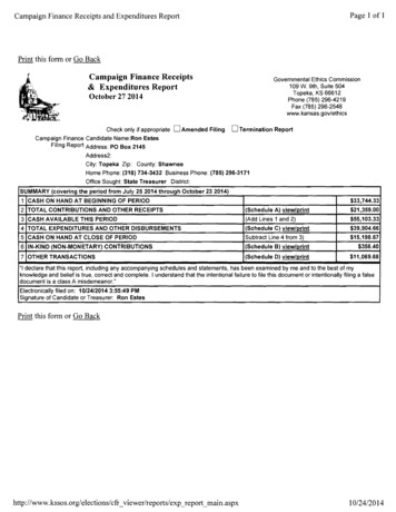
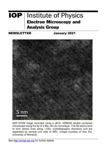
NEWSLETTERJanuary 2021ADF-STEM image recorded using a JEOL ARM200 double correctedmicroscope along the tip of a Nb0.1W0.9S2 monolayer. The Nb atoms tendto form atomic lines along 100 crystallographic directions and areseparated by several unit cells of WS2. (image courtesy of Xue Xia,University of Warwick)See http://emag.iop.org for further details
EMAG Group newsletterJan 2021CONTENTSEMAG COMMITTEE4LETTER FROM THE CHAIR5FORTHCOMING EMAG EVENTS7EMAG2021REMOTE HYPERSPY WORKSHOPSUPERSTEM SUMMER SCHOOLEMAG ANNUAL GENERAL MEETING 2021NEWS78889IN MEMORIAMSUPERSTEM FACILITY UPDATESEPSIC FACILITY UPDATESELECTRON MICROSCOPY - RESEARCH AND TECHNOLOGICAL PLATFORM ATWARWICK UNIVERSITYRESEARCH HIGHLIGHTS BY YOUNG SCIENTISTS910111214ENHANCING THE PERFORMANCE OF HYBRID PIXEL DETECTORS FOR HIGH-ENERGYTEM USING HIGH-Z SENSORS14ULTRAFAST NANO-IMAGING OF THE ORDER PARAMETER IN A STRUCTURAL PHASETRANSITION16MEETING REPORTS18EMAG2020: MICROSCOPY ENABLED BY DIRECT ELECTRON DETECTIONVIRTUAL EARLY CAREER EUROPEAN MICROSCOPY CONGRESS (EMC) 2020MICROSCOPY INFRASTRUCTURES ACCESS SCHEMESUPERSTEMEPSICESTEEM 3LEEDS EPSRC NANOSCIENCE AND NANOTECHNOLOGY FACILITY (LENNF)HENRY ROYCE INSTITUTEIOP RESEARCH STUDENTS CONFERENCE FUND2181921212122222426
EMAG Group newsletterJan 2021BURSARIES REPORTS (2020)26EMS MEMBERSHIP26ADDITIONAL FUTURE MEETINGS OF INTEREST26Focus on Microscopy 2021 - FOM2021EBSD 2021PICO 2021 - Sixth Conference on Frontiers of Aberration CorrectedElectron MicroscopyQEM 2021 (5th edition) - Quantitative Electron MicroscopyADVANCED WORKSHOP ON CRYO-ELECTRON TOMOGRAPHY32627272727
EMAG Group newsletterJan 2021EMAG COMMITTEEChairDr Andy BrownSchool of Chemical and Process Engineering, University of Leeds, Leeds, LS2 9JTTel: 0113 343 2382 Email: A.P.Brown@leeds.ac.ukSecretary & Honorary TreasurerProf Ana SanchezDepartment of Physics, University of Warwick, Coventry, CV4 7ALTel: 0247 615 1372 Email: a.m.sanchez@warwick.ac.ukOrdinary MembersDr Miryam Arredondo, Queen’s University Belfast m.arredondo@qub.ac.ukDr Laura Clark, University of Leeds, L.Clark@leeds.ac.ukMr Michael Dixon, Hitachi Europe, Michael.Dixon@hht-eu.comDr Sarah Harper, JEOL Ltd, sarah.harper@outlook.comDr Jo Sharp, University of Sheffield, j.sharp@sheffield.ac.ukDr Donald MacLaren, University of Glasgow, donald.maclaren@glasgow.ac.ukDr Cornelia Rodenburg, University of Sheffield, c.rodenburg@sheffield.ac.ukDr Larry Stoter, Retired, larry.stoter@gmail.comProf Jun Yuan, University of York, jun.yuan@york.ac.uk4
EMAG Group newsletterJan 2021LETTER FROM THE CHAIRDear Friends and Colleagues,Well, what a year! It is unfortunate that most of the news was not fake however,the COVID-19 enforced move to online activity has been kind to EMAG and forthat I would like to thank you our members for your engagement and positivityunder the rapidly changing circumstances.Our focussed EMAG workshop for 2020 was due to be in Glasgow, coveringMicroscopy enabled by direct electron detection. We duly moved this eventonline, prepared a compressed format (aimed to fit with the pressures of workingfrom home) and held our breath! We need not have worried, come the 6th Julywe had 280 delegates registered with typically more than 200 in-attendance attalks (7 invited speakers, 22 contributed scientific flash talks, 8 short trade talksand 10 online posters plus a Zoom pub quiz on the evening of the 7 th July). Wefeel the meeting was of high scientific quality with good, lively engagement ofparticipants and, for us, set the benchmark of what is possible with onlinemeetings. Thanks therefore go to Donald MacLaren for all the hard work inarranging and re-arranging this meeting as conference chair and to the IoP teamfor all their support in the organisation. There is a full meeting report by Donaldin this Newsletter (page 18).Online meetings are clearly here to stay and bring many benefits such as reducedtravel for example, see the excellent report on the Virtual Early Career EuropeanMicroscopy Congress meeting by Tamsin O’Reilly (Queen’s University Belfast;page 19). On this note, EMAG 2021 will be online, again as part of the RMS’sMMC conference series in July 2021 (www.mmc-series.org.uk). Abstractsubmission is to be confirmed but we have a fantastic group of invited speakersalready lined up (www.mmc-series.org.uk/conference/emag-2021). I encourageyou all to submit your recent work and look forward to ‘seeing’ many of you there!Student engagement remains a real feature and goal of EMAG meetings and Iwould like to draw your attention to reports from our prize-winning contributors tothe EMAG 2020 workshop; Kirsty Paton (University of Glasgow; page 14) andThomas Danz (University of Göttingen; page 16).I would like to welcome our new committee member for 2020, Dr Jo Sharp(University of Sheffield). Special thanks are also due to our outgoing co-optedcommittee member, Prof Sarah Haigh (University of Manchester). Sarah hasmade a fantastic contribution to EMAG over the years, finishing by taking on therole of editor for the Journal of Microscopy Special Issue covering EMAG 2019 atMMC 19. We deliberately chose to reflect recent trends and publish in a journal5
EMAG Group newsletterJan 2021rather than as a conference proceedings, and are pleased with the result of 21published articles /279/3). Wethank Sarah for all the hard work and hope you enjoy reading the Issue. We willbe aiming for a similar approach to a Special Issue for EMAG 2021 at MMC 21.Last but not least, I’d like to thank Ana Sanchez for putting together this excellentnewsletter. We have updated the list of physical science facing MicroscopyInfrastructure Access Schemes in the UK as we feel this remains a useful listingfor EMAG members (page 21). As always, we welcome your contributions forfuture issues or please let us know if we have missed anything – you can alwaysstay up to date with us on Twitter (@IoP EMAG)!Best wishes for 2021 and stay safe,Andy BrownUniversity of Leeds, EMAG Chair6
EMAG Group newsletterJan 2021FORTHCOMING EMAG EVENTSEMAG2021Call for PapersAbstract submission deadline TO BE CONFIRMED.Submit through .htmlOrganised by the Institute of Physics's Electron Microscopy and AnalysisGroup (EMAG), the 2021 EMAG Conference will be part of mmc2021.The EMAG2021 conference will be held online on 5-8th July 2021Confirmed Invited Speakers Prof. Ido Kaminer (Technion – Israel Institute of Technology, Israel) Dr. Demie Kepaptsoglou (SuperSTEM laboratory, Daresbury, UK) Dr. Emanuela Liberti (University of Oxford, UK) Dr. Lorena Ruiz-Perez (Imperial College London, UK) Dr. Steven R. Spurgeon (Pacific Northwest National Laboratory, USA) Dr. Andy Stewart (Department of Physics and Bernal Institute, Universityof Limerick, Ireland) Dr. Toma Susi (University of Vienna, Faculty of Physics, Austria)Abstract submission TO BE CONFIRMED (https://www.mmc-series.org.uk/).7
EMAG Group newsletterJan 2021Remote Hyperspy WorkshopDate TBC. ePSIC, Harwell, UKThis workshop is aimed to be an introduction to the Hyperspy python package forthe analysis of multidimensional data (primarily in electron microscopy). Topicswill include an introduction to Hyperspy and analysis of spectroscopic, scanningdiffraction and atomic resolution data. Due to COVID-19, we are aiming to runthis year’s workshop fully remotely. Participants will need to have their owncomputer and will be assisted over Zoom. Further details will be released shortly,but please contact mohsen.danaie@diamond.ac.uk if interested in attending.SuperSTEM Summer SchoolIn light of the further public health and travel restrictions worldwide, it would beunreasonable to expect we would be in a position to deliver the essential handson training that the SuperSTEM summer school normally offers this year. Theevent, initially scheduled for the summer of 2020, will now be taking place insummer 2022. Please check the facility’s website regularly for further updates, orsign up to receive regular updates: https://www.superstem.org/signupEMAG Annual General Meeting 2021No AGM of the EMAG group was held in 2020 as all our meetings were online!This may well remain the case for much of 2021 and IoP do not insist we hold anAGM however we will aim to have a short online meeting as part of EMAG 2021at mmc21. Assuming we do indeed go ahead with this an agenda and moredetails will be circulated nearer the time. If you cannot attend the AGM but haveany issues you would like to be raised at the meeting, please contact the honorarysecretary (a.m.sanchez@warwick.ac.uk).8
EMAG Group newsletterJan 2021NEWSIn memoriamDr Ziyou Li (29.09.1959 – 18.12.2020)It is with sadness to report the passing of Dr. Ziyou Li. She was a Reader inPhysics at University of Birmingham. She was well-known to members of theEMAG group, serving as a committee member from 2011-2018. She and Yulong were the local organizers for the successful EMAG2011 meeting held inBirmingham. Her research interests range from adsorption studies on surfacesto three-dimensional structure imaging of ultra-small nanoclusters and catalyticnanoparticles using techniques including quantitative scanning transmissionelectron microscopy. She was the first to apply quantitative atom countingtechniques for nanoparticle metrology. Her research has broad impact in clusterand catalytic science.Dr Kenneth Smith (20.03.1928 – 15.03.2020)We are sad to report that Ken Smith (K.C.A. Smith) passed away on 15.3.2020,a few days before his 92nd birthday - his name will be familiar to the oldergeneration of EMAG members. As you may be aware, the SEM as we know ittoday was developed in Charles Oatley’s lab in the Cambridge UniversityDepartment of Engineering in the years after WWII. The first instrument wasconstructed by Dennis McMullan; it was taken over and very considerablyimproved by Ken Smith who was also responsible for the first British HVEM in theCavendish Lab. Ken published an autobiographical article entitled “ElectronMicroscopy at Cambridge University with Charles Oatley and Ellis Cosslett: SomeReminiscences and Recollections” in Advances in Imaging & Electron Physics177 (2013)189–277.Prof. Sir John Meurig Thomas (15.12.1932-13.11.2020)It is also a great sadness to share the news that Professor Sir John MeurigThomas passed away on 13.11.2020. He was Director of the Royal Institution ofGreat Britain, a former head of the Department of Physical Chemistry and formerMaster of Peterhouse, University of Cambridge. Sir John was renowned for hiswork in the science of catalysts and solid-state chemistry and received aknighthood in 1991. Sir John was among the first to use electron microscopes tostudy structure/properties relationship in materials. He applied TEM imagingtechniques to analyse organic crystals, nanoporous materials and improve theircatalytic properties. His research also had a major impact in heterogeneouscatalysis and surface science.9
EMAG Group newsletterJan 2021SuperSTEM Facility updatesNew instrumentation – hybrid-pixel detectors for EELSCovid-19 permitting, the electron energy loss spectrometers (EELS) of theSuperSTEM2 and SuperSTEM3 microscopes will be retro-fitted in Spring 2021with new hybrid-pixel detectors optimised for EELS at low- to mid- primary beamacceleration voltage (40-100 kV). These will enable virtually noise-free low-signaldetection at high data rates.The monochromated SuperSTEM3 instrument will be equipped with aNion/Dectris ELA camera, specifically developed in collaboration with Nion Co.to exploit the 4 meV capabilities of the IRIS spectrometer, while the specificationsof the SuperSTEM2 detector are still being finalised. Check www.superstem.orgfor news and further updates.New instrumentation – Triple-beam FIB and high-resolution analytical lowvoltage TSEMSuperSTEM is in the process of commissioning a new highly specified triplebeam FIB/SEM/Ar instrument. The bespoke Hitachi NX5000 will be dedicatedto the fabrication and finishing of high-quality, uniformly thin specimens. Thanksto the third beam (Ar /Xe ) for low-damage highly-uniform milling, a 7-axissample stage for advanced positional control and an additional side-entry cryostage for direct milling at low temperatures, this new nanofabrication platform willuniquely address the very demanding requirements for samples with minimalsurface damage, roughness, and amorphisation. These are particularly strictgiven the facility’s focus on atomic-level characterisation at mid- to lowacceleration voltage, necessitating fabrication capabilities that go beyond typicalFIB-SEM instrumentation.The NX5000 will also provide the ability to observe the as-prepared samplelamellae in the advanced SEM column using analytical capabilities such as theobservation of samples in transmission mode (‘transmission’ SEM or TSEM),along with chemical (high solid-angle EDXS) and crystallographic (EBSD)characterisation. This instrument will be available to the user community throughthe usual facility access procedures https://www.superstem.org/facility/access. Itwill be supported by a dedicated FIB scientist, Dr Aleksander Mosberg(abmosberg@superstem.org), who recently joined the team with a researchfocus on methodological and instrumentation developments in FIB science.10
EMAG Group newsletterJan 2021In close collaboration with Hitachi High-Tech Europe, the triple-beam FIB-SEMwill be complemented by an additional high-resolution analytical low-voltageTSEM – a Hitachi SU9000. This instrument, also equipped with state-of-the-artanalytical instrumentation (windowless, high solid angle EDXS), will expand thefacility’s capabilities for low-voltage TSEM down to 0.3 nm spatial resolution.ePSIC Facility UpdatesePSIC, a national user facility for electron microscopy, has made significantupgrades to its equipment over the last 12 months. These upgrades will allownew experimentation, opening up fast TEM at high voltages and fully anaerobicstudies. The specific upgrades include: A new Direct Electron DE-16 direct detection camera, installed on theARM 300. This camera has a 4K x 4K field of view with a readout time of 60 s atfull frame, or 4237 fps when reading out small areas (128 X 128 pixels). Theperformance of the DE-16 is particularly tailored for high energy electrons (200and 300 kV) and therefore will complement our existing MerlinEM camera thathas optimal performance at low energies (80 kV and below). High-kV experiments11
EMAG Group newsletterJan 2021including fast-TEM, 4D STEM and scanning electron diffraction can now beperformed on this new camera. In conjunction with the new DE camera, a new scan generator has alsobeen installed on the ARM 300. This scan generator allows arbitrary patterns tobe input. Experiments exploring new scan geometries are now possible (includingspiral and Lissajous patterns), in addition to sparse scans for compressedsensing approaches. The scan generator can also be used to trigger the new DE16 camera, enabling arbitrary patterned 4D STEM experiments.Equipment has been installed to allow anaerobic transfer of specimensat ePSIC. A vacuum transfer holder for JEOL TEMs allows anaerobic transferfrom a glove box to the microscope. In addition, a transfer chamber allowsanaerobic transfer to and from a glovebox to the JEOL FIB. It is now possible toperform FIB sample preparation of TEM samples with no exposure to air, openingup atomic resolution studies of materials such as lithiated batteries and hydrogenembrittled grain boundaries.Electron Microscopy - Research and TechnologicalPlatform at Warwick UniversityInstrumentation update: New FIBSEM for sample preparationA new multi-source Focus Ion Beam (FIB)-Scanning machine has been recentlyinstalled at the University of Warwick, funded by the university as part ofWarwick’s capital plan for central research facilities. Currently, researchers arepushing the boundaries of elemental analysis and high-resolution imaging withtransmission electron microscopy (TEM), and for these studies they require ultralow damage specimen preparation. Focused ion beam (FIB) milling and argon(Ar) ion beam milling are used for preparation of electron transparent specimensfor a diverse class of materials, including semiconductors, metals and ceramics.The new system acquired will provide significantly improved capabilities,specifically for low-energy operation to produce damage-free, clean specimensonly a few nm in thickness for high resolution TEM studies. This is part ofWarwick’s central facility for electron microscopy and is available not just toresearchers at Warwick, but more widely to academic and commercial usersthroughout the UK, e.g. with Warwick Analytical Science Centre funding(EP/V007688/1), which runs until 2024.Our new FIB is a Tescan Amber FIBSEM that has two columns. First, a 1-30 kVelectron column with a Field Emission Gun enabling the taking of high-resolution(1.5 nm) Scanning Electron Microscopy images (SEM). Second a 0.5- 30kV ioncolumn with a Ga ion source to allow focused ion beam (FIB) cutting andimaging of samples with a multiple Gas Injector System (GIS) for depositing a12
EMAG Group newsletterJan 2021variety of different materials. There is also an Oxford Instruments EDS system toenable elemental analysis and element mapping of samples. The FIB allows forvery precise cutting of samples, with the SEM able to image the process in realtime to give good control. A micromanipulator is available to allow the user to pickup small sample objects. This opens up many possibilities, including picking upindividual particles a few micrometres in size, making a TEM specimen from aspecific site with nm precision and obtaining a model system through repeatedcutting and imaging.SpecificationFIB FIB resolution of 2.5 nm at 30 keVAccelerating voltage 0.5 to 30 kVMagnification 30 to 300000xProbe current: 1 pA–100 nASEM 1.5 nm at 1 keV0.9 nm at 15 keVAccelerating voltage 50V to 30 kVDual in-column SE and BSE detectionEDSOxford Instruments Aztec system13
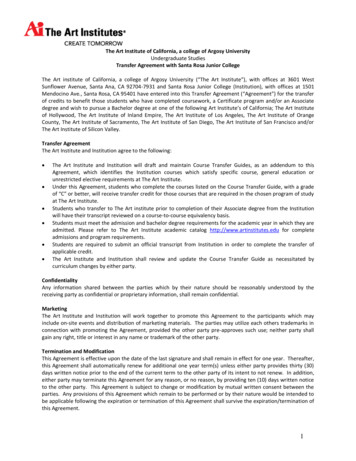
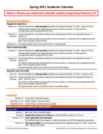
EMAG Group newsletterJan 2021RESEARCH HIGHLIGHTS by young scientistsEnhancing the Performance of Hybrid PixelDetectors for High-Energy TEM Using High-ZSensorsBy Kirsty A. Paton, University of GlasgowDirect electron detectors (DEDs) are facilitating innovations in all branches ofelectron microscopy, so it was fitting that EMAG 2020, which focused on DEDs,was innovative in terms of its format, becoming a virtual conference due to theCOVID-19 pandemic. While certainly a different experience, the virtual formatnevertheless facilitated the same kind of discussions and socialising that onewould expect of a traditional, in-person conference. I was delighted to have theopportunity to share my work investigating the advantages of novel sensormaterials for hybrid pixel detectors (HPDs) and thrilled to be jointly awarded beststudent presentation for my talk.HPDs are a type of DED that consist of an application-specific integrated circuit(ASIC) bump-bonded to a sensor. The on-ASIC signal-processing circuitry makessuch devices capable of kHz (and above) frame-rates and MHz electron countrates. This makes them especially suitable for techniques such as 4D scanningtransmission electron microscopy, and high-speed filming of dynamicalprocesses in transmission electron microscopy. However, high-energy ( 120keVelectrons) scatter over multiple pixels in Si sensors that are sufficiently thickenough to absorb incident electrons and protect the ASIC. Consequently, highenergy electrons are counted by multiple pixels, blurring the images recorded.Using sensors made of high-Z materials should enhance the spatial resolution ofHPDs for high-energy electrons, as characterised by the detector modulationtransfer function (MTF), due to the increased stopping power of such materials.However, there has been speculation that increased backscatter from high-Zmaterials may have a negative effect on detector efficiency, as quantified by thedetective quantum efficiency (DQE). In my work, I have compared theperformance of the Medipix3 ASIC bonded to a 500 µm Si (Z 14) sensor and a500 µm GaAs:Cr (Z 32) sensor using electrons in the energy range of 60–300keV. For high-energy electrons, the GaAs:Cr device surpasses the performanceof the Si detector both in terms of MTF and DQE, with the enhancement inperformance being greatest for 200 keV electrons.However, high-Z sensor materials such as GaAs:Cr are compoundsemiconductors and feature defects that Si sensors do not have. These defectsdistort the electric field lines that define the detector’s pixels, such that the pixelshave a variety of different shapes and sizes rather than composing a regular,homogeneous array. Figure 1 shows that the variation in intensity these defects14
EMAG Group newsletterJan 2021introduce into the images recorded by the GaAs:Cr device can be corrected byapplying a flat-field correction, though this does not correct geometric distortionsdue to the pixels having different shapes. Overall, this work confirms high-Zsensors have advantages for experiments that require high-energy electronswhile motivating further research into other high-Z sensor materials such as CdTeand CZT (Z 50) and ways of correcting for the defects in high-Z sensor materials.Figure 1: An image of a standard calibration sample recorded by a GaAs:Crdevice using 300 keV electrons both (a) without and (b) with a flat-field correctionapplied. In (c) and (d), close-ups of the region highlighted in (a) and (b) are shown,demonstrating that although the flat-field correction has been effective in reducingthe influence of the defects in the recorded image and much of the variation inintensity, residual artefacts remain.Note: A preprint based on the work presented at EMAG 2020 is available atarxiv.org/abs/2009.14565.15
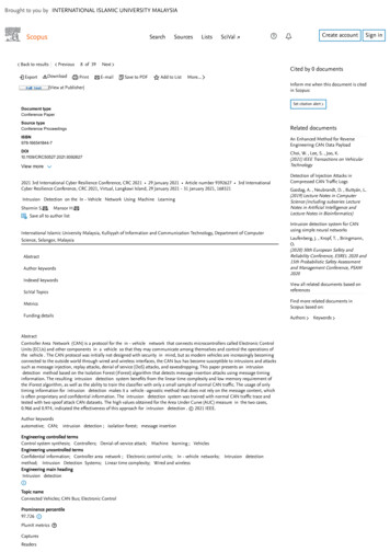
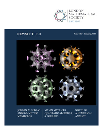
EMAG Group newsletterJan 2021Ultrafast nano-imaging of the order parameter in astructural phase transitionBy Thomas Danz, University of GöttingenThe EMAG 2020 conference certainly is an event to remember – not only wasthis my first virtual conference in an exceptional year, but there were a largenumber of compelling contributed and invited talks as well, showcasing the newopportunities enabled by direct electron detection technology. I had the greatpleasure to present my PhD work in the form of a flash talk and poster, which wasawarded “Best Student Presentation”.My work is an example of an application where direct electron detectioncontributes to the emergence of new possibilities in the field of ultrafasttransmission electron microscopy. In an ultrafast transmission electronmicroscope (UTEM), we combine a conventional TEM with a laser pump/electronprobe scheme in order to obtain simultaneous nanometre spatial andfemtosecond temporal resolution. One intriguing perspective of this technique isthe possibility to not only investigate means of optical control over femto- topicosecond dynamics in correlated materials, but also track the spatial evolutionof optical excitation in structured materials or nanoscale devices.Fig. 1. (A) Simplified schematic of the experimentalsetup. (B and C) Ultrafast electron micrographs of thespecimen before and after time-zero, showing NCCDW domains (bright) and IC CDW domains (dark).Along these lines, my research presented at the EMAG 2020 conferencedemonstrates real-space imaging of charge-density wave (CDW) phases in thecorrelated material 1T-TaS2 using a tailored ultrafast dark-field electronmicroscopy approach (see Fig. 1A). In the experiment, a free-standing, singlecrystalline 1T-TaS2 thin film is pumped out of the nearly commensurate (NC)16
EMAG Group newsletterJan 2021CDW phase at room temperature towards the high-temperature incommensurate(IC) CDW phase using a spatially structured laser field distribution.Almost exclusive NC CDW contrast in the real-space images is obtained byfiltering a number of 72 individual, low-intensity superstructure reflections in theback-focal plane of the microscope using a tailored aperture array. Additionally,image quality and signal-to-noise ratio is distinctly increased by the use of a directelectron detection camera instead of a conventional CCD. Specifically, thisapproach allows us to observe the formation, stabilization, and relaxation of CDWdomains in 1T-TaS2 after optical excitation on their intrinsic femtosecond tonanosecond timescales, yielding access to the order parameter of the structuralphase transition with 5 nm spatial resolution (see Figs. 1B and C).Allowing for sensitivity to further degrees of freedom in complex materials, theseresults will hopefully pave the way for other types of ultrafast investigation. Thiswork has just been published in .17
EMAG Group newsletterJan 2021MEETING REPORTSEMAG2020: Microscopy enabled by direct electrondetectionBy Dr. Donald MacLaren, University of GlasgowWith some trepidation, our annualconference, originally planned to beheld in Glasgow over 6th-8th July, wasmoved to become the IoP's first multiday online meeting. The meetingspread over the course of 3afternoons, with an evening socialevent and hotly-contested online quiz.The conference attracted over 280registered delegates - more thandouble the expected numbers - andindividual sessions attracted around200 attendees, many international.EMAG conferences alternate betweenthelargeRMSco-sponsoredMicroscience Microscopy Congress(mmc) events and smaller, less formalmeetings that offer an opportunity tofocus on a specific theme. The theme of this year’s conference was 'MicroscopyEnabled by Direct Electron Detection' and the ambition was to capture thetremendous science now being enabled by modern detector technologies. It wasfitting that a conference focused on improved technologies and methodologiesfor seeing the world should be timed for the year 2020, a number that issynonymous with perfect vision.Our IOP conference team, headed by Claire Garland and Keenda Sisouphanhwere early proponents of the move to an online format and approached theorganisation with remarkable competence and gusto. A compressed format wasadopted to accommodate the pressures of home-working; and each sessioncomprised an invited talk and a series of contributed 'flash' talks, sponsorpresentations and virtual posters from academia and industry. We are grateful forthe strong industrial support received – this has always been a staple of EMAGconferences and is an essential component of a successful event.The conference truly show-cased the ‘state of the art’ in modelling, development,use and analysis of direct electron detectors within electron microscopy.Distinguished international speakers spanned the breadth of the field, including:ptychography of beam-sensitive materials (Pete Nellist, University of Oxford, UK);18
EMAG Group newsletterJan 20214D-STEM (Colin Ophus, Lawrence Berkeley National Laboratory, USA); EELS(Mitra Taheri, Johns Hopkins University, USA); Lorentz imaging (StephenMcVitie, University of Glasgow, UK), Momentum-resolved STEM (Knut MullerCaspary, Ernst Ruska-Centre for Microscopy and Spectroscopy with Electrons,Germany) and SEM-based diffraction (Carol Trager-Cowan, University ofStrathclyde, UK). Each session was lively, engaging and enthusiasticallyembraced by the online audience, with the online format affording greateropportunity for questions. Student contributions were particularly strong and theeventual prizes, to Thomas Danz of the University of Göttingen and Kirsty Patonof the University of Glasgow, were very well deserved.This was the first such online event run by the IOP on this scale and judged agreat success by all who attended. The initial trepidation was unwarranted! At thetime, we had envisaged the online format being a one-off event; but theenthusiasm for the conference indicates that the most successful online aspectsare here to stay.Virtual Early Career European Microscopy Congress(EMC) 2020By Tamsin O’Reilly, Queen’s University Belfast.EMC is held every fouryears and is one of thelargest European stages forcross-disciplinary research.Many felt disappointmentwhen the original meet inCopenhagen had to becancelled.Fortunately,EMS and RMS organised a virtual platform for the event, giving early careerresearchers an opportunity to share their research with the community. The eventwas held from 24th-26th November and had an action-packed schedule with threefull days of impressive talks and poster presentations. The event had fourconference sessions, they were: Life Sciences – from molecules to complexsystems, Physical Sciences Applications, Ultra structural imaging in life sciencesand Physical Sciences Tools and Techniques.Early Career EMC was a well-attended meeting, with over 150 contributions fromresearchers all over Europe. The congress was split across two meeting rooms,with dedicated oral presentation and poster sessions and included short talksfrom sponsors that showcased the newest range of products available. In the lifesciences sessions, ‘from molecules to complex systems’ covered a range ofinteresting topics including high resolution imaging of sub-cellular events and19
EMAG Group newsletterJan 2021dynamic interactions in organisms. ‘Ultrastructural imaging’ brought us great talkson cryo-electron and correlative microscopy techniques, whilst also delving intothe world of Artificial Intelligence. In physical sciences, ‘tools and techniques’covered exciting techniques such as in-situ and in-operando environmentalmicroscopy as well as ultrafast microscopy and spectroscopy. Not to be outdone,the ‘applications’ symposium highlighted new exciting developments ofmicroscopy techniques and methodologies in materials science, including myown contribution to one of the poster sessions! Although the event was virtual,there was an active participation from the audience after talks and posters, whichwas helped by the excellent organis
School of Chemical and Process Engineering, University of Leeds, Leeds, LS2 9JT Tel: 0113 343 2382 Email: A.P.Brown@leeds.ac.uk Secretary & Honorary Treasurer . Sir John was among the first to use electron microscopes to study structure/properties relationship in materials. He applied TEM imaging techniques to analyse organic crystals .