Transcription
Envisioning CardiacElectrophysiology with ArtificialIntelligence
Context andChallengeAn estimated 17 million people die of cardiovasculardiseases (CVDs) every year worldwide. CVD covershypertension, sudden cardiac arrest, arrhythmia/rhythmdisturbance, stroke, peripheral artery disease, and manymore. Arrhythmias constitute a major problem, whereinthe heart beats either too quickly or too slowly or with anirregular pattern [1]. This indicates the malfunctioning ofthe hearth’s electrical system. Clinical symptoms includingshortness of breath, dizziness, sudden weakness, flutteringin the chest, lightheadedness, and fainting, are indicationsfor malfunctioning of the heart.As technology advances, various industries are adoptingtechnologies such as digital transformation, internet ofthings (IoT), artificial intelligence (AI), nanotechnology,and so on within their product/service portfolio and thethe medical device industry is no exception.The focus of this paper is to discuss software-basedsolutions incorporating AI within EP systems that canimprove overall system performance, improve thetherapeutic outcomes, reduce procedural time, and assistthe electrophysiologist during the procedure.An electrophysiology (EP) study is a test to assessa person’s cardiac electrical activity. It helps theelectrophysiologist to diagnose and determine the preciselocation and nature of arryhthmias. The test is performedby inserting catheters and wired electrodes to measureelectrical activity through blood vessels that enter theheart. The two main goals of a cardiac EP study are (1)to accurately diagnosis the conduction-disturbancemechanism and (2) to determine the best line of treatmentfor the conduction-disturbances. Treatment followinga cardiac EP study could range from ablation therapy topharmacologic to device to surgical intervention basedon the nature of the findings. With healthcare providerslooking for a holistic solution, cardiac EP systems haveevolved into diagnostic and therapeutic systems withradiofrequency ablation (RFA) to treat the arrhythmia foci.2
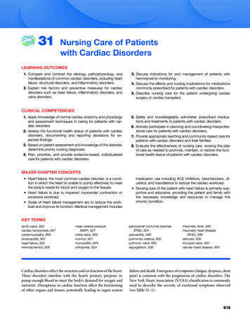
Evolution of EP SystemThe first EP system that was developed consisted of a recording system as its main component. A recording systemrecords the patient’s cardiac electrical activity, leveraging the surface electrocardiogram (ECG) and intracardiacelectrograms (EGMs). The recording system has evolved over the years, adding more intracardiac channels with theadvent of multi-electrode array catheters (MEAs) to improve accuracy in terms of location and timing references. This isbecause EGMs provide greater consistency and reliability. For intracardiac catheters, fluoroscopy was used to navigatethrough the anatomy. This helped electrophysiologists locate where the catheter was residing within the heart.EP testing and RFA have evolved as curative measures for a variety of arrhythmias, starting with straightforward to morecomplex ones enabled by technological advancements substantiated by the development of complex three-dimensional(3D) electro-anatomical mapping (EAM) systems. Complex arrhythmias are often associated with significant underlyingstructural cardiac abnormalities, such as congenital, ischemic, and post-surgical heart diseases. Heart anatomy oftenposes challenges to the electrophysiologist to accurately localize the site where the abnormalities originate. This requiresthe extensive clinical experience of the electrophysiologist and is very time-consuming. Also, a conventional fluoroscopicsystem has limited spatial resolution and may involve prolonged fluoroscopy and high ionizing radiation doses to patientsand staff, this is where the development of complex 3D EAM systems integrated with non-fluoroscopic mapping systemsbring significant value for the cardiac EP systems.First GenerationEP Recording(Surface Level)Surface level electrocardiogram(ECG) measures and monitorings like 12 lead ECGsSecond GenerationEP Recording(Intracardiac Level)Intervention catheters to measure intracardiac electrogram under the fluoroscopyPresent GenerationEP Recording(Mapping)Next GenerationAI Based ClinicalSupport Systems2D/3D map with 4 electrodes with 300 arrays options for mappingElectro Anatomical Mapping (EAM)Fusion of EAM with cardiac anatomy map segmented from CT/MRIPrediction of dose and duration of RF energyArrythmic area localizationFusion of EAM with cardiac map automaticallyImage stabilityOptimized procedure workflowMapping techniques have evolved significantly over the past few years, starting with 8 electrodes for 2D mapping tomore than 64 electrodes, enabling 300 points to capture the heart’s electrical activity. A large number of electrodeshelp to map narrow patches and even small scars in the heart’s electrical circuit, which eventually helps to improve theablation procedures. Though investigation and cardiac ablation of straightforward arrhythmias are currently based onoptimal knowledge of arrhythmia mechanisms of the cardiac anatomy, Investigation of complex arrhythmias is done using3D electro-anatomical navigation systems as they can optimally integrate both the anatomical and electrophysiologicalfeatures of a given arrhythmia for a patient.3
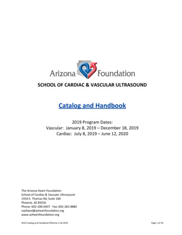
EAM fused with the cardiac anatomy data (gathered from computed tomography (CT) or magnetic resonance (MR)scans), makes it possible to accurately determine the location of the origin of the arrhythmia, define cardiac chambergeometry in 3D, delineate areas of anatomic interest, and allow catheter manipulation and positioning withoutfluoroscopic guidance.There are few challenges with these techniques that can be of potential consequences for the efficacy of catheterassisted EP procedure such as,Registration errors during mappingThe dependency of accuracy upon the skillset of the technologistConformational changes within cardiac chambers as a result of different loading conditions between the time ofimaging and interventionTranslational changes due to patient movementCardiac and respiratory motion could all lead to discrepancies between structural data and electrical substrateNewer systems addresses few of these challenges by correction for cardiac motion, respiration compensation, andincreasing stability to catheter locations to name a few. This can be substantiated by leveraging machine learningtechniques, including recent deep-learning approaches, for gaining new insights from the vast amounts of complexspatio-temporal information generated by various procedures to enable more accurate diagnosis and personalizedtreatment plans. The high-level procedure flow with the latest technologies incorporated is shown in Figure 1.Software Application3D Cardiac EP MapCardiac EPRecording System3D ElectroAnatomical Mapping3D Fused MapSemi-automated/Manual Registrationand FusionAblation ProcedureSelectionPatientAblation SystemImage ViewerUser InterfacePACS SystemCardiac CT/MR3D Anatomic MapGeneration(Segmentation andSurface Rendering)3D Cardiac Surface MapFigure 1: EP procedure flow with latest technologyIn summary, the EP procedure system has evolved from 2D recordings to 3D non-fluoroscopic mapping systems withfurther scope for improvements thanks to technological advancements. New emerging technologies such as artificialintelligence (AI) have promising growth within the healthcare industry, and in the EP domain. The subsequent sectionsdescribe the future focus areas of EP systems and AI-powered solution approaches.4
Future Focus Areas in EP SystemsThe future focus areas within the EP system are emphasized below. We have identified these based on our marketresearch and our domain expertise from over 20 years of building solutions for the medical devices industry. Use casesaround these focus areas are detailed out in the subsequent section.Ablation TargetsLocalizationProcedure WorkflowOptimizationOptimizing RFAblationAblation targets localization toprecisely localize the sitewhere abnormalities areoriginating:Conformational changes withincardiac chambers as well asstability of mapping/diagnosticcatheters due to variousconditions like cardiac andrespiratory artefacts impactcomputing targets of ablation.Optimized procedure workflowto reduce procedural time andimprove efficacy:EP procedures involve recordingof high quality electrograms inan environment with significantelectromagnetic interferenceand noise where communicationis difficult between teammembers, that effectivelyimpacts the procedural timeand engagement of physicians.Optimized force, energy, andlesion detection to prevent theperforation of tissues duringRF ablation:RF ablation has its ownchallenges if not performedadequately with care, such ascollateral damage and perforation, ulcer formation and risk ofthrombus formation.5
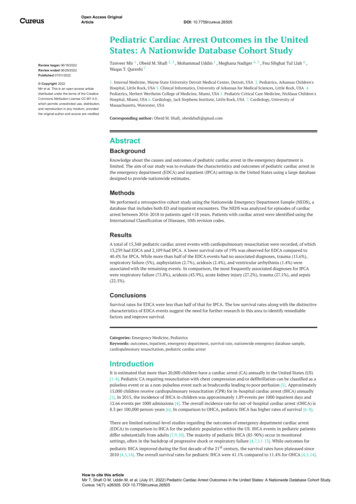
Use Cases and AI-poweredSolutionsDeveloping an AI-powered clinical decision support system to achieve accurate intra-procedural guidance will helpoptimize the workflow of ablation procedures as shown in Figure 2, which has been one of the major challenges facedby the present EP systems. This will be achieved by predicting the diagnostic catheters to use during mapping andrecommendations on ablation catheters and ablation parameters.Operating Room12Preparatory Tests12 Lead SurfaceECGDistal VesselAssessmentPatientPatient’s ClinicalHistorySupportSystem 1AutomaticRecommendationson DiagnosticCatheters3EP Recording andMappingIntra CardiacElectrogramRecordingMapping and 3DImage ConstructionSupportSystem 2AblationAblation ofAbnormal TissuesAutomaticRecommendationson AblationParametersEMRParameters ExtractionManual DecisionMaking - 1EPCardioOtherspecialtiesDiscussion owParameters umber ofelectrodes), size(French),shape (balloon,curve, lasso, basket)Standard EPprocessManual DecisionMaking - 2EPCardioOtherspecialtiesRecommendations:Catheter type, RFpayload/energy tobe delivered,temperature, forceto be applied and RFdurationDiscussion sion supportStages inEP workflowFigure 2: Intra-procedural guidance workflow (Manual vs AI-assisted)In the proposed clinical decision support system, an AI- based model can be implemented by using appropriatesupervised Machine Learning (ML) algorithms. In an abstract context, a model f is shown in equation (1), where, Ø, is oneor more parameters, x is input, and y is output.f(x,Ø) y(1)6
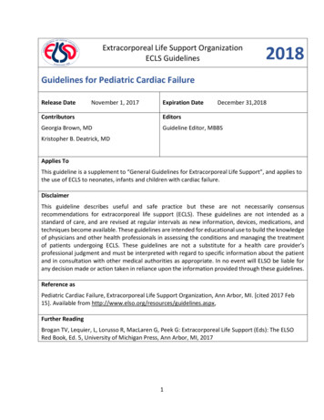
This ML model is prepared by choosing the appropriate combination of classifier and sample splitting methods. The mostcommonly used classifiers are support vector machine, random forest, and feed-forward neural networks. The samplesplitting methods can be but not limited to are 10-fold cross-validation, Monte Carlo cross-validation, and bootstrapping[2]. As an example, we can use the model with a random forest ensemble classifier with a Monte Carlo cross-validationprocedure to classify the study samples. After running the model for significant iterations (based on actual availabledata), the study sample can be divided into training and testing subsets. The objective of training the model is to seek Øsuch that for a set of training data {x} with corresponding outcomes {y}, the given loss function is minimized [3]. A simpleloss function might be the ε which computes the differences between the predicted outputs and the actual outputs andis given in equation (2).ε Ni 1f(xi , Ø)-yi(2)Recommendation on Diagnostic CathetersTypical RFA procedure workflow consists of three broad-level activities: i) preparation, ii) recording and mapping, andiii) ablation (Figure 2). Total procedure time is about three to six hours with at least 30 to 45 minutes for procedurepreparation [5]. The major step involved in procedure preparation is the selection of mapping and diagnostic catheters touse. Currently, catheter selection is a manual and trial-based process, demanding experience and skill of EP clinicians andcommunication among clinicians in real-time. Having an AI-assisted clinical decision support system facilitates catheterselection in real time, thus reducing the time from preparation and improving overall procedural time. The detailed architectural flow is presented in Figure 3.Operating RoomAI-powered Decision Support System 1Preparatory Test FeaturesClinical HistoryPatient age, body mass index, past clinicalhistory and risk factorsEMRSurface ECGType and area of arrhythmia (s), QRS interval,R-R interval, chamber to be studied, and more.Distal Vessel AssessmentFemoral venous access or internal jugular veinaccess, vessel size, vessel access difficulty andpatient comfortReal-timeOfflineReal-time featurecollectionKnowledge base Ground truthFeatures datapre-processingAI / ML algorithmSignificantfeatures selectionInferenceLive featuresPredictedrecommendationsEPClinician review and final recommendationCatheter Configuration (number ofelectrodes), size (french), shape (balloon,curve, lasso, basket)PredictedparametersPatientFigure 3: Solution approach of AI-powered decision support for diagnostic catheter selection7
Benefits of this solutionPatientImproved patient comfort the patient need not undergo multiple catheter trials during mappingReduced healthcare cost and overall procedure time resulting in lower clinician and operating room charges,CliniciansReduced overall procedure time leads to saving of engagement time for cliniciansManufacturerIncrease in market share: Reducing overall procedure time and improved clinical outcomes (for both patients andclinicians) helps to take advantage over competitors and improve overall market share/sales.Recommendation on ablationThe RFA procedure is associated with many risks, such as perforation, creation of atrial esophageal fistula, or thrombusformation, failure to create durable lesions will result in the recurrence of many complex ablations. Accurate and effective ablation procedures will minimize these associated risks. Having an AI-assisted clinical decision support system helpsto accurately select ablation parameters such as the selection of ablation catheter, regions to be ablated, RF payload,temperature, and force to be applied while applying RF energy, thereby improving the efficacy of the ablation process.The detailed architectural flow is presented in Figure 4. In the proposed solution approach for measuring tissue/muscleheat information and microbubble formation in cardiac muscles, intra-cardiac echocardiography (ICE) [4] data are considered.Operating RoomAI-powered Decision Support System 2Features of Intra-Cardiac Recording and MappingClinical HistoryPatient age, body mass index, andassociated risk factorsEMRRecordingTargets of ablation, type of arrhythmia, tissuestrength, softness, and temperature, catheterirrigation capacityMappingLocal activation delay, peak-peak voltage, meanCFE, etc.Real-timeOfflineReal-time featurescollectionKnowledge base Ground truthFeatures datapre-processingAI / ML algorithmSignificantfeatures selectionInferenceLive featuresPredictedrecommendationsEPClinician review and final recommendationCatheter type, RF payload/ energy,temperature, required force, and RFdurationPredictedparametersPatientFigure 4: Solution approach of AI-powered decision support for ablation parameters selection8
Benefits of this solutionPatient outcomeWith improved efficacy of the ablation procedure, the probability of the patient undergoing the ablation process multipletimes is eliminated, thus improving patient comfort and reducing healthcare costs.With improved accuracy, clinical side-effects are reduced, thus reducing risks and complications following the ablationprocedure.CliniciansReduced overall procedure time leads to saving of engagement time for cliniciansClinicians’ reputation and work satisfaction is improved as no/little chances of failureManufacturerReduction in overall health care cost with accurate ablation process helps to gain over competitors thus increasing marketshare/sells.ConclusionCardiac arrhythmias are a major global healthcare problem and there is significant scope forimproving their diagnosis and treatment. Improvements are achieved by implementing technologyadvancements, such as fusion of EP recording systems with mapping systems, fusion of CT/MRIimages with electrical mapping systems to create 3D reference models of the heart, and combiningmapping systems with ablation systems.The EP mapping system, combined with an ablation system, has great potential for improvement byusing modern machine learning techniques. These intelligent clinical decision support systems withappropriate machine learning techniques, offer a powerful approach towards personalized care byimproving effectiveness and accuracy of EP procedures.9
References[1] WHO report (2017), “Fact sheets on Cardiovascular Diseases”[2] Sengupta, P. P., Kulkarni, H., Narula, J., (2018), “Prediction of Abnormal Myocardial Relaxation from Signal ProcessedSurface ECG,” J Am Coll Cardiol, 71(15): 1650–1660.[3] Chris, D. C., Yumnah, M., Konstantinos, N. T., et al., (2019), “Rethinking multiscale cardiac electrophysiology withmachine learning and predictive modelling,” Computers in Biology and Medicine, 104: 339–351.[4] Hijazi, Z. M., Shivkumar, K., Sahn, D. J., (2009), “Intracardiac echocardiography during interventional andelectrophysiological cardiac catheterization. Circulation,” 119(4):587–96.[5] “FAQ: Electrophysiology Study and Catheter Ablation,”About the AuthorsTushar PednekarSr. Director, Medical SolutionsPradeep KolankariSr. Director, Medical SolutionsDigital Engineering andManufacturing ServicesDigital Engineering andManufacturing ServicesNayana ChaudhariSr. Manager, Medical SolutionsAnal R DharamshiSr. Manager, Medical SolutionsDigital Engineering andManufacturing ServicesDigital Engineering andManufacturing ServicesDr. Sapthagirivasan VivekanandhanManager, Medical SolutionsDigital Engineering andManufacturing Services10
AboutCapgeminiCapgemini is a global leader in consulting, digital transformation, technology and engineering services. The Group is at theforefront of innovation to address the entire breadth of clients’ opportunities in the evolving world of cloud, digital andplatforms. Building on its strong 50-year heritage and deep industry-specific expertise, Capgemini enables organizationsto realize their business ambitions through an array of services from strategy to operations. Capgemini is driven by theconviction that the business value of technology comes from and through people. Today, it is a multicultural company of270,000 team members in almost 50 countries. With Altran, the Group reported 2019 combined revenues of 17billion.Visit us atwww.capgemini.comPeople matter, results count.The information contained in this document is proprietary. 2020 Capgemini. All rights reserved.About Capgemini’sDigital Engineering andManufacturing ServicesCapgemini’s Digital Engineering and Manufacturing Services bringstogether deep domain expertise to lead the convergence of Physicaland Digital worlds through technology, engineering and manufacturingexpertise to boost our clients’ competitiveness. A recognized leader withover 10,000 engineers across the globe and 30 years of experience,Capgemini’s comprehensive portfolio of end-to-end solutions enablesglobal companies to unlock the true potential of their product portfoliosand manufacturing efficiencies.To learn more please contact:marketing.dems.global@capgemini.com
structural cardiac abnormalities, such as congenital, ischemic, and post-surgical heart diseases. Heart anatomy often poses challenges to the electrophysiologist to accurately localize the site where the abnormalities originate. This requires the extensive clinical experience of the electrophysiologist and is very time-consuming.