Transcription
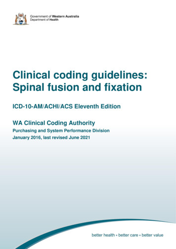
Theme SymposiumNEW HORIZONS IN SPINAL SURGERY: SPINE ARTHROPLASTYViral B. Shah*, Appaji Krishnan* and Sajan K. Hegde**From the: Spine Fellows*, Consultant Spinal Surgeon**, Department of Spinal Surgery,Apollo Hospitals, Chennai 600 006, India.Correspondence to: Dr. Sajan K. Hegde, Consultant Spinal Surgeon andHead, Spinal Surgery Unit, Apollo Hospitals, Greams Road, Chennai 600 006. India.E-mail: sajanhegde@vsnl.com(Presented at the Annual Meeting of Association of Spine Surgeons of India, Bangalore, January 2005)Background: Degenerative primary osteoarthritis is one of the commonest causes of pain in peripheraljoints. This is commonly seen in the knee and hip joints. Replacement of these worn out joints is now theestablished treatment, which gives stable pain free mobile joints. Arthrodesis or fusion of worn out joints isnow rarely performed. Degeneration of the intervertebral disc is a common cause of back as well as neckpain. Fusion of the degenerated and unstable spinal segments is an established procedure to relieve pain.But, at the same time this procedure eliminates motion at the fused level and also accelerates degenerationat the adjacent levels. The replacement of the worn out intervertebral disc with “Artificial Disc” is an emergingtechnology with the aim to maintain motion in the intervertebral unit and, reduce stress on the adjacent levelsand thereby decrease the incidence of degeneration at those levels. The "Artificial Disc" does not simplyportray another technique in spine surgery, but rather, is the first practical step in the evolution of motionretaining spine surgery.Purpose of the Study: To evaluate early results of total disc replacement in the cervical and lumbar spine.Materials and Methods: A prospective study has been designed with the strict patient selection criteria. Atotal of 18 artificial discs have been implanted. The SB Charite’ type III disc was used for lumbar spine (6patients) and the Bryan disc was used for the cervical spine (ten patients and 12 discs). The follow up rangedfrom three months to two years. All the patients were assessed by standard scoring systems.Results: We report excellent to good early results in all our patients at the end of two years. We did notencounter any complications related to the procedure or the implanted disc.Conclusion: The early results of total disc replacement in cervical and lumbar spine show a promising futureto avoid the limitations of spinal fusion. Clearly the long-term results are awaited in comparison to theestablished spinal fusion procedures.Key words: Adjacent level disc degeneration, Degenerated Disc Disease (DDD), Spinal Arthroplasty, SpinalFusion.INTRODUCTIONSpinal fusion is a widely accepted treatment for theDDD in the cervical as well as in the lumbar spine [1].The Interbody grafting technique promotes foraminaldistraction and restoration of normal alignment. Despitegood results, there are certain limitations of the fusionprocedures.Limitation of Spinal Fusion(i)Bone graft site pain.(ii)Prolonged post operative recuperation.(iii) Pseudoarthrosis-failure to fuse.Artificial Disc Replacement is the new technology forreconstruction of the degenerated disc, which eliminatesmany of the limitations of fusion procedures.(iv) Varied success rates from 48% to 89%.(v)Several biomechanical studies demonstrate significantincrease in the shear strain, intradiscal pressure and segmental motion at the adjacent level [2,3] after spinalfusion. Hilibrand, et al. reported that symptomatic degeneration with radiculopathy or myelopathy occurs at 26%rate at 10 years follow up [4]. Often these patients requireadditional procedures to address degeneration of adjacentlevel (Fig. 1). Baba H, et al reported long term follow up of146 patients and concluded that 25% of patients had discdegeneration at adjacent level following spinal fusion [5].Gore and Sepic reported 14% rate of adjacent levelradiculopathy at seven years follow up of 133 patients [6].Causes stiffness and decreased motion of spine.(vi) Increase stress to adjacent level which acceleratesdisc degeneration.89Advantages of the ADR(i) Preserve the normal physiological motion.Apollo Medicine, Vol. 2, No. 2, June 2005
Theme Symposium(a)(b)Fig.1a,b.Patient had C6-C7 level fusion for degenerated disc with radiculopathy before 5 years. She presented with severe discdegeneration and radiculopathy at C5-C6 level.(ii)(iii)(iV)(v)(vi)(vii)(viii)(ix)UHMWPE (Ultra High Molecular Weight Poly Ethylene)sliding core to allow unconstrained motion.Provide segmental stability.Restore disc height.Re-establish lordotic alignment.Reduce discogenic pain.Early return to pre-op occupation.No external braces.Decreased hospital stay.Distributes stress over the spine to reduce adjacentlevel disc degeneration.THE ARTIFICIAL LUMBAR DISCThe SB Charite’ Type III DiscOn 19th of September, 1984 the world’s first threecomponent artificial intervertebral disc that preservedmobility and function was implanted in a human. This discwas named the “SB Charite’ Modular Type DiscProsthesis” after its inventors and the clinic where it wasdeveloped (Fig. 2). Today SB Charite’ III prosthesis hashad extensive clinical use over 17 years in more than6,000 patients in 30 countries of the world. The device iscomposed of two oval cast cobalt-chrome endplates and aApollo Medicine, Vol. 2, No. 2, June 2005Fig. 2. The artificial lumbar disc prosthesis “The SB Charite’Type III”.90
Theme SymposiumIndications for Lumbar ADRContraindication for cervical ADR(i) Mono and bisegmental DDD with or without previoussurgery with long-term chronic mechanical low backpain (Primary discopathy).(ii) Post-discectomy syndrome (Secondary discopathy).(i) Infection(ii) Osteoporosis(iii) Mechanical instabilityMATERIAL AND METHODSContraindications for Lumbar ADRThe lumbar artificial disc replacement(i)Mechanical instability in form of Lysis, listhesis orresection of the facet joints.(ii) Radiculopathy due to disc prolapse (leg pain muchmore than back pain).(iii) Facet arthrosis with or without spinal stenosis.(iv) Infection.(v)Osteoporosis.(vi) Adhesive archnoiditis.(vii) Multiple abdominal surgery.(viii) Morbid obesity.We present the results of our patients who underwentlumbar arthroplasty. A total of six patients were operatedfrom October 2004 to May 2005. All patients werebetween 30-45 years age group. Four patients were femaleand two male. All patients had single level arthroplasty.Of these, three patients had L4-5 level and three patientshad L5-S1 level disc arthroplasty. Five patients hadprimary discopathy with chronic mechanical low backpain and one patient had previously been operated for discprolapse with persistent complaint of chronic low backpain.THE ARTIFICIAL CERVICAL DISCPre-operatively, all the patients were evaluated with Xrays and MRI of lumbar spine. Plain X-rays showed discspace reduction (Fig. 4). Provocative discography was donein multiple level disc degeneration. Provocation of pain during discography was considered as an important indicatorfor level selection. Patients were operated in the supine position. By a retroperitoneal approach, adequate anterior decompression and meticulous discectomy was done. The endplates were prepared and SB Charite’ III disc was implantedwith specific jig instruments (Figs. 5,6)The Bryan discVincent Bryan, an American Neurosurgeon inventedthe artificial cervical disc, which has been in clinical usesince 1997. The Bryan disc (Fig.3) is composed ofpolyurethane polymeric nucleus between two titaniumalloy shells with porous coated surfaces for boneingrowths. The Bryan disc has been utilized for single aswell as double level replacement in our institution.Indication for cervical ADRDegenerative disc disease associated withradiculopathy or myelopathy from C3-4 to C6-7 levels.Fig. 4. X-ray showing degenerated disc with space reductionat L5-S1 level (arrow), other levels show normal discheight.Fig. 3. The artificial cervical disc prosthesis “The Bryan Disc”.91Apollo Medicine, Vol. 2, No 2, June 2005
Theme SymposiumPost-operative complicationsNo complication related to implant or the procedurewas encountered.All the patients were mobilized from first post-op daywith soft lumbosacral brace support. The follow up rangedfrom two to ten months. Patients were reviewed after sixweeks and then at three and six months. During follow upflexion and extension X-rays were taken to documentfunctional movement.Follow up evaluation was done with(i) Visual analogue score (VAS).(ii) Functional range of movement.(iii) Return to prior work.Out of six, in five patients the visual analogue score forpain improved from eight to zero while one patientcontinues to have some low back pain though of muchreduced intensity. All patients demonstrated to haveaverage 7º of functional movements on dynamic X-rays.All the patients returned to their prior job within two toseven days of surgery.The cervical artificial disc replacementWe present an analysis of early clinical outcome of ourpatients who underwent cervical arthroplasty. TwelveBryan discs were implanted between 2003-2004. Out ofthese, eight patients had single level and two patients haddouble level arthroplasty (Figs. 7,8). All patients werebetween 30-45 years age group with equal gender ratio.Eight patients were from a high professional group(doctors, executives and software specialists). Clinicallynine patients had complaints of radiculopathy with neckpain. One patient had compressive cervical myelopathy.The duration of symptoms ranged from 5 to 30 months.The commonest level was C5-C6 disc (8 cases). Onepatient had previous fusion at the C6-7 level and then shedeveloped disc degeneration and radiculopathy at theadjacent C5-C6 level for which arthroplasty wasdone.Fig. 5. MRI of degenerated disc (arrow) at L5-S1 level incontrast to normal disc at other levels.Pre-operatively all the patients were evaluated withX-ray and MRI of cervical spine. A CT scan was done inall patients to accurately assess the size of the disc.Patients were operated in supine position. Adequateanterior decompression and meticulous discectomy wasdone, following which the Bryan disc was implanted withspecific jig instruments.Post operative complicationsOne patient developed hoarseness of voice, whichrecovered in three weeks. No other complication related tothe implant or procedure was encountered.Fig. 6. X-ray showing SB Charite' Type III disc prosthesis atL5-S1 level.Apollo Medicine, Vol. 2, No. 2, June 200592
Theme SymposiumFig. 7. X-rays showing single level Bryan disc prosthesis at C5-C6 level.Fig. 8. X-rays showing double level Bryan disc prosthesis at C5-6 and C6-7 level.93Apollo Medicine, Vol. 2, No 2, June 2005
Theme SymposiumAll the patients were mobilized from first post-op daywithout brace support. Neck movements were encouragedfrom second post-op day. The follow up ranged from sixmonths to 1½ years. Patients were reviewed at six weeksand then seen at three and six months. During follow upflexion and extension X-rays were taken to documentrange of movement.extremely important observation predicts satisfactorylong-term performance.Limitations of artificial disc ReplacementAs a new technology the ADR requires long termfollow up. Selection of the patient is of paramountimportance for the success of the procedure. Theimplantation of this prosthesis requires a thoroughunderstanding of the biomechanics and training in thetechniques. The high cost of implant is comparable withthe cost of the other total joint implants for knee and hipjoints.Follow up evaluation was done with(i)Visual analogue score(ii) Range of movement(iii) Return to prior workCONCLUSIONRESULTSThe long-term results of lumbar disc replacementgives a possibility to offer a motion retaining spinalsurgery and a possibility of being able to minimizeadjacent level disc degeneration. However, intermediateand long-term results are required for cervical arthroplastybefore we can safely say that spinal arthroplasty is theprocedure of choice in degenerative disc disease. Thebiomechanical studies have demonstrated that discreplacement creates less strain on the adjacent level thanfusion. Wear studies suggest that there may be lesspotential for aseptic loosening than large jointarthroplasty. Obviously the jury is still out on thistechnology, but early reports suggest the verdict may bepromising in carefully selected cases.Patients were assessed according to the modifiedOdom’s criteria. At the end of one year excellent resultswere reported in 90% of cases with good results inremaining 10% of cases.Wear AnalysisThe failure of total joint replacement has been largelyattributed to wear debris from polymer-bearing surfaces.Wear debris induces an intense cellular inflammatoryreaction, which results in then production of cytokinesthat cause resorption of bone. This process known asaseptic loosening, leads to destabilization and mechanicalfailure of the joint. The possibility of the similar processoccurring with polymer nucleus of both the variety of discprostheses has been studied.REFERENCESThe Lumbar SB Charite’ disc [7]1. Robinson RA, Smith GW. Anterolateral cervical discfremoval and interbody fusion for cervical disc syndrome.Bull Johns Hopkins Hospital 1955; 96: 223-224.The clinical evaluation of users of the SB Charite’discs have shown that the wear debris develop only byabrasion of the polymer surface. These abrasions donotoccur when parallel positioning of properly sizedendplates has been achieved. The abrasion occurs onlywhen the load is concentrated on a small region of the coredue to incorrect positioning or too small size of implant. Itis interesting to note that no signs of “polyethylenedisease” or periprosthetic osteolysis from such abrasionhave been detected in the tissues surrounding the artificialdisc, even where particles of polyethylene were found.This is most probably due to the absence of synovia in thearea where the prosthesis is positioned [8].2. Eck Jc, Humphreys SC, Lim TH, et al. Biomechanicalstudy on the effect of cervical spine fusion on adjacentlevel intra-discal pressure and segmental motion. Spine2002; 27: 2431-2434.3. Matsunaga S, Kabayama S, Yamamoto T, et al. Strain onintervertebral disc after anterior cervical decompressionand fusion. Spine 1999; 24: 670-675.4. Hilibrand AS, Carlson GD, Palumbo MA, et al.Radiculopathy and myelopathy at segment adjacent tothe site of a previous anterior cervical arthrodesis. J BoneJoint Surg (Am) 1999; 81: 519-528.5. Baba H, Furusawa, Imura, Tomita K. Long terms follow upof anterior cervical fusion after discectomy. Spine 1993;18: 2167-2173.The Cervical Bryan DiscAnderson, et al. [9] have published a pivotal study onthe wear characteristic of the Bryan disc in vitro in acervical spine simulator and in vivo biologic response ingoat and chimpanzee models. The authors concluded thatthe wear rate is low in vitro and they did not find anyinflammatory response in vivo models. This is anApollo Medicine, Vol. 2, No. 2, June 20056. Gore DR, Sepic SB. Adjacent level degenerationfollowing anterior cervical fusion. A long term study. Spine1984; 9: 667-671.7. Linf HD, Keller A. Biomechanics of total disc replacementin the “Artificial Disc”. Springer Verlag publication, 2004;pp 33-52.94
Theme Symposium8. Sibbit WZ Jr. The normal and the diseased joint. In:Bromer F, Worel RV (eds). Orthopaedics: Principles ofBasic and Clinical Sciences. CRC Press, Florida, pp 141163.9. Anderson PA, Ronlean JP, Bryan VE, Carlson CS. Wearanalysis of the Bryan cervical disc prothesis. Spine 2003;28: S186-S194.95Apollo Medicine, Vol. 2, No 2, June 2005
The Bryan disc Vincent Bryan, an American Neurosurgeon invented the artificial cervical disc, which has been in clinical use since 1997. The Bryan disc (Fig.3) is composed of polyurethane polymeric nucleus between two titanium alloy shells with porous coated surfaces for bone ingrowths. The Bryan disc has been utilized for single as