Transcription
Pharmaceutical Research, Vol. 21, No. 12, December 2004 ( 2004)Preparation of PLGA Microparticlesby an Emulsion-Extraction ProcessUsing Glycofurol as Polymer SolventAnne Aubert-Pouëssel1,Marie-Claire Venier-Julienne1,3, Patrick Saulnier1,Michelle Sergent2, and Jean-Pierre Benoît1Received April 13, 2004; accepted September 5, 2004Purpose. To develop biodegradable poly(lactic-co-glycolic acid)(PLGA) microparticles prepared by an original emulsion-extractionprocess, with glycofurol, a nontoxic excipient, as polymer solvent.Methods. The preparation of microparticles consisted in dissolvingpolymer in glycofurol. This solution was emulsified in a vegetable oil,and then amphiphilic agent was added into the emulsion to extractglycofurol and lead to microparticle formation. Physicochemical studies were carried out, and an experimental design was prepared inorder to elucidate the impact of the formulation composition on themicroparticle characteristics. Finally, encapsulation tests were madewith a model protein.Results. In a ternary diagram, a small feasibility area allowing particleformation was located. The resulting microparticles were sphericalwith a homogeneous, polymeric matrix structure. They exhibited avariable size from 3 to 15 m, which was controlled by the differentformulation parameters. Differential scanning calorimetry (DSC)analysis made it possible to detect their composition. Preliminaryresults showed that these particles were able to encapsulate a proteinmodel, lysozyme.Conclusions. This simple and convenient technique enabled us toobtain spherical, biodegradable microparticles from acceptable excipients. Moreover, the process conditions made possible the encapsulation of drugs, including proteins.KEY WORDS: diffusion process; glycofurol; microparticles; poly(lactic-co-glycolic acid); protein.INTRODUCTIONBiodegradable poly(lactic-co-glycolic acid) (PLGA)polymers show interesting properties for biotechnologythrough their biocompatibility and their authorization by theFood and Drug Administration (FDA) for drug delivery.Various polymeric drug delivery systems like microparticlesor nanoparticles have been developed using these polymersfor the delivery of a variety of drugs (1). However, the technology processes often use organic solvents to dissolve thewater-insoluble PLGA. Usually, halogenated solvents, suchas methylene chloride and chloroform, are used in the microencapsulation process. These solvents are rated as class 2 (“tobe limited”) according to the International Conference ofHarmonization (ICH). The permitted daily exposure (PDE)1INSERM U646, Ingénierie de la Vectorisation Particulaire, IBT, 10rue André Boquel, 49 100 Angers, France.2Laboratoire de Méthodologie de la Recherche Expérimentale, Université Paul Cézanne Aix Marseille III, avenue Escadrille Normandie-Niemen, 13 397 Marseille cedex 20, France.3To whom correspondence should be addressed. (e-mail: venier@univ-angers.fr)0724-8741/04/1200-2384/0 2004 Springer Science Business Media, Inc.Research Paperfor methylene chloride is 6.0 mg, and for chloroform the PDEis 0.6 mg (2). Furthermore, additional steps of desorptionhave to be considered to reduce the residual content of organic solvent in the resulting products. Even if a favorablebenefit-risk ratio is taken into consideration for microspheresaimed at treating serious pathologies, no potential toxicity istolerated for microspheres aimed at vaccination or minor disorders, where the risk is greater than the potential benefit.Moreover, the use of such solvents makes it difficult to respect safety conditions during production; thus, the development of halogenated solvent-free microencapsulation methods is desirable.Efforts to avoid the use of halogenated solvents in PLGAmicroencapsulation processes have been undertaken. For example ethyl acetate (3,4), and ethyl formate (5), both belonging to class 3 entitled “low toxic potential” and limited to 50mg/day, were used as alternative solvents. Equally, some encapsulation techniques, using supercritical fluids, especiallysupercritical carbon dioxide, were proposed in order to completely eliminate organic solvents or at least to minimize theiruse (6). Jain (1) described a technique based on phase separation using only oil phases (triacetin, Mygliol 812, etc.).However, in order to dissolve the polymer in the triacetinphase, a heating step (65 C) was required; this thermal treatment could both alter the polymer properties and also limitthe encapsulation of delicate drugs such as proteins.In the current work, excipients were chosen from thoseused in human medicine. Thus, in order to dissolve the polymer, a parenteral solvent, glycofurol, was used (7–9); it is acomponent of a marketed product (Mobic: meloxicam). Glycofurol has also been used in research as a vaccine adjuvant(10), and as a solvent in injectable polymer implants (11–13).In order to form an emulsion with the polymer solution, oliveoil was chosen. This common vegetable oil has been used asan excipient in long-acting injectable preparations. Finally,different non-ionic amphiphilic excipients were used both inthe extraction and washing steps: Labrafil M 1944 CS andpoloxamer 188, respectively. Labrafil has been used as a solvent in an injectable gel delivering contraceptive steroids(14); poloxamer 188 has been commonly used in various marketed products, especially in injectable preparations (Norditropin). The strong interest in halogenated solvent-free methods is driven by the potential for a reduction of toxicologicalrisk and an improved preservation of drug integrity, especially for proteins. Indeed, with the steady increase of recombinant protein-based pharmaceuticals due to the development of genetic engineering, there clearly existed a need fordeveloping effective polymeric delivery systems. Therefore,to test the ability of the proposed method to encapsulateproteins, preliminary studies with a model protein, lysozyme,were carried out.MATERIALS AND METHODSMaterialsUncapped PLGA was obtained from Phusis (PLGA 75/25) (Saint-Ismier, France). The mean molecular weight (Mw)was 21,000 (polydispersity index I ⳱ 1.7) as determined bysize-exclusion chromatography (polystyrene standard). Otherpolymers were used in a preliminary screening, such as PLGA2384
Preparation of PLGA Microparticles50/50 Resomer RG506, Mw ⳱ 75,000 (BI Chimie, France)and poly(lactic acid), Mw ⳱ 96,000 (Phusis Saint-Ismier,France). Glycofurol (tetraglycol or -[(tetrahydro-2-furanyl)methyl]- -hydroxy-poly(oxy-1,2-ethanediyl) was purchasedfrom Sigma (Saint Quentin Fallavier, France). Labrafil M1944 CS (oleoyl macrogol-6 glycerides), Lutrol F68 (poloxamer 188: polyoxyethylene-polyoxypropylene), andCrodamol EO (ethyl oleate) were kindly supplied by Gattefossé (Saint Priest, France), BASF (Levallois-Perret, France),and Croda (Trappes, France), respectively. Olive oil was purchased from Coopérative Pharmaceutique (Melun, France).Lysozyme is a monomeric globular protein with 129 aminoacids and an isoelectric point pI ⳱ 10.7. Lysozyme fromchicken egg white (95%), its substrate, Micrococcus lysodeikticus, and buffer components were purchased from SigmaAldrich (Saint Quentin Fallavier, France). A Bio-Rad DCprotein assay kit was obtained from Bio-Rad Laboratories(Marnes-la-Coquette, France).Feasibility DomainA ternary diagram was developed to consider the threemajor components used in the microparticle process (polymer-glycofurol solution, vegetable oil, and Labrafil M 1944CS). The feasibility zone was defined as an area that allowedformation of micrometric-sized, nonaggregated, and sphericalmicroparticles.Microparticle PreparationAn initial phase containing glycofurol and PLGA polymer was prepared at room temperature. The polymer concentration used in glycofurol was 1%, 2%, or 3% w/w. Thepolymer solution was emulsified in an olive oil phase undermechanical stirring for 5 min at 500 rpm (Heidolph RGH 500,Merck Eurolab, Strasbourg, France). This step was carriedout at 24 C in a thermostatic bath (Polystat 22, BioblockScientific, Illkirch, France). An amphiphilic agent, Labrafil M1944 CS, was added to the emulsion under mechanical stirring(500 rpm) through a syringe for 10 min to obtain particleformation. Deionized water (100 ml) was added to the resulting mixture and magnetically stirred for a further 10 min.Finally, the formed microparticles were filtered through an0.45- m filter (HVLP type, Millipore SA, Saint Quentin Yvelines, France), washed with 100 ml 1% Lutrol F68 solution,rinsed five times with 50 ml of deionized water, and freezedried (RP2V Serail SGD, Argenteuil, France) to obtain afree-flowing powder (15). The dried particles were stored at 4 C until required. The excipient proportions were chosenwith respect to the feasibility zone previously described; thetotal quantity of excipients (polymer solution, olive oil, Labrafil M 1944 CS) was fixed at 20 g.Experimental DesignIn order to determine the influence of the componentson microparticle size (mode, diameter) and particle distribution (peak width), a mixture experimental design wasplanned. Three variables, corresponding to the amount ofpolymer solution, olive oil, and Labrafil M 1944 CS, respectively X1, X2, and X3, were considered. These nonindependent variables were subjected to the constraint that their sumwas 100%. Due to the constraints of the component propor-2385tions to be used, the region of interest was limited to a parallelogram, located into an equilateral triangle in one plan.Whatever the formulation, the proportion of polymer in theglycofurol was fixed at 1% w/w. To determine the best formulation according to the targets, we needed to obtain information of the entire domain of interest. For this purpose, weused an empirical model, this being a reduced cubic model,whose proposed equation is as follows (where representsthe response and i the coefficients of the model): ⳱ 1X1 2X2 3X3 12X1X2 13X1X3 23X2X3 123X1X2X3To calculate the estimation of the coefficients, an experimental design with 13 experiments was carried out. Someexperiments were replicated in order to estimate the reproducibility of the experimental results. To minimize the effectof systemic errors, experiments were carried out in a randomfashion. Two responses were considered: the mode of theparticle diameter (Y1) and the peak width, which representedparticle distribution (Y2). All samples presented a monodisperse size distribution. If the mode classically characterizedparticle size, the peak width was preferred to the coefficientof variation, as it was more discriminating (it was measured asthe peak width at half height).From the experimental results, an estimation of the coefficients of the model was calculated using the least squaresmethod. Construction of the experimental design, estimationof the coefficients of the models, and statistical study wereachieved using the Nemrod software (16).Particle CharacterizationMorphology and SizeEach microparticle batch was observed by optical microscopy (BH2, Olympus, Tokyo, Japan). The surface and theinternal morphology of the microparticles were investigatedby using scanning electron microscopy (SEM; JSM 6310F,JEOL, Paris, France). Freeze-dried microparticles weremounted onto metal stubs using double-sided adhesive tape,vacuum-coated with a film of carbon (10-nm thick) using aMED 020 (Bal-Tec, Balzers, Lichtenstein). To characterizethe internal morphology, the particles were embedded inEpon resin, and 20-nm-thick sections were sliced off using acryocut at 26 C (Frigocut 2800, Reichert-Jung, Paris,France). Average particle size and their distribution were determined using a Coulter Multisizer (Coultronics, Margency,France). Microparticles were suspended in isotonic saline solution without surfactant and sonicated a few minutes prior toanalysis.DSC AnalysisDifferential scanning calorimetry (DSC) was performedwith a Mettler Toledo Star System (Mettler-Toledo, Viroflay,France). Approximately 10 mg of sample was placed in asealed aluminum crucible; experiments were replicated (n ⳱3). The measurements were carried out in the range of 150to 300 C at 10 C·min 1 under a nitrogen flow; two heatingcycles were carried out.
2386Protein Encapsulation EfficiencyEncapsulation tests were achieved with lysozyme as amodel protein. Lysozyme-loaded microparticles were prepared according to the previously described protocol. Protein(insoluble in glycofurol) was dispersed in the polymer solution (2% PLGA in glycofurol) to obtain encapsulation ratiosbetween 0.5% and 2.0% w/w (with respect to the amount ofPLGA). Entrapment efficiency was determined by using twotechniques allowing the estimation of both the total and theactive entrapped protein. Total protein was determined byusing a method reported by Yan et al., consisting of a basicdigestion of microparticles followed by a Bio-Rad proteinassay (17). Active entrapped protein was determined by measuring lysozyme activity in the presence of its substrate Micrococcus lysodeikticus following microparticle dissolution ina mixture of dimethyl sulfoxide and acidic solution. The biological activity of lysozyme was not impaired by the extractionmethod used as reported by Ghaderi et al. (18).RESULTS AND DISCUSSIONMicroparticle PreparationThe microparticle preparation process was based on ahydrophilic/lipophilic emulsion followed by an extractionstep. The polymer solution (composed of PLGA and glycofurol) was emulsified in an oily phase; then to induce theparticle formation, the oily phase properties were modified toallow the extraction of the polymer solvent. The first stage ofthe process was polymer dissolution in glycofurol at roomtemperature. PLGA (50/50 or 75/25) and PLA with variousmolecular weights from 14,000 to 96,000 Da were dissolved inglycofurol up to 10% w/w, beyond which rate gels wereformed. Low PLGA 75/25 concentrations were used (1%,2%, or 3% w/w) for improved polymer dissolution.Then, in a second stage, an emulsion was formed by dispersing the polymer-glycofurol solution into a nonsolublephase, selected after the screening of excipients used in human medicine. Two vegetable oils, olive and sesame, werechosen because they presented the advantage of forming withthe polymer-glycofurol solution a relatively stable emulsionwithout the use of surfactant. This was partly due to the verylow interfacial tension ( 1 mN · m 1) between polymerglycofurol solution and vegetable oil.Third, in order to extract glycofurol, an amphiphilicagent that was miscible with both vegetable oil and glycofurol,was selected. Crodamol EO (ethyl oleate) and Labrafil M1944 CS (oleoyl macrogol-6 glycerides) fulfilled these conditions. By allowing the polarity of olive oil to change, andhence making glycofurol soluble in this phase, we attemptedto promote the diffusion of glycofurol toward the oil, andhence to polymer desolvatation. Both selected excipientsachieved polymer desolvatation, however only Labrafil allowed the formation of spherical particles. To confirm theassumption of the glycofurol diffusion phenomenon, an experiment was performed using a hydrophilic dye (methyleneblue) mixed with glycofurol. After the formation of glycofuroldrops in a drop of olive oil, the addition of Labrafil promotedthe glycofurol diffusion toward the oil/Labrafil solution. Thismodel allowed us to visualize the process under static conditions. We could therefore suppose that this phenomenon wasAubert-Pouessel et al.increased in the dynamic conditions of the particle formationprocess. It should also be noted that the process up to thisstage does not require water. Working in anhydrous conditions is advantageous for some drugs, including proteins, byfavoring their stability (19). However, the resulting sphericalparticles suspensed in a mixture composed of oil, Labrafil,and glycofurol needed to be improved (morphology and homogeneity). Consequently, an additional step favoring glycofurol extraction was performed. It consisted of adding wateror propylene glycol to the mixture under stirring (10 min).This last step of glycofurol extraction allowed both suitableparticles to be obtained and a reduction in filtration time.Importantly, in the presence of water, but in the absence ofLabrafil, no particles were formed, proving the key role ofLabrafil in particle formation. We also studied the influenceof the volume of water added on particle size. Results showedthat size remained stable (about 5 m) with a narrow distribution whatever the volume of water, except for volumesbelow 75 ml (Fig. 1). These low volumes appear to be insufficient to complete particle formation, leading to their agglomeration and increasing their final size.Finally, at the end of the whole process, the microparticles were recovered via filtration and washed. Washing withwater alone was not able to remove efficiently the oil residue(observation of powder aggregates using microscopy andCoulter Multisizer). Therefore, it was necessary to use a surfactant solution composed of Lutrol to obtain a free-flowingpowder.Further improvements in the process were made to improve reproducibility. It included the study of different parameters affecting particle formation, which were refinedlater. The stabilization of the temperature appeared to beimportant: at temperatures below 24 C, particle formationwas incomplete. The temperature had important effects oncomponent viscosity, solubility, and ease of exchange, factorsat the heart of the process. Moreover, Labrafil partially crystallizes at temperatures below 20 C. Therefore, for reasons ofdrug thermal stability, the minimum temperature was fixed at24 C. Also, it was found that particle formation was not possible when the stirring rate was below 250 rpm. Finally, byusing different stirring rates and polymer concentrations, itFig. 1. Particle size evolution according to the volume of added waterduring microparticle preparation.
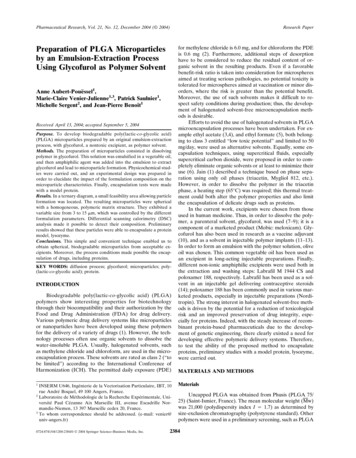
Preparation of PLGA Microparticles2387Fig. 2. (A) Illustration of the feasibility zone composed of a discontinuous domain and a continuousdomain represented by the gray parallelogram. (B) Illustration of the experimental design (13 designexperiments including 4 test points) inside the interest zone: the continuous domain.was found that the particle size could be modified. For instance, between 250 and 1000 rpm, particle sizes varied from10.0 to 4.8 m at a constant concentration of polymer (2%).Between 1% and 3% w/w polymer concentration, particlesize varied from 5.6 to 13.2 m (mode) at a constant stirringrate (500 rpm).To confirm the microparticle formation mechanism, weneeded to show that particles had their origin in the polymer-glycofurol drops. Thus, we compared the size of polymerglycofurol solution drops with that of the microparticles. Unfortunately, the size distribution of the drops could not bemeasured due to technical limitations, so average diameterswere measured instead by observation under an optical microscope. Results showed a slightly inferior size of microparticles compared to the drop size, with around 6 m and 10 m, respectively. This size reduction confirmed the diffusionTable I. Experiment Design and Corresponding Results for the Surface Response AnalysisWeight fraction of excipient (%)Measured response ( m)Response estimated by the model ( m)Experimentno.Glyco PolyX1Olive oilX2LabrafilX3ModeY1Peak widthY2ModeY1Peak 8.135.635.635.635.636.525.886.685.458910111213
Aubert-Pouessel et al.2388Table II. Validation of the Model Predictions (Three Test Points)Weight fraction of excipient (%)Glyco Poly0.04000.03910.0390Y1: mode ( m)Y2: peak width ( onfidence interval ⳱ YA/estimated t /2,N p [s2 (1 dA)], where dA ⳱ function of variance at point A, S2mode ⳱ 1.001 (11 df), and S2peak⳱ 0.896 (11 df). t: Student’s variable; : significance level (0.05); N: number of data points; p: number of coefficients in the model (N-p:number of degrees of freedom)widthof the glycofurol from the drops leading to polymer desolvatation.Determination of the Feasibility ZoneA ternary diagram was established in order to determinethe experimental conditions that yielded microparticles. Theformation of microparticles strongly depended on the proportions of the three components (polymer solution, olive oil,Labrafil M 1944 CS). The ternary diagram was rationally ex-plored to define the feasibility zone. It was defined by a parallelogram with the following coordinates: 2.5% to 7.0% of polymer-glycofurol solution, 70% to 80% of vegetable oil (olive orsesame oil), and 13.0% to 27.5% of Labrafil (Fig. 2A). Thefeasibility zone contained two domains: a discontinuous and acontinuous domain. In the discontinuous domain, both sphericalor nonspherical objects such as fragments or fibers were obtained. On the other hand, in the continuous zone, only spherical microparticles were formed. To study the influence of components on microparticle size, we focused on this continuousFig. 3. Graphical representation of the proposed model for the two responses: (A, C) the microparticlediameter (mode) (Y1) and (B, D) the microparticle distribution (peak width) (Y2). (A) and (B) representthe contour plots allowing the prediction of the size and the distribution of the microparticles. (C) and(D) represent the respective influence of each component on mode diameter and peak width. Xi is theproportion of the considered component
Preparation of PLGA Microparticleszone, which was defined by the co-ordinates 2.50% to 4.38% ofpolymerglycofurol solution, 73.25% to 80.00% of olive oil,and 15.63% to 24.25% of Labrafil (Fig. 2B).Experimental DesignWithin the previously defined domain, a response surface analysis was carried out to predict the effect of excipientson particle size. Table I shows the results in terms of diameter(mode) and particle distribution (peak width) correspondingto the 13 experiments carried out. To predict these two responses within the experimental domain, a reduced cubicpolynomial model was designed. A statistical analysis basedon the lack of fit (differences between the measured responses and those calculated by the model (Table I) allowedus to accept the proposed models for the two responses. Totest the model predictions, three additional experiments werecarried out afterwards. Experimental test point values wereincluded in the confidence interval for a given statistical significance ( ⳱ 0.05) (Table II). The models could be used topredict the responses in each point of the domain of interest,and contour plot of the two responses are graphically represented in Fig. 3. Mode diameters were from 5 to 10 m (Fig.3A), which correspond to a mean diameter of 7 to 14 m.Peak widths were between 4 and 8.5 m (Fig. 3B). Whensuperposing both graphical representations, it was possible todefine a zone where particles presented the best homogeneity(peak width 5.5 m) corresponding to a particle modeⱕ6 m.The effect of each component is shown in Figs. 3C and3D to enable comparisons to be made between their respective influences on mode diameter and peak width. The graphrepresents the variation of the responses when we shift alonga line drawn between the center of the domain of interest andthe pure component (top of the ternary diagram). From thecenter of the domain (point G), when the proportion of theconsidered component (Xi) varies into the limits defined bythe domain, the proportions of the other components vary ina constant ratio (20). Considering both responses, the polymer-glycofurol solution appeared to be the most critical component. When the proportion of the dispersed phase was increased, particle diameter strongly decreased. A range ofvariation of 2% led to a variation of 3 m of particle diameter(Fig. 3C). This result implied that perfect control of theamount of polymer-glycofurol solution was required for therobustness of the process. The vegetable oil and the surfactant were influential to a lesser extent. For a range of variation of 6.7% and 6.8%, the size variation was 2 and 1 m foroil and Labrafil, respectively. The major effect of glycofurolon particle size and on homogeneity could be explained bythe importance of the emulsion step. During the emulsionprocess, two contrary phenomena occur and influence the sizeof the drops: the shearing of the drops and their coalescence.By increasing the polymer-glycofurol solution volume (dispersed phase), the probability of breaking up drops is lessimportant than coalescence, leading to larger droplets (21,22).Unfortunately, our results were at the opposite of this observation. It should also be noted that we did not evaluate dropsize but the particle size. Particle formation requires an additional step of solvent diffusion. This process is governed by2389Fick’s law; in this way, a higher ratio of dispersed phase tocontinuous phase leads to longer solvent diffusion time andalso to a longer drop solidification time. This phenomenonwas described by Tuncel (23). Therefore, by increasing thepolymer-glycofurol solution volume (dispersed phase), thecontinuous phase allows a low and sustained diffusion of thesolvent leading to smaller particles.Fig. 4. Scanning electron micrographs (SEM) showing: (A, B) thesurface morphology of microparticles and (C) the internal morphology of microparticles after section (the white lines correspond to theblade tracks of the cryocut).
Aubert-Pouessel et al.2390Table III. Thermal Events Observed During DSC Analysis for Components Going into Microparticle Formulation and Physical Mixtures(Temperature in C)ComponentsSingle componentsPLGA (P)Glycofurol (G)Labrafil (L)Olive oilDispersed phaseP-95/G-5Continuous ous phase t)EndothermalpeakExothermalpeak 80/ 65/ 47/ 24/ 1 4 95/128 74/ 60/ 40/220 384646184 6 5 8 41 95/ 42/133 95/ 47/13045 819132 7182Tg: glass transition.Microparticle CharacterizationMicroscopyOptical microscopy showed well-formed, spherical particles. Their size distribution was in accordance with formulations based on emulsions. Under SEM analysis, microparticles exhibited a smooth skin without pores. Particles wereformed by a dense and homogeneous structure, supportingthe hypothesis of particle formation (i.e., the desolvatation ofthe drops of polymer solution) (Figs. 4A–4C).DSC AnalysisUnloaded microparticles as well as pure and mixed components were studied (Table III). All of the particle batches(n ⳱ 5) showed similar thermal events: a glass transitionbetween 32 C and 34 C, an endothermal peak between 5 Cand 7 C, and an exothermal peak at around 180 C. In orderto identify these thermal events, single components werestudied. PLGA (75/25) exhibited a glass transition at around46 C. Glycofurol exhibited two exothermal peaks at around 96 C and 130 C. Olive oil and Labrafil exhibited a multitudeof peaks related to the nature of their mixture. For olive oil,they included a crystallization and a fusion peak at 38 C and 4 C, respectively. Finally, Labrafil showed a series of crystallization-fusion peaks between 80 C and 0 C. By comparing the thermograms, we could observe that no thermal eventbelonging to a single component corresponded with thoseobserved with microparticles. A study was then carried out toshow the evolution of the thermal events according to theproportions of the components in the mixtures.First, we studied the dispersed phase, a binary mixture ofpolymer-glycofurol by increasing PLGA proportions. Theprogressive increase of PLGA led to a shift of the glycofurolevents, allowing a rough determination of glycofurol inPLGA (i.e., its second exothermal peak increased from 128 Cto 184 C for a PLGA:glycofurol ratio varying from 0:100 to95:5, respectively). PLGA did not exhibit any glass transitionup to a 50:50 PLGA:glycofurol proportion. But, at 60:40(PLGA:glycofurol), PLGA suddenly recovered its initialPLGA glass transition at 46 C. This value then remained unchanged from 60:40 to 95:5 PLGA:glycofurol. Therefore, considering microparticles, we can assume that the presence of anexothermal peak (182 C) indicates glycofurol residues in microparticles at levels up to 5% w/w with respect to the amountof PLGA. However it does not explain the decrease of PLGATg (from 46 C to 32 C).Second, we studied the continuous phase of olive oil,Labrafil (Table III). Olive oil and Labrafil (L) were mixed indifferent proportions. Density measurements of the mixturedemonstrated that olive oil-Labrafil mixtures behaved as realsolutions (results not shown). DSC results showed multiplethermal events that only partially reflected each of both components. The addition of glycofurol to the olive oil-Labrafilmixture was also studied. Glycofurol did not change the thermal events, regardless of the added amount, suggesting anabsence of interaction with the other components.Finally, different physical mixtures were made with thepolymer and the other components. The results obtained witha mixture of 90% polymer and a minimum amount of othercomponents (due to technical limitations: 5% of glycofurol,and 5% of [oil-90/Labrafil-10]) showed thermal events closeto those observed with microparticles: a fusion peak at 8
vent in an injectable gel delivering contraceptive steroids (14); poloxamer 188 has been commonly used in various mar-keted products, especially in injectable preparations (Nordi- . Merck Eurolab, Strasbourg, France). This step was carried out at 24 C in a thermostatic bath (Polystat 22, Bioblock Scientific, Illkirch, France). An amphiphilic .