Transcription
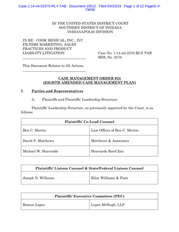
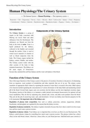
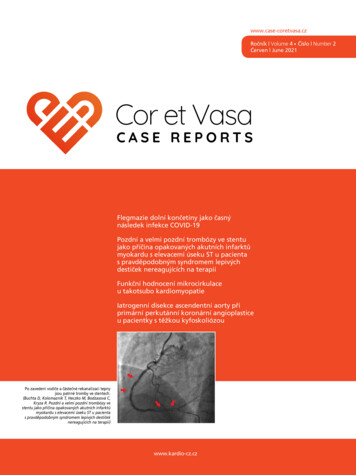
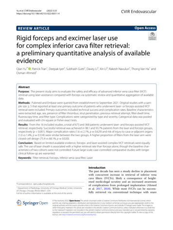
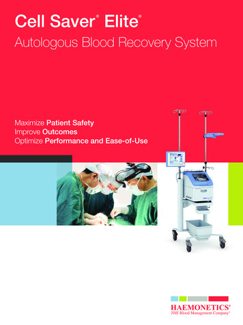
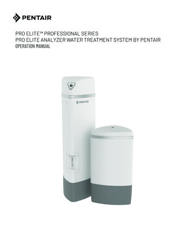
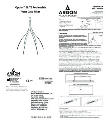
Option ELITEVena Cava FilterOption ELITE RetrievableVena Cava FilterInstructions For UseCatheter Sheath Introducer5 Fr. ID (6.5 Fr. OD) / 100cm lengthFigure 1: Option ELITE Filter SystemKit ContentsA.B.C.D.E.Catheter Sheath IntroducerAngiographic Vessel DilatorPusher with Deployment MarkerOption ELITE Filter in CartridgeSheath CapSterile. Sterilized with ethylene oxide gas. Nonpyrogenic. Radiopaque. For single use only. Do not autoclave.Not for Sale in the US.Figure 2: Option ELITE FilterI. Device DescriptionThe Option ELITE Vena Cava Filter is designed for the prevention ofrecurrent pulmonary embolism via percutaneous delivery in the inferiorvena cava (IVC).The Option ELITE Vena Cava Filter 100cm System is designed for IVCfilter insertion, delivery, deployment and placement via the popliteal andantecubital approach.The self-centering Option ELITE Filter is laser cut from nickel – titaniumalloy (Nitinol) tubing. The Option ELITE Filter (Figure 2) consists of shapememory Nitinol struts emanating from a central location and is designed for optimal clot capture. Retentionanchors (retention hooks) are locatedat the caudal portion of the filter. These anchors are intended for filter fixation to the vessel wall. The Option ELITE Filter is intended to be usedin caval diameters up to 32mm. A retrieval hook is centrally located at the cranial extremity.The constrained Option ELITE Filter is flexible and expands to the internal diameter of the IVC upondeployment. The Option ELITE Filter imparts an outward radial force on the luminal surface of the vena cavato ensure proper positioning and stability. The Option ELITE Filter is designed to prevent pulmonary embolismwhile maintaining caval patency through central filtration.The introduction kit is comprised of a filter housed in a filter cartridge, Catheter Sheath Introducer (5F ID),Angiographic Vessel Dilator with an open end, (Figure 3) and a Pusher with deployment marker (Figure 4).The Angiographic Vessel Dilator has side holes and 2 radiopaque markers, separated by 32mm (betweenthe marker bands), that provide linear measurement of the inferior vena cava and assists in angiographicvisualization when radiopaque contrast is delivered. The pusher advances the filter through the Catheter SheathIntroducer up to the deployment marker, and is then used to fix the filter in place during uncovering. The locationof the distal end of the Catheter Sheath Introducer can be controlled by rotating the entire device to position theCatheter Sheath Introducer in the center of the vena cava.The Filter Cartridge houses the Option ELITE Filter. The body of the Cartridge has text and colored arrowsprinted on it that identify assembly orientation, femoral is printed in green (Figure 5A) and jugular is printedin blue (Figure 5B). The arrow of the desired access site will point into the Catheter Sheath Introducer hub. TheAngiographic Vessel Dilator is designed to provide angiographic visualization and linear measurement of thevasculature when used in conjunction with the delivery of radiopaque contrast media to the vena cava.Figure 3: Angiographic Vessel Dilator TipArgon Medical Devices, Inc.1445 Flat Creek RdAthens, TX 75751Tel: 1 (903) 675-9321Tel: 1 (800) 927 4669www.argonmedical.comEmergo Europe2797Prinsessegracht 202514 AP The HagueThe Netherlands 31 70 345 8570Figure 4: Pusher with Deployment MarkerOption is a Trademark of Argon Medical Devices, Inc.Manufactured under one or more of the following U.S.patent: 7,704,266, 8,100,936, 8,162,972 and 8,715,313Argon Medical Devices, Inc.1445 Flat Creek RdAthens, TX 75751Tel: 1 (903) 675-9321Tel: 1 (800) 927 4669www.argonmedical.comOption is a Trademark of Argon Medical Devices, Inc.Manufactured under one or more of the following U.S.patent: 7,704,266, 8,100,936, 8,162,972 and 8,715,313UK Representative:Argon Medical Devices UK Ltd.Tel: 01283 538538Fax: 01283 538400Emergo EuropeWeb: https://argonmedical.eu.com/Prinsessegracht 202797Registered Office: Number1, Hague2514 AP TheThe NetherlandsEastgate Business Centre, 31 70 345 8570Eastern Avenue, Burton,Staffordshire, DE13 0ATRegistered No. 03543457VAT No. GB 706 4913 37P/N: 2017-0205-00 Rev. 0520DFigure 5A: Femoral Approach Cartridge OrientationFigure 5B: Jugular Approach Cartridge OrientationII. Indications For UseThe Option ELITE Vena Cava Filter 100cm System is intended for the prevention of recurrent pulmonaryembolism (PE) via placement in the vena cava in the following conditions: Pulmonary thromboembolism when anticoagulant therapy is contraindicated Failure of anticoagulant therapy in thromboembolic diseases Emergency treatment following massive pulmonary embolism where anticipated benefits of conventionaltherapy are reduced Chronic, recurrent pulmonary embolism when anticoagulant therapy has failed or is contraindicated
German:Sie finden das Symbolglossar elektronisch unterwww.argonmedical.com/symbolsGreek:Το γλωσσάρι των συμβόλων βρίσκεται στην ηλεκτρονικήδιεύθυνση www.argonmedical.com/symbolsEl glosario de símbolos, en formato electrónico, se encuentra troninen symbolikirjasto on osoitteessawww.argonmedical.com/symbolsFrench:Le glossaire des symboles est situé électroniquement sur :www.argonmedical.com/symbolsCroatian:Rječnik simbola nalazi se na adresiwww.argonmedical.com/symbolsHungarian: A szimbólumok szójegyzéke elektronikusanmegtalálható itt: www.argonmedical.com/symbolsItalian:Il glossario dei simboli è disponibile in formatoelettronico all’indirizzo www.argonmedical.com/symbolsLithuanian: Elektroninis simbolių žodynas pateiktas adresuwww.argonmedical.com/symbolsLatvian:Simbolu glosārijs elektroniskā formā irpieejams: www.argonmedical.com/symbolsDutch:De lijst met symbolen staat in elektronische vorm opwww.argonmedical.com/symbolsNorwegian: Symbolordlisten befinner seg elektroniskpå www.argonmedical.com/symbolsPolish:Słownik symboli w postaci elektronicznej jest dostępny nastronie internetowej www.argonmedical.com/symbolsPortuguese: O glossário de símbolos está localizado eletronicamenteem ник символов в электронном виде находится поадресу www.argonmedical.com/symbolsSlovak:Slovníček symbolov je dostupný v elektronickej podobena adrese www.argonmedical.com/symbolsРечник симбола се налази на edish:Symbolordlistan är elektronisk och finns påsidan www.argonmedical.com/symbolsTurkish:Semboller sözlüğü elektronik olarak www.argonmedical.com/symbolsadresinde bulunmaktadırСписок символів в електронній формі див. на etnamese: Bảng chú giải biểu tượng dạng điện tử có tạiwww.argonmedical.com/symbols
nhằm để mô tả chung về sản phẩm vào thời điểm sản xuất và không cấu thành bất kỳ bảo đảm rõ ràng nào.Nhà Sản Xuất và Nhà Phân Phối sẽ không chịu trách nhiệm về bất kỳ thiệt hại trực tiếp, ngẫu nhiên hoặc do hậuquả nào do tái sử dụng sản MBOLI/JELEK/SIMBOLI/SUTARTINIAI MBOLLER/СИМВОЛИ/BIỂU ó/Definizione/Sąvokos ачення/Định NghĩaFemoralБедренаFemorálníVena eFemoralnoFemoralisFemoraleŠlauniesFemorālā ая �тегновийPhần OLI/JELEK/SIMBOLI/SUTARTINIAI MBOLLER/СИМВОЛИ/BIỂU ó/Definizione/Sąvokos ачення/Định NghĩaJugularЮгуларнаJugulárníVena ā я ремнийCổEnglish:The symbols glossary is located electronically кът на символите се намира в електронен видна адрес www.argonmedical.com/symbolsCzech:Slovníček symbolů v elektronické podobě senachází na www.argonmedical.com/symbolsDanish:Listen over symboler findes elektronisk påwww.argonmedical.com/symbolsGerman:Sie finden das Symbolglossar elektronisch unterwww.argonmedical.com/symbolsGreek:Το γλωσσάρι των συμβόλων βρίσκεται στην ηλεκτρονικήδιεύθυνση www.argonmedical.com/symbolsEl glosario de símbolos, en formato electrónico, se encuentra troninen symbolikirjasto on osoitteessawww.argonmedical.com/symbolsLe glossaire des symboles est situé électroniquement sur :The Option ELITE Filter may be removed according to the instructions supplied in the Section IX, entitled“Optional Procedure for Filter Retrieval” in patients who no longer require a filter. Retrieval of the filter can only beperformed by the jugular approach.The Angiographic Vessel Dilator is designed to provide angiographic visualization and linear measurement of thevasculature when used in conjunction with the delivery of radiopaque contrast media to the vena cava.III. ContraindicationsThe Option ELITE Filter should not be implanted if any of the following conditions are present:1. Patient has an inferior vena cava diameter larger than 32mm.2. Patient is at risk for septic embolism.3. Patient has confirmed bacteremia.4. Patient has a known hypersensitivity to nickel or titanium alloys.5. Pregnant patient when radiation from fluoroscopic imaging may endanger the fetus. Risks and benefitsshould be carefully assessed.6 The jugular orientation should not be used for the popliteal approach and the femoral orientation shouldnot be used for the antecubital approach. Only use the proper orientation for the cartridge for the intendedapproach. Use of the incorrect cartridge orientation for either approach may result in upside-downdeployment that may cause severe adverse events in patients.There are no known contraindications for use of the Angiographic Vessel Dilator.IV. Warnings:Contents supplied STERILE using an ethylene oxide (EO) process. Do not use if sterile barrier is damaged. For single product and patient use only. Do not re use, reprocess or re-sterilize. Reuse, reprocessing or resterilization may compromise the structural integrity of the device and/or lead to device failure, which inturn may result in patient injury, illness or death. Reuse, reprocessing or re-sterilization may also create a riskof contamination of the device and/or cause patient infection or cross-infection, including, but not limitedto, the transmission of infectious disease(s) from one patient to another. Contamination of the device maylead to injury, illness or death of the patient. Accordingly, the Manufacturer or its Distributors will not beresponsible for any direct or consequential damages or expenses resulting from reuse, reprocessing or resterilization of any of the components in the Option ELITE Filter introduction kit. Non-clinical testing has demonstrated that the Option ELITE Filter is MR Conditional. A patient with theOption ELITE Filter can be safely scanned immediately after placement under the followingconditions:– Static magnetic field of 3 Tesla– Spatial gradient magnetic field of 720 Gauss/cm– Maximum whole body averaged specific absorption rate (SAR) of 3.0 W/kg for 15min of scanningIn non-clinical testing, the Option ELITE Filter produced a temperature rise of less than or equal to 1.70C ata maximum whole body averaged specific absorption rate (SAR) of 3.0 W/kg for 15 minutes of MR scanningin a 3.0 Tesla General Electric Healthcare MR scanner. The SAR calculated using calorimetry was 2.8 W/kg. MRimage quality may be compromised if the area of interest is in the exact same area or relatively close to theposition of the Option ELITE Filter. Therefore, it may be necessary to optimization of MR imaging parametersto compensate for the presence of this metallic implant. When injecting contrast medium through the Angiographic Vessel Dilator, do not exceed the maximumpressure rating of 800 psi. After filter implantation, any catheterization procedure requiring passage of a device through the filter maybe impeded. The Option ELITE Filter is supplied loaded in a cartridge indicating the appropriate orientation for poplitealand antecubital approaches. Never reload a fully ejected filter into the Cartridge as this could affect itsshape and function and could result in incorrect filter orientation for the selected access site. Never reloada (partially) ejected filter into the cartridge as this could affect its shape and function. Accordingly, theManufacturer or its Distributors will not be responsible for any direct, incidental or consequential damagesresulting from replacement of the Option ELITE Filter in the cartridge. The Option ELITE Filter should only be used by physicians who are trained in diagnostic and percutaneousinterventional techniques, such as placement of vena cava filters. Accordingly, the Manufacturer or itsDistributors will not be responsible for any direct or consequential damages or expenses resulting from useby untrained personnel. Persons with allergic reactions to nickel-titanium alloys (Nitinol) may suffer an allergic response to thisimplant. Never advance the guidewire, introducer sheath/dilator or deploy the filter without fluoroscopic guidance. If large thrombus is observed at the initial delivery site, attempt filter delivery through an alternative site. Asmall thrombus may be bypassed with the guidewire and introducer. Never redeploy a malpositioned or retrieved filter. For the standard procedure, once the Option ELITE Filter is advanced into the sheath, do not retract then readvance the Pusher, which may cause premature deployment of the filter. For the standard procedure, once the Pusher delivery marker enters the metal tube of the Filter Cartridge, thefilter must be fully deployed and it cannot be re-sheathed. For the Over-the-Wire procedure, once the Dilator delivery marker enters the metal tube of the FilterCartridge, the filter must be fully deployed and it cannot be re-sheathed. The Option ELITE Vena Cava Filter 100cm System is intended for a popliteal and antecubital approach. Thejugular orientation should not be used for the popliteal approach and the femoral orientation should not beused for the antecubital approach.For Optional Filter Retrieval: Excessive force should not be used to retrieve the filter. Retrieval of the filter should not be attempted if thrombus is present in the filter, IVC or deep veins. Retrieval of the filter is possible only from the jugular approach. Before attempting retrieval of the filterfrom the jugular access site, verify that the filter retrieval hook is oriented in a cephalad direction – i.e.pointed toward the jugular access site. The retrieval hook at the cephalad end of the filter is the location forendovascular snare engagement. Retrieval of the filter should only be performed by physicians who are trained in percutaneous interventionaltechniques. Never redeploy a retrieved Filter. Please refer to Section IX labeled “Optional Procedure for Filter Retrieval”.V. PrecautionsPhysicians should be properly trained prior to using the Option ELITE Vena Cava Filter.Store in a cool, dark, dry place.Do not use if package is open or damaged.Use prior to “Use By” date.Do not autoclave or resterilize.Do not continue to use any component damaged during the procedure.If strong resistance is met during any stage of the procedure, discontinue the procedure and determine thecause before proceeding. The Option ELITE Filter has been tested and qualified with the accompanying or recommended accessories.The use of any other accessory could result in complications and/or an unsuccessful procedure. Anatomical variances may complicate Filter insertion and deployment. Careful attention to these Instructionsfor Use can shorten insertion time and reduce the likelihood of difficulties.
Spinal deformations: It is important to exercise care when contemplating implantation in patients withsignificant kyphoscoliotic spinal deformations because the inferior vena cava may follow the general courseof such anatomic deformations.VI. Potential ComplicationsProcedures requiring percutaneous interventional techniques should not be attempted by physicians unfamiliarwith the possible complications. Complications may occur at any time during the implantation, indwellingperiod, or at the time of or following filter retrieval. Possible complications may include, but are not limited to,the following: Vena cava or other vessel injury or damage, including rupture or dissection, possibly requiring surgical repairor intervention Injury or damage to organs adjacent to vena cava, possibly requiring surgical repair or intervention Vena cava stenosis or occlusion Incorrect positioning or orientation of the filter Filter migration/movement Extravasation of contrast media Vasospasm or decreased/impaired blood flow Bleeding or hemorrhagic complications that require transfusion or medical intervention (e.g., IV fluids,medication) Thromboembolic events, including DVT, acute or recurrent pulmonary embolism or air embolism, possiblycausing end organ infarction/damage/failure Infection, possibly requiring medical or surgical intervention (e.g. antibiotics or incision and drainage) Respiratory insufficiency or failure Cardiac arrhythmia Myocardial infarction or coronary ischemia Cerebrovascular accident or other neurologic event Renal insufficiency or failure Reaction to contrast media/ medication Hematoma, possibly requiring medical intervention or surgical revision Other vascular access site injury, including, bruising, AV fistula, or pseudoaneurysm Neurological deficit associated with vascular access, possibly requiring nerve intervention or neurologyconsultation Device breakage or failure or inability to retrieve implanted device as described in IFU, possiblyrequiring another intervention or treatment modality to complete procedure DeathThese events may be serious in nature, and may require hospitalization or intervention to address the condition.The Option ELITE Filter MUST be placed using either the Standard Percutaneous Procedure or the Overthe-wire Percutaneous Procedure.VII. Standard Percutaneous Procedure for Filter ImplantationPre-implant cavography is required: To confirm the patency and visualize the anatomy of the vena cava. To mark the level of the renal veins. To locate the highest level of any thrombus which may be present. To determine the desired level for filter deployment and to mark the position with respect to the vertebralbodies. To confirm that the diameter of the vena cava (AP projection) at the site where the filter is to be deployed is lessthan or equal to the maximum authorized diameter (refer to section I Device Description).1. Select a suitable venous access site, on either the right or left side, depending upon the patient’s sizeor anatomy, operator’s preference or location of venous thrombosis.2. Prep, drape and anesthetize the skin puncture site in standard fashion.3. Remove the components of the introduction kit from the package using sterile technique.4. Wet the operator-selected guidewire (max .038”) with sterile heparanized saline or suitable isotonic solution.5. Flush the Catheter Sheath Introducer and Angiographic Vessel Dilator with heparanized saline or suitableisotonic solution.6. Close the side-port after flushing by rotating the stopcock.7. Insert the Angiographic Vessel Dilator through the Catheter Sheath Introducer, snapping it into place at thehub. Flush with heparanized saline or suitable isotonic solution.8. Puncture the access site using the Seldinger technique.9. Holding the needle in place, insert the guidewire through the needle and into the vessel. Gently advance theguidewire to the desired location.Caution: Do not withdraw a PTFE-coated guidewire through a metal cannula as this may damage theguidewire coating.10. Holding the guidewire in place, remove the needle over the guidewire11. Advance the Catheter Sheath Introducer together with the dilator over the guidewire and into the IVC.12. Position the Catheter Sheath Introducers’ radiopaque tip and the marker bands of the Angiographic VesselDilator in the inferior vena cava below the renal veins in preparation for an angiographic overview of the IVC.13. Remove the guidewire.14. Inject contrast media through the Angiographic Vessel Dilator to determine the diameter of the inferior venacava at the intended implantation site below the lowest renal vein, using its marker bands as a reference. Thedistance between the two marker bands, inside edge to inside edge, is 32mm.Caution: Do not use with Ethiodol* or Lipiodol contrast media, or other such contrast media thatincorporate the components of these agents.Caution: Do not exceed 800 psi when injecting.15. Reintroduce the guidewire.16. Advance the Catheter Sheath Introducer tip to the desired location in the IVC.17. Detach and withdraw the Angiographic Vessel Dilator with the guidewire from the Catheter SheathIntroducer by unsnapping the snap-fit at the hub.Caution: To avoid damage to the Catheter Sheath Introducer tip, do not withdraw the dilator until theCatheter Sheath Introducer tip is at the desired location in the IVC.18. Aspirate from the sideport extension to remove any potential air.19. Determine which end of the cartridge (containing the filter) is to be placed into the hub of the CatheterSheath Introducer.Note: The selected access site will determine the cartridge insertion orientation. The orientation isidentified on the body of the cartridge, femoral is green (used for popliteal approach) and jugular is blue(used for antecubital approach). The arrow of the desired access site will pointinto the Catheter SheathIntroducer hub.20. Place the appropriate end of the cartridge into the hub of the Catheter Sheath Introducer until a snap isachieved (Figure 6).Figure 6: Cartridge Insertion Into Sheath Hub (popliteal shown)*Ethiodol is a trademark of Guerbet S.A.
Một nghiên cứu đơn nhánh, tiến cứu, đa trung tâm và không chọn ngẫu nhiên được thiết kế để thu thập dữ liệuvề độ an toàn và tính hiệu quả của Lưới Lọc Tĩnh Mạch Chủ Rex Medical Option khi thực hiện cả thiết bị vĩnhviễn và có thể thu hồi. Một trăm (100) bệnh nhân đã đặt lưới lọc. Có 52 bệnh nhân nam và 48 bệnh nhân nữ ghidanh. Độ tuổi trung bình là 59,1 16,7 tuổi (khoảng tuổi: 18-90). Năm mươi (50) bệnh nhân đã nhận lưới lọcOption như là biện pháp dự phòng (50%), có 15% bệnh nhân mắc bệnh nghẽn mạch huyết khối. Năm mươi(50) bệnh nhân đã nhận lưới lọc Option do mắc bệnh nghẽn mạch huyết khối tiến triển (50%) có biến chứngcủa thuốc chống đông máu, chống chỉ định sử dụng thuốc chống đông máu hoặc không thể dùng thuốc chốngđông máu. Ba mươi hai (32) bệnh nhân đã ghi danh có bệnh trạng Ung Thư có từ trước (32%). Ba mươi sáu(36) bệnh nhân đã được thu hồi lưới lọc thành công. Bốn mươi bảy (47) bệnh nhận được xem là các bệnh nhânđược cấy lưới lọc Vĩnh Viễn khi họ hoàn tất 6 tháng đánh giá theo dõi. Mười bảy (17) bệnh nhân tử vong do bệnhtrạng có từ trước hoặc gian phát (ví dụ như Ung Thư). Dựa vào quyết định độc lập của Giám Sát Y Tế, không cótrường hợp bệnh nhân tử vong nào được cho là do các thủ thuật cấy ghép hoặc thu hồi thiết bị lưới lọc.Các thủ thuật cấy ghép không có biến cố, với 100% bệnh nhân đạt Thành Công Kỹ Thuật Đặt . Trong quá trìnhtheo dõi xuyên suốt 6 tháng, hai bệnh nhân (2,0%) cho thấy có tình trạng bị di chuyển lưới lọc nhẹ (23 mm), vượtquá giới hạn quy định 20 mm. Ba bệnh nhân (3,0%), tất cả đều bị Ung thư một trạng thái tăng tính đông ngay từban đầu, đã biểu hiện chứng tắc nghẽn tĩnh mạch chủ có triệu chứng. Bốn bệnh nhân biểu hiện các cơn thuyêntắc phổi, được xác định là rõ ràng và có liên quan đến lưới lọc, với tỷ lệ là 4,0%. Tỷ lệ quan sát tình trạng thuyêntắc phổi, tắc nghẽn tĩnh mạch chủ có triệu chứng và di chuyển lưới lọc nhất quán với tài liệu đã xuất bản. Khôngcó sự cố thuyên tắc hoặc nứt gãy lưới lọc.Ba mươi chín (39) bệnh nhân đã cố gắng thu hồi. 36 trong số 39 bệnh nhân (92,3%) đã đạt Thành Công KỹThuật Thu Hồi. Ba mươi chín (39) bệnh nhân đã cố gắng thu hồi trong bốn mươi hai (42) thủ thuật. Thành CôngKỹ Thuật Thu Hồi đã đạt được ở 36 trong số 42 thủ thuật (85,7%). Tỷ lệ Thành Công Kỹ Thuật Thu Hồi đượcquan sát thấy trong nghiên cứu này xảy ra ở phạm vi thuận lợi hơn của tài liệu đã xuất bản. Trong ba trường hợp,không thể thu hồi lưới lọc, do không có khả năng gắn lưới lọc hoặc tháo lưới lọc ra khỏi thành tĩnh mạch chủ.Thời gian cấy ghép trung bình là 67,1 50,4 ngày (phạm vi: 1,0 - 175,0 ngày). Sau khi tiếp cận tĩnh mạch, thủthuật thu hồi không gây ra biến cố bất lợi nào, cho thấy sự an toàn của thao tác thu hồi lưới lọc ở những bệnhnhân không còn cần lưới lọc tĩnh mạch chủ nữa.Tóm lại, việc lắp đặt và thu hồi lưới lọc Option có thể được thực hiện an toàn với tỷ lệ thành công về mặt kỹthuật và lâm sàng tương đối cao. Đối với bệnh nhân không còn có nguy cơ mắc thuyên tắc huyết khối, lưới lọcOption có thể được cấy ghép trong vài tháng và sau đó được thu hồi một cách an toàn. Dữ liệu cho thấy sựan toàn và tính hiệu quả của việc lắp đặt và thu hồi hệ thống lọc Option ở nhóm bệnh nhân liên quan về mặtlâm sàng.XI.21. Insert the lead wire of the Pusher into the Cartridge.Note: No resistance should occcur while advancing the pusher wire through the cartridge.If resistance is felt, withdraw the pusher wire and and reinsert.22. Slowly advance the filter using the pusher until the leading edge of the delivery marker on the pusher ispositioned just proximal to the end of the filter cartridge.Note: Once the Option ELITE Filter is advanced into the sheath, do not retract then re-advance thePusher which may cause premature deployment of the filter.Note: The delivery marker indicates that the filter is at the distal tip of the Catheter Sheath Introducerbut still fully contained within the sheath (Figure 7).Note: If filter advancement difficulties arise when using a tortuous vessel approach, stop filteradvancement prior to the curve. Advance the sheath to negotiate the curve and then continue toadvance the filter. Perform filter release (or deployment) under continuous fluoroscopy.Verify the intended filter location in the inferior vena cava is correct prior to releasing the filter from theCatheter Sheath Introducer.Figure 7: Advance Pusher Until Deployment Marker is Adjacent toCartridge (popliteal shown)Note: Check both A/P and Lateral views under angiographic visualization for optimal placement.23. To deploy the Option ELITE Filter, fix the Pusher in position, then pull the sheath back over the pusher touncover the filter (Figure 8).24. Ensure that the Option ELITE Filter is fully released and deployed.25. Carefully remove the Filter Cartridge along with the Pusher, ensuring that the pusher wire does not interferewith the deployed filter.Figure 8: Filter Deployment Using Uncover Technique (popliteal shown)Khước Từ Trách Nhiệm Bảo Hành và Giới Hạn Biện Pháp Khắc PhụcKhông có bảo đảm rõ ràng hay ngụ ý nào, bao gồm nhưng không giới hạn ở bất kỳ bảo đảm ngụ ý nào về khảnăng tiêu thụ hoặc phù hợp với mục đích cụ thể, đối với Nhà Sản Xuất hoặc (các) sản phẩm của Nhà Phân Phốiđược mô tả trong ấn phẩm này. Trong mọi trường hợp Nhà Sản Xuất hoặc Nhà Phân Phối của Nhà Sản Xuất đềukhông chịu trách nhiệm về bất kỳ thiệt hại trực tiếp, ngẫu nhiên hoặc do hậu quả nào ngoài thiệt hại được luật cụthể quy định rõ ràng. Không ai có quyền ràng buộc Nhà Sản Xuất hoặc Nhà Phân Phối của Nhà Sản Xuất đối vớibất kỳ tuyên bố hoặc bảo đảm nào trừ khi được quy định cụ thể trong tài liệu này.Mô tả hoặc thông số kỹ thuật trong tài liệu in ấn của nhà sản xuất và nhà phân phối, bao gồm ấn phẩm này, chỉnhằm để mô tả chung về sản phẩm vào thời điểm sản xuất và không cấu thành bất kỳ bảo đảm rõ ràng nào.Nhà Sản Xuất và Nhà Phân Phối sẽ không chịu trách nhiệm về bất kỳ thiệt hại trực tiếp, ngẫu nhiên hoặc do hậuquả nào do tái sử dụng sản phẩm.26. Place the Sheath Cap on the Catheter Sheath Introducer.27. Perform a control cavagram prior to terminating the procedure. Verify proper filter positioning.28. Remove the Catheter Sheath Introducer by placing compression on the vessel above the puncture site, slowlywithdrawing the Catheter Sheath Introducer.29. Discard the introduction kit and packaging materials.Note: After use, the introduction kit and packaging materials may be a potential biohazard.Handle and dispose of in accordance with accepted medical practice and with applicable local, stateand federal laws and regulations.VIII. Over-the-wire Percutaneous Procedure for Filter ImplantationSYMBOLS/BIỂU TƯỢNGDefinition/Định NghĩaFemoralĐùiJugularCổPre-implant cavography is required: To confirm the patency and visualize the anatomy of the vena cava. To mark the level of the renal veins. To locate the highest level of any thrombus which may be present. To determine the desired level for filter deployment and to mark the position with respect to the vertebralbodies. To confirm that the diameter of the vena cava (AP projection) at the site where the filter is to be deployed isless than or equal to the maximum authorized diameter (refer to section I Device Description).1. Select a suitable venous access site, on either the right or left side, depending upon the patie
Option ELITE Vena Cava Filter Instructions For Use Catheter Sheath Introducer 5 Fr. ID (6.5 Fr. OD) / 100cm length Figure 1: Option ELITE Filter System Kit Contents A. Catheter Sheath Introducer B. Angiographic Vessel Dilator C. Pusher with Deployment Marker D. Option ELITE Filter in Cartridge E. Sheath Cap Sterile.