Transcription
Mutational Pattern in Multiple Pulmonary Nodules Are Associated With Early Stage LungAdenocarcinomaDong, Shao-wei; Li, Rong; Cheng, Zhiqiang; Liu, Dong-cheng; Xia, Jinquan; Xu, Jing; Li,Shixuan; Wang, Jian; Yue, Yongjian; Fan, Yingrui; Cao, Yundi; Dai, Lingyun; Wang, Jigang;Zhao, Pan; Wang, Xin; Xiao, Zhangang; Qiu, Chen; Wang, Guang-suo; Zou, ChangPublished in:Frontiers in OncologyPublished: 01/02/2021Document Version:Final Published version, also known as Publisher’s PDF, Publisher’s Final version or Version of RecordLicense:CC BYPublication record in CityU Scholars:Go to recordPublished version (DOI):10.3389/fonc.2020.571521Publication details:Dong, S., Li, R., Cheng, Z., Liu, D., Xia, J., Xu, J., Li, S., Wang, J., Yue, Y., Fan, Y., Cao, Y., Dai, L., Wang, J.,Zhao, P., Wang, X., Xiao, Z., Qiu, C., Wang, G., & Zou, C. (2021). Mutational Pattern in Multiple PulmonaryNodules Are Associated With Early Stage Lung Adenocarcinoma. Frontiers in Oncology, 10, iting this paperPlease note that where the full-text provided on CityU Scholars is the Post-print version (also known as Accepted AuthorManuscript, Peer-reviewed or Author Final version), it may differ from the Final Published version. When citing, ensure thatyou check and use the publisher's definitive version for pagination and other details.General rightsCopyright for the publications made accessible via the CityU Scholars portal is retained by the author(s) and/or othercopyright owners and it is a condition of accessing these publications that users recognise and abide by the legalrequirements associated with these rights. Users may not further distribute the material or use it for any profit-making activityor commercial gain.Publisher permissionPermission for previously published items are in accordance with publisher's copyright policies sourced from the SHERPARoMEO database. Links to full text versions (either Published or Post-print) are only available if corresponding publishersallow open access.Take down policyContact lbscholars@cityu.edu.hk if you believe that this document breaches copyright and provide us with details. We willremove access to the work immediately and investigate your claim.Download date: 17/06/2022
ORIGINAL RESEARCHpublished: 19 February 2021doi: 10.3389/fonc.2020.571521Mutational Pattern in MultiplePulmonary Nodules Are AssociatedWith Early Stage LungAdenocarcinomaEdited by:Haichang Li,The Ohio State University,United StatesReviewed by:Feng Wang,Affiliated Hospital of NantongUniversity, ChinaHsian-Rong Tseng,University of California, Los Angeles,United States*Correspondence:Chang Zouzou.chang@szhospital.comGuang-suo Wangwgswy01@163.comChen Qiuszchester@163.com†These authors have contributedequally to this workSpecialty section:This article was submitted toPharmacology of Anti-Cancer Drugs,a section of the journalFrontiers in OncologyReceived: 07 September 2020Accepted: 08 December 2020Published: 19 February 2021Citation:Dong S-w, Li R, Cheng Z, Liu D-c,Xia J, Xu J, Li S, Wang J, Yue Y, Fan Y,Cao Y, Dai L, Wang J, Zhao P,Wang X, Xiao Z, Qiu C, Wang G-s andZou C (2021) Mutational Pattern inMultiple Pulmonary Nodules AreAssociated With Early Stage LungAdenocarcinoma.Front. Oncol. 10:571521.doi: 10.3389/fonc.2020.571521Frontiers in Oncology www.frontiersin.orgShao-wei Dong 1,2†, Rong Li 3†, Zhiqiang Cheng 4†, Dong-cheng Liu 1, Jinquan Xia 1,Jing Xu 4, Shixuan Li 5, Jian Wang 5, Yongjian Yue 6, Yingrui Fan 3, Yundi Cao 3,Lingyun Dai 7, Jigang Wang 7, Pan Zhao 1, Xin Wang 8, Zhangang Xiao 9, Chen Qiu 6*,Guang-suo Wang 5* and Chang Zou 1,2*1Clinical Medical Research Centre, The First Affiliated Hospital, Southern University of Science and Technology, ShenzhenPeople’s Hospital, Shenzhen, China, 2 Shenzhen Public Service Platform on Tumor Precision Medicine and MolecularDiagnosis, Shenzhen, China, 3 Department of Oncology, Taikang Xianlin Drum Tower Hospital, Nanjing University School ofMedicine, Nanjing, China, 4 Department of Pathology, The First Affiliated Hospital, Southern University of Science andTechnology, Shenzhen People’s Hospital, Shenzhen, China, 5 Department of Thoracic Surgery, The First Affiliated Hospital,Southern University of Science and Technology, Shenzhen People’s Hospital, Shenzhen, China, 6 Department of Respiratoryand Critical Medicine, Shenzhen People’s Hospital, Second Clinical Medical College of Jinan University, Shenzhen, China,7 Department of Geriatrics, The First Affiliated Hospital, Southern University of Science and Technology, Shenzhen People’sHospital, Shenzhen, China, 8 Department of Biomedical Sciences, City University of Hong Kong, Hong Kong, Hong Kong,9 Key Laboratory of Medical Electrophysiology of Ministry of Education, School of Pharmacy, Southwestern MedicalUniversity, Luzhou, ChinaThe clinical significance of mutation in multiple pulmonary nodules is largely limited bysingle gene mutation-directed analysis and lack of validation of gene expression profiles.New analytic strategy is urgently needed for comprehensive understanding of genomicdata in multiple pulmonary nodules. In this study, we performed whole exome sequencingin 16 multiple lung nodules and 5 adjacent normal tissues from 4 patients with multiplepulmonary nodules and decoded the mutation information from a perspective of cellularfunctions and signaling pathways. Mutated genes as well as mutation patterns shared inmore than two lesions were identified and characterized. We found that the number ofmutations or mutated genes and the extent of protein structural changes caused bydifferent mutations is positively correlated with the degree of malignancy. Moreover, themutated genes in the nodules are associated with the molecular functions or signalingpathways related to cell proliferation and survival. We showed a developing pattern ofquantity (the number of mutations/mutated genes) and quality (the extent of proteinstructural changes) in multiple pulmonary nodules. The mutation and mutated genes inmultiple pulmonary nodules are associated with cell proliferation and survival relatedsignaling pathways. This study provides a new perspective for comprehension of genomicmutational data and might shed new light on deciphering molecular evolution of earlystage lung adenocarcinoma.Keywords: pulmonary nodules, whole-exome sequencing, mutation, functional analysis, pathway analysis1February 2021 Volume 10 Article 571521
Dong et al.Mutational Patterns in Pulmonary Nodules(Gene Ontology, www.geneontology.org) analysis indicates thatmutated genes tend to converge into cell proliferation andsurvival related signaling pathways in malignancy incomparison with adjacent normal samples. This convergingtendency seems to be more obvious in IA than in MIA.Moreover, the levels of protein structural changes caused bydifferent mutations as well as that of converged signalingpathways are higher in IA than in MIA. The levels of proteinstructural changes and converged signaling pathways are alsohigher in all malignant lesions than in adjacent normal tissues,indicating these protein molecules and pathways may beresponsible for the malignant evolution of LUAD. Somespecific driver genes in cancer development such as EGFR,mutations of this gene are found both in cancer lesions andadjacent normal samples, despite of more severe extent ofmutations in malignancy, suggesting driver gene mutationsneed to be further validated particularly for clinical application.Besides, in comparison with The Cancer Genome Atlas database(TCGA), most of the mutated genes in MIA and IA identified inthis study are genes functionally related to the development oflung cancers. Our findings, by function-directed analysis of WESdata from MPNs, that a group of functionally convergedmutations rather than a single driver gene mutation might bethe triggering onset of the origination and malignant progressionof lung adenocarcinoma. This study will provide new insightsand new strategies to decipher the early development oflung cancer.INTRODUCTIONLung adenocarcinoma (LUAD) is the most frequent subtype oflung cancer. Even with regularly performed and thoroughradiological and cytological screening in high risk populations,LUAD is usually diagnosed in its middle and late stage (1). Noveltherapeutic strategies have been under active research anddevelopment to conquer LUAD, but the survival rate ofpatients has unfortunately kept at a low level (1). Theemerging of improved cancer screening methods such as liquidbiopsy holds great promise for reversing this serious situationsince most of solid tumors are curable in the early stage (2).Unfortunately, this strategy is still limited by the incompleteunderstandings of the complicated molecular mechanisms ofcancer origination and malignant progression as well as intratumoral heterogeneity.Multiple pulmonary nodules (MPNs) contribute to themajority of newly diagnosed LUAD (3). Pathologically, themalignant evolution of early stage LUAD comprises fourcontiguous steps: atypical adenomatous hyperplasia (AAH), insitu pulmonary adenocarcinoma (AIS, also named asbronchioloalveolar carcinoma, BAC), minimally invasiveadenocarcinoma (MIA), and invasive adenocarcinoma (IA), allof which could be well exemplified in some patients with MPNs(4). It is well characterized that AAH represents the precancerous lesion of LUAD and AIS is the localizedadenocarcinoma without any invasion. MIA and IA aredifferentiated by the tumor size. MIA is defined as single ormultiple invasive neoplasia less than 5 mm and restrained instage of IA, while IA represents invasive tumor larger than5 mm (5).In view of different pathological grades and clinical stages ofindividual adenoma loci in the same genetic background,MPNs represent an ideal model for biological characterizationof molecular evolution of the early stage LUAD. A number ofefforts have been made in depicting the molecular diagramof MPNs by using genome-based approaches, e.g., wholegenome sequencing, whole exome sequencing (WES) andmethylation analysis (6–9). However, the theoretical andclinical significance of these reported mutations are largelylimited by the lack of paired validation studies due to the rarityof malignant cells as well as the paucity of neoplasia in MPNssamples (10). In addition, since the origination and malignantprogression of solid tumors is a multifaced and multiple-steporchestration, these findings, mostly based on single genechange-directed analysis, needs to be carefully interpreted andreevaluated especially for clinical application.To better understand the molecular evolution of MPNs, weperformed WES in 21 tissues dissected from 4 patients withMPNs and analyzed the somatic mutations using a cell functionand pathway converging-directed strategy. Mutated genes e.g.,KTM2C and EGFR, and patterns of these mutations weresuccessfully identified and characterized. No ubiquitousmutated genes or mutation loci were discovered in allmalignancy or all patients. Interestingly, more mutated geneswere found in IA samples than in MIA samples. KEGG (KyotoEncyclopedia of Genes and Genomes, www.genome.jp) and GOFrontiers in Oncology www.frontiersin.orgMATERIALS AND METHODSPatients and Clinical InformationTumor samples and their matched normal tissues were obtainedfrom four patients from Shenzhen people’s hospital from 2017 to2019. The clinical information such as age/gender/smokinghistory was listed in Supplementary Table 1. These patientsdid not receive preoperative chemotherapy or radiotherapybefore surgery. Tumor sizes ranged from 0.2 to 3.0 cmaccording to pathology reports. All patients were free of extrathoracic metastasis. Three of the patients were non-smokers, andone patient had a history of cigarette smoking. The images of CTand hematoxylin-eosin staining were reviewed by experiencedpathologists to determine the histopathological subtype and wereshown in Supplementary Figure S1. The present study wasapproved by the Ethics Committee of Shenzhen people’shospital. All study procedures were performed according to theDeclaration of Helsinki ethical principles. Informed consentswere obtained from the patients.Sample PreparationGenomic DNA from formalin-fixed paraffin-embedded (FFPE)samples was extracted using QIAamp DNA FFPE Tissue Kit(Qiagen, Valencia, CA). DNA from fresh tumor tissue and bloodwas extracted by DNAeasy Blood and Tissue kit (Qiagen,Valencia, CA). Genomic DNA was fragmented into 250 bpby M220 Focused-ultrasonicator (Covaris, Inc. Woburn, MA)2February 2021 Volume 10 Article 571521
Dong et al.Mutational Patterns in Pulmonary Nodulesfollowed by whole genome library preparation using KAPAHyper Prep Kit (KAPA Biosystems, Wilmington, MA). Exomecapture was performed using the SureSelect Human All Exon V6panel (Agilent Technologies, Santa Clara, CA).for Interaction Datasets) database (14), and the PPIN in eachtypes of samples were illustrated using Cytoscape (15).StatisticsAll the statistics are performed by RStudio (www.rstudio.com)using R version 3.6.1, and a p-value cutoff at 0.05 is used in allthe analysis.Whole Exome Sequencing (WES)WES was performed by Geneseeq Technology Inc. (Nanjing,China) using an Illumina HiSeq4000 system. The adapters inFASTQ files were cleaned using Trimmomatic-0.36 (11). Thealignment was performed using Burrows-Wheeler Aligner (12),and GRCh37 (hg19) downloaded from Ensembl website was usedas reference. The exome mutations in pulmonary nodules andpara-cancer (PAR) samples were compared with mutationsobtained from peripheral blood mononuclear cells (PBMCs), andonly somatic specific mutations were retained for further analysis.RESULTSCharacterization of PulmonaryNodule SamplesA total of 16 nodules with different stages of neoplasia and fivepara-cancer (PAR) tissues from four patients diagnosed asmultiple pulmonary nodules (MPNs) were collected duringsurgery. After DNA extraction, whole exome sequencing(WES) was performed on these samples and peripheral bloodmononucleated cells (PBMCs) of patients. The somaticmutations were retrieved after the filtration against germ-linemutations through a comparison with PBMC mutations, asillustrated in Figure 1A.Among these 21 samples, two were in atypical adenomatoushyperplasia (AAH) stage, four in minimally invasiveadenocarcinoma (MIA) stage, 10 in invasive adenocarcinoma(IA) stage, and five were PAR tissues. The histologicalcharacteristics of samples together with their IDs from eachstage were presented in Figure 1B. The locations of all the lesionswere indicated in the CT images and the histological image ofeach tissue were shown in Supplemental Figure 1.Converging AnalysisConverging index is defined as follows: 1) take any given twosamples S1 and S2 with mutated genes from some pathways,KEGG for example 2) the numbers of mutated pathways(pathways with mutated genes) were calculated as P1 and P2;3) the number of overlapped mutated pathways between twosamples (mutated pathways occurred in both samples) iscounted as P; and 4) the converging index is calculated as (P/P1)*(P/P2). The converging index in GO biological pathwaysand GO molecular functions is calculated using thesame method.The ranking of converged pathways/functions was guided bythe probability of Poisson distribution, which is calculated usingthe number of mutations involved in certain pathways in PARand IA samples. The Poisson distribution is performed in R 3.6.3using ppois() function. The KEGG pathway information ofhuman proteome was downloaded from the KEGG website(www.genome.jp) and the human GO biological pathway andmolecular function information were retrieved from the Uniprotwebsite (www.uniprot.org).Mutational Analysis of Whole ExomeSequencing ResultsWES was performed on these 21 samples from four patients aswell as their PBMCs. The somatic mutations in each sample weresummarized in Figure 1C. Most of the mutations are samplespecific, with a small portion of mutations shared in more thanone sample (scattered lines in Figure 1C). The number ofmutations varies among different samples, with the leastnumber of seven mutations (Pa1-1; Pa-2; Pa2-P) and thehighest number of 214 mutations (Pa4-1). The average numberof mutations in PAR, AAH, MIA, and IA samples are 49.4, 31.5,29.5, and 53.0, respectively, and the average number of mutatedgenes in PAR, AAH, MIA, IA samples are 44.2, 25.5, 24.8, and43.8, respectively, as listed in Figures 2A, B. These resultsindicated that there is no significant difference in the numberof mutations or mutated genes between AAH and MIA samples,but there is a dramatic increase in the number of both mutationsand mutated genes in IA samples as compared to the restof samples.For mutations on individual gene, missense mutation is themost common one across all types of samples (SupplementalFigure 2). As for the mutation types, single nucleotidepolymorphism (SNP) is the most common type of mutationsacross all the samples (Supplemental Figure 3), which isconsistent with the prevalence of missense mutations; referringEGFR Protein Structural AnalysisEGFR protein structure was retrieved from the Protein DataBank (www.rcsb.org) using entry 1M17. The protein structuresand mutated residues were illustrated by Pymol software (ThePyMOL Molecular Graphics System, Version 1.2r3pre,Schrödinger, LLC.) using “cartoon” and “surface” functions.Comparison With TCGA Mutation DataThe genomic mutation information in 32 types of cancers wasretrieved from the TCGA (The Cancer Genome Atlas) (13) viaXena platform from University of California Santa Cruz. Thenumber of mutations in samples were compared with the TCGAgenomic mutation data in each types of cancer, and a pairedWilcoxon test was performed to evaluate the difference betweeneach types of samples.Protein-Protein Interaction Network(PPIN) AnalysisThe protein-protein interaction information of mutated geneswas retrieved from BioGrid (The Biological General RepositoryFrontiers in Oncology www.frontiersin.org3February 2021 Volume 10 Article 571521
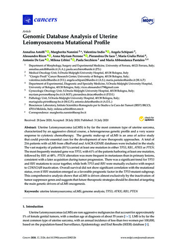
Dong et al.Mutational Patterns in Pulmonary NodulesBACFIGURE 1 Pulmonary nodule sample selection and whole exome sequencing. (A). Schematic illustration of research design. Twenty-one tissue samples andPBMC samples were collected. After DNA extraction and whole exome sequencing (WES), somatic mutations in different tissues were further analyzed;(B) Histological characteristic representation of samples and patient IDs with histopathological stages; (C) Summary of WES results across all samples. Eachdifferent-colored line represents a different type of mutations, and the scattered lines represent the shared mutations between different samples.(KMT2C is treated as specific in patient 3, and hence not countedhere), only occurs in 37.5% of pulmonary nodules (6 out of 16samples). The low concurrence of mutated genes indicates thehigh genetic diversity of pulmonary nodules, and hence we tooka step back and tried to explore the commonality amongpulmonary nodule samples from a perspective of signalingpathways and cell functions.to SNV (Single Nucleotide Variant) classes in SNPs, C– T andG– A conversions are the top two common SNV types, andthese two types of conversions occur most in IA samples(Supplemental Figure 4).Hotspot Mutated Gene Analysis inPulmonary NodulesThe frequency of mutated genes in all nodules (AAH, MIA, and IAsamples) were further counted and ranked, and the hotspot genes(mutated genes occurred in more than 2 pulmonary nodulesamples) were listed in Figure 2C. Among these 19 mutatedhotspot genes, KMT2C (with both missense and nonsensemutations) gene mutation has the highest overall frequency,however, this mutation was only found in the Pa3 samples(including PAR samples); EGFR gene mutation, with two types ofmutations (missense and in frame deletion mutations), occurs in allfour patients and two types of pulmonary samples (MIA and IA);PRB2 gene mutation, with two types of mutations (missense andnonsense mutations), occurs in two of the patients and two types ofpulmonary samples (MIA and IA). Interestingly, some of thesehotspot genes with mutation type changes in different types ofsamples were found and will be further discussed in thefollowing sections.An interesting phenomenon, as discovered in our study, aswell as in many other studies on pulmonary nodules, is the lowconcurrence of mutated genes between different samples. Forexample, mutated EGFR, the gene with the highest frequencyFrontiers in Oncology www.frontiersin.orgConverging Tendency Towards Functionsor Pathways Related to MalignantTransformation in Pulmonary NodulesTo explore whether there is a converging tendency of mutatedgenes in samples at different nodules, we introduced aconverging index, which is defined as the overlapping ofsignaling pathways or cell functions, including KEGGpathways, GO Biological Pathways (GO BP), and GOMolecular Functions (GO MF), in which the mutated genesare involved between two samples (detailed in Materials andMethods). The higher converging index between two samples,the higher probability of two samples with same pathways orcell function got affected (pathway or cell function related genesgot mutated). To compare the converging status betweensamples at different stages, we calculated the convergingindex for KEGG, GO BP, and GO MF between sampleswithin each nodules and the results are shown in Figure 3A(there are only two samples in AAH stage hence only oneconverging index was generated).4February 2021 Volume 10 Article 571521
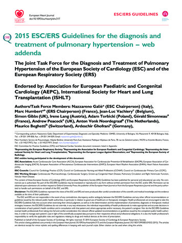
Dong et al.Mutational Patterns in Pulmonary NodulesABCFIGURE 2 The mutation status of samples from different nodules. (A) List of average numbers of mutations/mutated genes in samples from different stages;(B) Violin plot representing the mutations (left) and mutated genes (right) in different samples. Each dot represents the number of mutations (non-synonymousmutations, left) and mutated genes (right) in different samples. (C) Chart listing hotspot mutated genes occurring in more than two pulmonary nodules. Each coloredrectangle represents a mutated gene with different colors indicating different mutation types.lesions, indicating that the cell-cell adhesion ability has beenaffected in the pathogenesis of LUAD and might contribute tometastasis. Among the top 12 converged GO molecularfunctions, many of them are related to regulation oftranscription, which is in consistent with GO BP results. Inaddition, GO:0004713, Protein tyrosine kinase activity andGO:0004714 Transmembrane receptor protein tyrosine kinaseactivity, that are directly involved in cancer-related signaling,have also been affected in pulmonary neoplasia. These findingsimplicate that multiple cell function disorders in these early stagelesions might trigger and/or accelerate the developmentof LUAD.In all three types of pathway or cell function terms (KEGG,GO BP, and GO MF), the average converging indexes of lesionsare higher than those of PAR tissues, except for the indexes inAAH samples, which might be caused by the limitation of samplenumbers. Moreover, the indexes increase with the developmentalstages of pulmonary nodules (AAH MIA IA).To further explore the converged status of nodular samples atdifferent stages, we retrieved all converged functions/pathways,ranked them based on the enrichment levels (detailed inMaterials and Methods) (Figure 3B). Among the top 12converged KEGG pathways, most of them are cancer related orimportant signaling pathways such as FoxO/MAPK/Calciumpathways; among the top 12 converged GO biologicalpathways, most of them are involved in transcriptionregulation and protein phosphorylation, the aberrant functionsof these pathways have been shown directly related to lungcancer development in many studies (16). Moreover,GO:0098609 Cell-cell adhesion, which is directly related toepithelial–mesenchymal transition (EMT), is also converged inFrontiers in Oncology www.frontiersin.orgEffects of Mutation on the Function of theProteins Encoded by Hotspot GenesAside from converging tendency of mutated genes in cancerrelated cellular functions and pathways, we also investigated theeffect of mutation on the function of hotspot genes at the proteinlevel. The changes of mutation types in pulmonary nodules were5February 2021 Volume 10 Article 571521
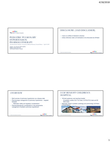
Dong et al.Mutational Patterns in Pulmonary NodulesABFIGURE 3 Converging status of samples from different nodules. (A) The converging ratio of KEGG pathways (left), GO BP (GO biological pathways, middle), andGO MF (GO molecular functions, right) in samples from different nodules. The converging ratio is defined in Materials and Methods with higher converging indexrepresenting the higher tendency of mutated genes converging into the same pathway or function; (B) The converged KEGG pathways (left), GO BP (middle), andGO MF (right) with the number of mutated genes in samples from different stages (PAR, grey; AAH, pink; MIA, cyan; IA, red) and their names (bottom). Theconverged items were ranked based on the probability calculated by Poisson distribution using the number of mutated genes in PAR samples and IA samples.compared with mutations in PAR tissues listed in Figure 4A. Forexample, in EGFR protein, there is a point mutation (L858R) inPAR tissues and IA samples; another mutation type, a deletion of5 amino acids (746-750Del), occurs in both MIA and IA samples.746-750Del locates in Erlotinib binding region (highlighted inred, Figure 4B), and the deletion of this beta3-alphaC loopregion has been previously reported in Erlotinib-resistantpatient (17).In PRB2 protein, there is a point mutation of K88R in PARtissues and IA samples; a nonsense mutation (R1098*) occur inMIA and IA samples. The nonsense mutation R1098* is apremature stop codon which might affect the function of PRB2protein, and might relate to the development of pulmonary nodules.Missense mutation might also cause dramatic structuralchanges. For example, in OR10G9 protein, a missensemutation (K3N) occurs in IA samples, leading to conversion ofa positively charged amino acid (Lysine) to a polar amino acidFrontiers in Oncology www.frontiersin.org(Asparagine). In PRB2 protein, a missense mutation (Q314K)occurs in IA samples, leading to conversion of a polar amino acid(Glutamine) to a positively charged amino acid (Lysine). InPRDM9 protein, two missense mutations occur in MIA and IAsamples (V594D; S814R) leading to conversion of a non-polaramino acid (Valine) to a negatively charged amino acid (Asparticacid) and a polar amino acid (Serine) to a positively chargedamino acid (Arginine). In SLC35G6 protein, a missensemutation occurs in MIA samples (A263D), leading toconversion of a non-polar amino acid (Alanine) to a negativelycharged amino acid (Aspartic Acid). In TCHH protein, amissense mutation occurs in MIA samples, (K1394E) leadingto conversion of a positively charged amino acid to a negativelycharged amino acid (Glutamic Acid). In TXNDC2 protein, a newmissense mutation (G207S) occurs in IA samples, leading toconversion of a nonpolar amino acid (Glycine) to a polar aminoacid (Serine).6February 2021 Volume 10 Article 571521
Dong et al.Mutational Patterns in Pulmonary NodulesABFIGURE 4 The mutation change in the same gene from different pulmonary nodules. (A) List of genes with different mutation in para-cancer tissues (PAR) andpulmonary nodule tissues (AAH, MIA and IA); (B) The 3D surface structure of EGFR in complex with Erlotinib. The different types of mutations are highlighted in pinkand red colors.IA samples. The complexity of PPI network could contribute tothe explanation of development of pulmonary nodules.Pulmonary Nodule Mutated GenesTo explore the association of cancer related genes and mutatedgenes in different pulmonary nodules screened in WES, weretrieved the mutational data of these genes in 32 types ofcancers from TCGA database, and compared the averagenumber of mutations per gene among different types ofcancers. Among 32 types of cancers, IA-related mutated geneshave the highest number of mutations per gene in UCEC(Uterine corpus endometrial carcinoma). LUSC (Lungsquamous cell carcinoma) and LUAD (Lung adenocarcinoma)ranks among the top #5 and #7, respectively of these 32 types ofcancers, indicating the IA-related mutated genes have a highermutation rate in lung cancers comparing to other types ofcancers (Figure 5A). Among the four stages of nodules,pulmonary nodule related genes have a higher mutation rate inall types of cancers in comparison with PAR-related genes, andthe mutation rate increases with the development of pulmonarynodules (IA MIA, IA AAH, AAH PAR) (Figure 5B).To explore the functional importance of pulmonary nodulerelated mutated genes, we calculated the number of each mutatedgene involved in KEGG pathways (Figure 5C), GO BP (Figure5D), and GO MF (Figure 5E). The rationale behind this is thatthe higher the number of genes involved in the pathways/functions, the more important role one gene could play, andhence the mutation could make bigger impact on cell activities.In general, the average numbers of mutated genes involved inpathways/functions are higher in pulmonary nodules than thatof PAR-related mutated genes, suggesting the increasedfunctional importance of mutated genes in pulmonary nodules.We further constructed the protein-protein interaction (PPI)networks generated by mutated genes from different pulmonarynodules (Figure 5F) and compared them with each other (thePPI network of AAH-related genes was not shown due to limitednumber of mutated genes). The PPI network generated by IArelated mutated genes is more complex when compared withMIA or PAR nodules, indicating a complex function affected inFrontiers in Oncology www.frontiersin.orgDISCUSSIONMPNs is defined as more than one solid nodules in the lung of asingle patient. Pathologically, MPNs are commonly identified asAAH, AIS, MIA, and IA. They represent an ideal model ofdeciphering the clonal evolution of early stage lungadenoca
different mutations as well as that of converged signaling pathways are higher in IA than in MIA. The levels of protein structural changes and converged signaling pathways are also higher in all malignant lesions than in adjacent normal tissues, indicating these protein molecules and pathways may be responsible for the malignant evolution of .