Transcription
Hjetland et al. BMC Geriatrics(2021) SEARCH ARTICLEOpen AccessAmbient bright light treatment improvedproxy-rated sleep but not sleep measuredby actigraphy in nursing home patientswith dementia: a placebo-controlledrandomised trialGunnhild J. Hjetland1,2,3* , Eirin Kolberg1, Ståle Pallesen4,5, Eirunn Thun1,5, Inger Hilde Nordhus1,6,Bjørn Bjorvatn4,7 and Elisabeth Flo-Groeneboom1AbstractBackground: Up to 70% of nursing home patients with dementia suffer from sleep problems. Light is the mainzeitgeber to the circadian system and thus has a fundamental impact on sleep-wake behaviour. Low indoor lightlevels in nursing homes have been reported, and in combination with age-related reductions in light sensitivity,insufficient light exposure is likely to contribute to sleep problems in this population. Increasing daytime lightexposure using bright light treatment (BLT) may represent a feasible non-pharmacological treatment for sleepproblems in nursing home patients with dementia.Methods: The present study reports on sleep outcomes, which are the primary outcomes of the DEM.LIGHT trial(Therapy Light Rooms for Nursing Home Patients with Dementia– Designing Diurnal Conditions for Improved Sleep,Mood and Behavioural Problems), a 24-week cluster-randomised placebo-controlled trial including 8 nursing homeunits and 69 resident patients. The intervention comprised ambient light of 1000 lx and 6000 K from 10:00 to 15:00,with gradually increasing and decreasing light levels prior to and following this interval, using ceiling mountedlight-fixtures and light emitting diode technology. The placebo condition had continuous standard light levels(150–300 lx, 3000 K). Sleep was assessed at baseline and follow-up at week 8, 16, and 24, using the proxy-ratedSleep Disorder Inventory (SDI) and actigraphy (Actiwatch II, Philips Respironics). Mixed linear models were used toevaluate intervention effects, adjusting for relevant covariates such as age, gender, number of drugs, severity ofdementia, eye disease, and estimated light exposure.(Continued on next page)* Correspondence: Gunnhildjohnsen.Hjetland@fhi.no1Department of Clinical Psychology, Faculty of Psychology, University ofBergen, Bergen, Norway2City Department of Health and Care, City of Bergen, NorwayFull list of author information is available at the end of the article The Author(s). 2021 Open Access This article is licensed under a Creative Commons Attribution 4.0 International License,which permits use, sharing, adaptation, distribution and reproduction in any medium or format, as long as you giveappropriate credit to the original author(s) and the source, provide a link to the Creative Commons licence, and indicate ifchanges were made. The images or other third party material in this article are included in the article's Creative Commonslicence, unless indicated otherwise in a credit line to the material. If material is not included in the article's Creative Commonslicence and your intended use is not permitted by statutory regulation or exceeds the permitted use, you will need to obtainpermission directly from the copyright holder. To view a copy of this licence, visit http://creativecommons.org/licenses/by/4.0/.The Creative Commons Public Domain Dedication waiver ) applies to thedata made available in this article, unless otherwise stated in a credit line to the data.
Hjetland et al. BMC Geriatrics(2021) 21:312Page 2 of 15(Continued from previous page)Results: Sleep as measured by the SDI was significantly improved in the intervention group compared to thecontrol group from baseline to week 16 (B 0.06, 95% CI -0.11 - -0.01, p .05) and from baseline to week 24 (B 0.05, 95% CI -0.10 - -0.01, p .05). There was no effect according to the SDI at week 8 and no significant effects interms of actigraphically measured sleep.Conclusions: Proxy-rated sleep improved among nursing home patients with dementia following 16 and 24 weeksof BLT. These improvements were not corroborated by actigraphy recordings.Trial registration: ClinicalTrials.gov Identifier: NCT03357328. Registered 29 November 2017 – Retrospectivelyregistered.Keywords: Sleep, Dementia, Nursing home, Actigraphy, Sleep disorder inventoryBackgroundDementia denotes a group of disorders characterised byprogressive neurodegenerative and/or vascular damage,with accompanying impairments of cognition and disturbances of mood and behaviour [1]. These disorders constitute a severe burden at the individual and societallevel [2], making them a social health priority of theWorld Health Organization [3].Disrupted sleep constitutes a major challenge in relation to dementia, as it affects up to 70% of the patients[4]. As sleep is essential for normal functioning, its disruption may have severe negative effects on cognitive,emotional, and physical functioning [5]. In the contextof cognitive impairment, sleep disruption places a significant burden on caregivers [6]. Related events such asnight-time wandering and confusion may increase therisk of falls and injuries [7, 8]. Importantly, sleep problems are associated with increased depressive symptoms[9–11], reduced functional status (ability to perform normal daily activities) [12], and cognitive decline [13]. Dueto limited staff in nursing homes, pharmacological treatment (e.g., antidepressants, benzodiazepines, zhypnotics, and antipsychotics) are often resorted to forrelieving sleep disturbances [14]. Medications are, however, often associated with side-effects such as sedationand risk of falling [14, 15], and there is generally limitedevidence for their efficacy in these populations [16].Thus, identifying safe and effective treatments for disturbed sleep is of crucial importance.As dementia progresses, brain systems involved insleep-wake regulation are increasingly affected [5, 17].Importantly, this includes neurodegeneration of thesuprachiasmatic nucleus (SCN) of the hypothalamus, themain circadian pacemaker, which generates 24-hrhythms in hormone levels, body temperature, andsleep-wake behaviour. As a result, people with dementiaoften display a fragmented sleep pattern with severalsleep and wake periods occurring throughout the 24-hday [17, 18]. Disrupted sleep may be further worsenedby factors such as reduced social interaction, inactivity,and medications [19, 20]. In addition, light exposureplays a major role in circadian regulation, and insufficient exposure has consequently been associated withsleep problems in dementia patients [21, 22].Light is detected by the intrinsically photosensitive retinal ganglion cells of the eye (ipRGCs; neurons in theretina containing the light sensitive photopigment melanopsin) and relayed directly to the SCN. The effects oflight exposure on the circadian system depend on thetiming, duration, illumination level (lux), and spectralcomposition (i.e., the colour of the light), as well as lightexposure history [23]. The ipRGCs are maximally sensitive to short wavelengths ( 480 nm) [24, 25], hencepolychromatic light with high amounts of short wavelengths, i.e., blue light, therefore elicits stronger circadian responses than light with large amounts of longwavelengths (red-yellow) [26, 27]. The colour appearance of light is defined by correlated colour temperature(CCT), measured in Kelvin (K). Daylight has a largeamount of blue light, with a CCT of about 5700–7700 K,depending on atmospheric conditions [28]. In comparison, standard light bulbs typically deliver more yellowlight, with lower CCT (2700–3000 K).With increasing age, lens yellowing and excessive pupilconstriction (senescent miosis) reduce the amount oflight reaching the retina [29, 30]. Although some evidence has suggested compensatory mechanisms preserving light sensitivity [31], lens yellowing has beenassociated with self-reported sleep disturbances [32].Further, Alzheimer’s disease is associated with severalpathological changes in the visual system, including lossof ipRGCs [33]. Several studies have reported light levelsin nursing homes far below what is considered necessaryfor circadian entrainment (synchronising of an organisms’ rhythms to recurring environmental cues) [17, 34–37]. For example, in a study of seven nursing homes inthe Netherlands, Sinoo et al. [37] found that 65–96% ofthe light measurements fell below 750 lx. In a recentstudy including 15 Norwegian dementia unit livingrooms [38], median vertical illumination was below 300lx, even in summer. In contrast, daylight ranges from6500 to 130,000 lx depending on weather conditions [39,
Hjetland et al. BMC Geriatrics(2021) 21:31240]. In sum, age and dementia-related changes to theeye and low indoor illumination in relevant institutionssuggest that dementia patients are rarely exposed to lightlevels sufficient to entrain the circadian clock. Therefore,increasing light exposure, i.e., bright light treatment(BLT), constitutes a promising non-pharmacologicaltreatment for disrupted sleep in people with dementia.Traditionally, BLT has been administered using lightboxes with high illumination white light (2500–10,000 lxat distances from 10 to 50 cm) for 30 to 120 min eachday. This requires the patient to sit relatively still andface the box. Several studies have reported within-groupimprovements of sleep outcomes using this type of BLT[41–47], although the results are not consistent [48–50].An issue with light boxes is that dementia patients are inneed of continuous supervision to receive sufficient lightexposure [42], which is often too demanding for thestaff. Recent advances in light emitting diode (LED)technology have allowed for the manipulation of bothlight illumination and its spectral composition. Thistechnology is able to deliver light of high CCT and illumination and may be used to change the ambient lightof entire rooms. Researchers have in this realm typicallyused light ranging from 6500 K and 1200 lx to 13,000 Kand 400 lx [51]. By this way of administering light, thepatients may move around freely, and the need for staffto ensure treatment adherence is eliminated. In addition,such systems may be programmed to provide light contrasts between midday (high illumination short wavelength light) and early mornings and evenings (lowerillumination and “warmer”, longer wavelength light).Although BLT has been evaluated as a treatment for sleepproblems and emotional and behavioural symptoms in dementia populations for two decades, conclusive results aboutits efficacy are lacking. Recent meta-analyses, with 6 to 11studies included, have reported only small effects (Hedges’ gof 0.25–0.30) of BLT in dementia [52, 53]. Generally, fewrandomised controlled trials have been conducted and thereare considerable variations in methods and designs, such asthe timing, duration, and means of delivering BLT. Hence,there is a need for more high-quality studies [51].In the present study we report the primary outcomesfrom the DEM.LIGHT trial aiming to evaluate the effects of a ceiling-mounted dynamic ambient BLT solution on sleep in nursing home patients with severedementia. We hypothesised that the BLT conditionwould improve sleep measured by actigraphy and aproxy-rated sleep scale across the 24-week treatmentperiod, as compared to a control group receiving conventional light with standard and constant light levels.MethodsThe present paper is based on data from the 24-weekcluster randomised placebo-controlled trial “TherapyPage 3 of 15Light Rooms for Nursing Home Patients with Dementia–Designing Diurnal Conditions for Improved Sleep, Moodand Behavioural Problems”, the DEM.LIGHT trial(ClinicalTrials.gov Identifier: NCT03357328). This trialevaluated the effect of a BLT solution on sleep, circadianrhythmicity, mood, behaviour, and function in nursinghome patients with dementia. The present paper reportson the sleep outcomes of the DEM.LIGHT trial. Theother outcomes will be reported in subsequent papers.The DEM.LIGHT trial was conducted from late September 2017 to early April 2018, in Bergen, Norway (60 3’N,5 3’E). The intervention period lasted for 24 weeks anddata were collected at baseline, week 8, week 16, andweek 24. The present study adheres to the CONSORTguidelines [54].ParticipantsThe Department of Health and Care, City of Bergen,Norway, supported the study by providing informationabout eligible nursing home dementia units (i.e., nursinghomes not involved in other trials or quality of care projects, and where ceiling light instalment was possible).The management of eligible nursing homes was contacted by the two researchers leading the data collection (GJH, EK). Site visits were conducted to provideinformation about the study and assess if light instalment was possible. In total, 14 nursing homes wereapproached, of which 8 units from eight nursing homeswere included. Nursing home units and patients were recruited between September 2016 and August 2017. Theeligibility criteria were discussed with the resident medical practitioner of each nursing home before all the eligible patients at the unit were invited to participate.Inclusion criteria were as follows: The patient had to be60 years of age or older, in long-term care ( 4 weeks),have dementia in accordance with DSM-5 criteria andeither sleep/circadian rhythm disturbances, BPSD, or severely reduced activities of daily living. Exclusion criteriawere blindness or other reasons the patient was unableto benefit from light; the patient was taking part in another trial; a condition that contra-indicated the intervention; an advanced, severe medical condition and/orexpected survival of less than 6 months; psychosis or asevere mental disorder; or other aspects that could interfere with participation. Due to the long follow-up of 6months, it was not possible to maintain stable doses ofmedications during the course of the study. The numberof psychotropic medications were, however, controlledfor in the analyses.ProceduresThe interventionA LED ceiling-mounted bright light solution, deliveredby Glamox AS (supplier of professional lighting
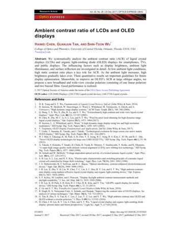
Hjetland et al. BMC Geriatrics(2021) 21:312solutions), was installed in the common rooms of thefour intervention units. Glamox engineers calculated thenumber of LED units needed in each common room(Glamox, 1 x C95 48 CCT 6500 K MP 47 W / 4702 lm),accounting for the number and direction of windows.The light panels were programmed to provide a cycle ofgradually higher illumination and CCT during the dayand then lower illumination and CCT in the evening(see Fig. 1), with the highest light levels of 1000 verticallux and 6000 K from 10:00 to 15:00. The light equipmentwas installed prior to the start of the study and lightlevels were maintained at standard levels (approximately100 lx and 3000 K) before the study commenced so thatstaff and participants were familiarised with the newlight units. The intervention light sequence was activatedfollowing baseline data collection. The control panel forthe light was locked with a personal identification number only known to the researchers. The contact information of the researchers was provided in case of anyproblems with the light setup.The placebo control condition was created by replacing the light bulbs in the existing fittings with conventional 3000 K light bulbs in all common rooms (CFLAURA UNIQUE-D/E LL 18 W/830 G241–2 in threecommon rooms and CFL AURA UNIQUE-L LL 18 W/830 2G11 in one), thus largely maintaining the preexisting light levels in these units. These light bulbs wereinstalled immediately following the baseline data collection. After the intervention was activated and the placebo lights were installed, the daytime light levels weremeasured by the researchers in all eight common roomsusing the GL Spectis 1.0 T Flicker spectrometer (GLOptic). Spectral analysis was performed in GL Spectrosoft. Vertical measurements were recorded to approximate corneal illumination (120 cm above the floor), andhorizontal measurements were recorded at the typicalheight of reading or other visual tasks (80 cm above thefloor). Effective illuminances were calculated accordingto the α-opic illuminance model using the irradiancetoolbox by Lucas et al. [55].Page 4 of 15OutcomesAll questionnaires used in the present study were completed by daytime nursing staff that knew the patientswell, i.e., regular staff working directly with the patients.Research staff involved in the DEM.LIGHT trial guidedthe nurses in using the assessment tools. The questionnaires were completed once at each data collection timepoint: either the same week the patients wore the actigraphs or the following week. Data regarding sociodemographic characteristics, diagnoses, and medicationstatus were collected from medical records. One of theresearchers was granted access to extract these datafrom a centralised journal system used in nursing homesby Bergen Municipality.Sleep outcomesChanges in sleep parameters comprised the primary outcome of the DEM.LIGHT trial. Sleep was proxy-rated bythe nursing staff using the Sleep Disorder Inventory(SDI) [12]. The SDI assesses seven symptoms that arescored in terms of frequency (0–4), severity (0–3), andcaregiver distress (0–5). The SDI total score was calculated as the sum of the products of the frequency and severity ratings of each of item 1–7 (range: 0–84). TheSDI has previously been demonstrated to correspondwell to actigraphy outcomes in this population [56], aswell as in home-dwelling dementia populations [12]. Atotal score of five or more indicates the presence of clinically significant sleep disruption [56].Actigraphs (Actiwatch II, Philips Respironics) wereused to provide an objective estimation of sleep. Actigraphs are wrist-worn devices that measure activity continuously over days or weeks [57]. In accordance withprevious studies on nursing home patients, the actigraphs were placed on the dominant or most mobilewrist [58–61] as this increases the chances of detectingmovement in patients who are often immobile or lethargic. Based on the activity data, each epoch was scored assleep or wake by the Actiware software, version 6.0.9(Philips Respironics). The threshold for wakefulness wasFig. 1 An illustration of the intervention light sequence. The light changed gradually across 30 min between each condition. Each nursing homecould choose if they wanted to turn the lights off or to maintain 100 lx and 2500 K throughout the night (21:00–07:00). The patients generally didnot spend time in the common room during the night, and whether or not the light was on in the common room depended on the needs ofthe staff during the night
Hjetland et al. BMC Geriatrics(2021) 21:312set to medium and epoch length to 1 min, as recommended by Camargos et al. [61].Determining the true bedtime and wake-time in nursing home patients is challenging, as these patients frequently spend more than 12 h in bed (e.g., [58, 60]). Weinitially planned to score the rest intervals based on apredetermined scoring protocol; however, there wererarely clear indications of bedtime and wake time, andthe nurses did not consistently press the event buttons.Therefore, a fixed rest interval was set from 22:00 to 06:00, in accordance with previous studies, which ensuredcapturing the main sleep episode of the vast majority ofparticipants [22, 34, 48, 62]. A fixed daytime interval wasalso set from 10:00 to 18:00. This interval correspondsto other studies in dementia populations, and waschosen to avoid including periods when the participantswere in bed [63]. Activity data were collected for sevenconsecutive days at baseline, and at follow-up after 8, 16,and 24 weeks. Patients had to have at least five nights ofrecordings to be included in the analyses. The followingactigraph outputs were extracted: Sleep efficiency (SE;the percentage of time spent asleep in the rest interval),TST rest (total sleep time in the rest interval), 24-h TST,daytime TST (TST in the daytime interval), and wakeafter-sleep-onset (WASO; the time spent awake aftersleep onset) in the rest interval. Since TST rest and SEare perfect linear functions of each other in a fixed restinterval, including both in the analyses would be redundant and TST rest was only used as a descriptivevariable.Other outcomesIn addition to sociodemographic data, several secondaryoutcomes were used to describe the population at baseline and as control variables in the analyses. The MiniMental State Examination (MMSE) assesses the level ofcognitive impairment (range: 0–30), where a low scoreindicates worse cognitive function [64]. The FunctionalAssessment Staging (FAST) [65] rates the severity of dementia according to seven stages, where a score of 6–7indicates severe dementia. The Charlson ComorbidityIndex (CCI) assigns weights to 17 comorbidities to assess the burden of comorbid disease, where a higherscore is associated with a higher mortality risk [66]. Toassess activities of daily living (ADL), the instrument byLawton and Brody was used [67]. This scale includes sixitems (composite score range 0–30), where a lower valueindicates better functioning and independence. Thenursing home version of the Neuropsychiatric Inventory(NPI-NH) [68, 69] is a proxy-rated tool that assesses“Behavioural and Psychological Symptoms of Dementia”by assigning frequency and severity ratings to 12 symptoms (delusion, hallucination, agitation, depression, anxiety, euphoria, apathy, disinhibition, irritability, aberrantPage 5 of 15motor behaviour, night-time behaviour and eatingdisturbance).The staff at each unit were urged to report any changein patient health or behaviour in relation to the intervention. Potential adverse events and tolerability were alsomonitored at each data collection visit.Adherence to treatmentTo assess adherence to treatment, a questionnaire wasadministered to the nursing staff, where they were askedto estimate the time each patient spent in the commonroom during different epochs of the day (average duringthe last 8 weeks). The epochs corresponded with thelight cycle of the intervention, so that the day was splitinto 07:00–10:00, 10:00–15:00, 15:00–18:00, and “after18:00”. The nursing staff provided a time estimate foreach epoch in terms of hours and minutes. They werealso instructed to report the number of days when thepatient was not present in the common room, and informed that these days should not be included in theabove-mentioned estimate. For the purpose of this study,the time estimates corresponding to the epoch with thehighest light levels (10:00–15:00) were included in theanalyses.Sample size and powerExpecting moderate effect sizes (Cohen’s d .50) for theactigraphy outcomes, a .05 alpha level (two-tailed), andthe power set to .80, the power-analysis showed that 64participants from a minimum of 8 clusters were neededin order to detect differences between conditions.Expecting a 20% dropout, the aim was to recruit 80 participants [70, 71].RandomisationAfter eight nursing home units were included, clusters(each unit constituted one cluster) were randomised tothe intervention condition (1) or the control condition(0) using random group assignment in SPSS. Randomisation and enrolment were completed by the researchgroup.BlindingAlthough the placebo effect might not be an issue inthose with severe dementia, staff might be affected, creating bias such as the Hawthorne effect [72, 73]. Hence,potential changes in staff routines and behaviour in response to the treatment may affect outcomes, making acontrol condition necessary. Because staff and participants could not be kept blind to the intervention, as thelight setup comprised an obvious change in the commonroom, new light bulbs were fitted in the control units tomimic an intervention and ensure similar light levelsacross the control units. The participants and nursing
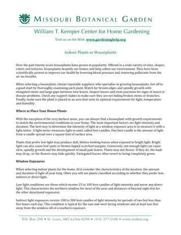
Hjetland et al. BMC Geriatrics(2021) 21:312staff were consequently unaware of condition assignment and all included units were located at differentnursing homes to minimise threats to internal validity interms of performance and detection bias [74]. After theend of the trial, the study design was explained to thestaff, who were then asked to answer a questionnaireasking whether they thought they were in the control orintervention group.Page 6 of 15using correlation analysis for continuous data and comparing groups for categorical data using Student’s t-test.Differences in light levels between the intervention andthe placebo condition, as well as differences in light exposure time were evaluated using Student’s t-tests andMann-Whitney U tests in SPSS for Windows, version 25(IBM).Missing dataStatistical analysesWe initially planned to analyse data using ANOVA, butowing to some missing data due to non-compliance toactigraphy and because some patients passed away ormoved to another facility during the study, we evaluatedthe intervention effects by means of linear mixed modelsusing the lme4 package in R [75, 76]. This analysismethod takes into account all available data, performsbetter than alternatives when there are missing data, andestimates fixed effects while adjusting for correlationdue to repeated measurements on each subject [77, 78].Linear mixed models using restricted maximum likelihood estimation were used to assess all outcomes.Group (intervention vs. control), time (treated as categorical with levels baseline, 8 weeks, 16 weeks, and 24weeks), and the group-by-time interaction were includedas fixed effects in the model. The models were fittedwith random intercepts at the patient level to accountfor intra-participant correlation of the outcomes, andrandom slope was included if model fit improved.Models were selected based on best fit according to likelihood ratio tests.A range of factors are likely to impact the response toBLT, and a range of predetermined prognostic variableswere tested as covariates. AIC and BIC were examined,and likelihood ratio tests were performed using theAnova package in R. Furthermore, covariates that improved fit were added to the model. The following list ofcovariates were tested: Age, gender, number of psychotropic medications, CCI-scores, FAST-scores, MMSEscores, eye disease, whether the patient passed away ormoved during the study (drop-out), and average time inthe common room during the day (between 10:00 and15:00). Melanopic lux was also tested as a covariate because the light levels varied within the interventiongroup (ranging from 675 to 1050 mean vertical melanopic lux) and control group (56 to 261 melanopic lux)(Table S1).Preliminary analyses showed that the SDI data werehighly skewed, consequently the scores were transformed to achieve normal distribution by adding a constant of 0.5 and using a Box Cox transformation, whichresulted in a lambda of 0.6.Further, the relationship between sleep, demographicand secondary outcomes at baseline was investigated byIf single questionnaires were missing data on 20% ormore of items on the SDI, they were excluded from theanalyses. For questionnaires missing less than 20%, andwhere data were missing completely at random, imputations were made using expectation maximisation (EM)in SPSS.ResultsSeventy-eight patients were evaluated for eligibility, ofwhich 69 were recruited to the study (see the flow chartin Fig. 2). Figure 3 shows the number of participants included in each analysis at each time point and reasonsfor missing data (not counting data missing due to deathor moving away). Of the 69 patients who were enrolled,the mean age was 84 years, and 68% were female. Themean MMSE score was 6.4 (SD 6.7) (Table 1).Table 2 shows the mean light levels in the interventionand control condition, respectively. All light metricswere significantly higher in the intervention group thanthe control group (all p’s .05 and Bonferroni adjustedp .008). The supplemental Table S1 shows the lightmeasurements separately for each unit. There was somevariability in light levels across intervention units, andone unit in particular had lower than intended verticalillumination (722; SD 69; photopic lux and 675; SD 62; melanopic lux). Also, one control unit had higher illumination than the other control units, with 368 (SD 99) photopic lux and 261 (SD 125) melanopic lux. Allother control units had between 134 and 271 photopiclux and between 56 and 143 melanopic lux. Both horizontal and vertical measurements and photopic lux, melanopic lux, and CCT values are provided in Table 2 aswell as in supplementary Table S1. Across the intervention period, participants in the intervention group spenta mean of 3 h and 24 min (SD 1 h 30 min), and thecontrol group spent 3 h and 0 min (SD 1 h 42 min) inthe common room during the time of day with the highest light levels (10:00–15:00), as estimated by staff(Table 3). There were no significant differences betweenthe groups regarding time spent in the common room atany time point.The primary outcome of this randomised controlledtrial (RCT) was sleep. The linear mixed model analysesfor actigraphically measured sleep outcomes showed nostatistically significant differences between patients who
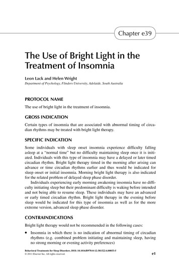
Hjetland et al. BMC Geriatrics(2021) 21:312Page 7 of 15Fig. 2 Showing the flow of participants from eligibility assessment to study completion. Note: Main cause for moving was a deterioration ofsomatic health requiring the patient to live in a somatic wardreceived BLT compared to those who received standardlight (see Table 4).For the SDI, the regression coefficient for the time*group interaction, representing the group difference inchange from baseline, was .06 (95% CI .11 to .01; p .020) at week 16 and .05 (95% CI .10 to .01; p .028)at week 24, indicating relatively better sleep in the intervention group (i.e., less disrupted sleep) than the controlgroup at these time points. The average number of psychotropic drugs was included as a covariate in this model.There was no significant effect at week 8 (Tab
light-fixtures and light emitting diode technology. The placebo condition had continuous standard light levels (150-300lx, 3000K). Sleep was assessed at baseline and follow-up at week 8, 16, and 24, using the proxy-rated Sleep Disorder Inventory (SDI) and actigraphy (Actiwatch II, Philips Respironics). Mixed linear models were used to