Transcription
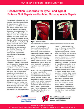
LARGE ROTATOR CUFF REPAIR PROTOCOLThe intent of this protocol is to provide the clinician with instruction, direction, rehabilitative guidelines andfunctional goals for all rotator cuff repair procedures. It is not intended to be a substitute for clinical decisionmaking regarding the progression of a patient’s post-operative course based on physical exam/findings andindividual progress. The physiotherapist must exercise their best professional judgment to determine how tointegrate this protocol into an appropriate treatment plan. The general treatment for a variety of shoulderprocedures involves protection of the repair, stretching/mobilizing tight or restricted structures, strengthening therotator cuff and strengthening and retraining the scapular musculature.This particular protocol divided into 4 phases and the timeline can vary from 4 months to 1 year: Phase 1: Passiverange of motion; Phase II: Active assisted active range of motion; Phase III: Resisted exercises/strengthening;Phase IV: Advanced strengthening/dynamic stability. Therefore, decisions to advance patients through the phasesof rehabilitation should be based on achieving the appropriate level of soft tissue healing, as well as clinicalpresentation and response to treatment. As an individual’s progress is variable and each will possess various preoperative deficiencies, this protocol must be individualized for optimal return to activity. Some exercises may beadapted depending on the equipment availability at each facility. There may be slight variations in this protocol oradditional restrictions placed by the surgeon post-operatively depending on findings at the time of the surgery. If aclinician requires assistance in treatment progression please contact the referring physician or the physiotherapydepartment.1GENERAL CLASSIFICATION OF ROTATOR CUFF TEAR SIZESmall: 1cm in lengthMedium: 1-3 cmLarge: 3-5 cmMassive: 5 cmAlso, tears are described as either partial or full thickness depending on the amount of tissue damage. Partial tearsdo not go all the way through the cuff, although a large surface area may be involved either on the bursal side or,more commonly, on the articular side of the tendon(s). Full tears are completely through the tendon(s) (similar toa button-hole on a shirt) creating a gap/hole in the cuff.GENERAL CONSIDERATIONS FOR ROTATOR CUFF REPAIR21. Quality of tissue and integrity of repair (stronger tissue if 50 years old). This includes the quality of thetendon, muscular tissue, and bone. Rehabilitation for the patient with good or adequate tissue would be aslightly more aggressive program, whereas the patient with poor tissue quality would follow a moreconservative approach.2. Acute vs. chronic tears/duration. Longer duration of symptoms has been correlated with histological changes3in the muscle that are often progressive and irreversible and potentially increase the difficulty of repair. As aresult, active ROM can be more difficult to achieve with chronic tears.43. Trauma vs. degenerative tear (traumatic tears tend to have better outcomes)4. Tear size (large/massive tear or 1 tendon repair difficult to achieve full ROM, caution with AROM and resisted4exercises with chronic/large tears). Functional outcome is directly related to size of the tear . Therefore, therate of progression for post-surgical rehabilitation should vary based on the size and extent of the tear. Therate of progression following rotator cuff repair surgery is often determined by the amount of retractionpresent prior to repair, with the more retracted tendon requiring a slower rehabilitation course because of ahigher postoperative failure rate.25. First vs. revision surgery (revisions can be more prone to fibrosis and pain)6. Use pain as in indicator of progression. Pain should decrease over time.7. The early focus of physiotherapy is on achieving ROM before emphasizing strengthening. Early PROM of GHjoint is essential to prevent capsular adhesions and fibrosis. This is done with muscles in a shortened position.(Supraspinatus repair: avoid passive IR, Hor Add, Ext. SubScapularis repair: avoid passive ER, Hor Add, Ext). It1
has be shown the greatest improvement in strength recovery are during the first 6 months after surgery but to5, 6reach near-maximum strength recovery it can take up to 1 year. Recovery of strength is correlated to tearsize: a) small and medium tears strength recovery almost complete during the first year, b) large and5massive tears much slower and less consistent.STRESS/STRAIN AND ROM ON HEALING ROTATOR CUFF TISSUE7Tendon-to-bone healing is slow after injury/surgery as tendons have lower oxygen uptake than skeletal muscle . Ithas been shown in animal studies that healing begins with the formation of a fibro-vascular tissue interface8, 910between tendon and bone. The bone grows into the interface tissue and gradually, collagen fibre continuity is11created between the tendon and bone . It requires at least 12 weeks of healing to allow adequate pull-out12strength of the repair. As a result, strengthening should be postponed until this general timeline.Following rotator cuff repair surgery, a post-operative abduction pillow brace supporting the shoulder 30 abduction is used, for a minimum of 2-6 weeks, as there is documented evidence that there is less strain on the13repaired supraspinatus tendon in that range vs. arm at side. Furthermore, strain is lowest in the scapular and13coronal plane vs. the sagittal plane. Generally, passive external rotation is restricted to 60 with the arm at 30 elevation in the scapular or coronal plane to avoid excessive tension on the repair. Since active and passive ROMexercises can significantly increase strain on the repair site, they should be used with caution in certain ranges on ahealing rotator cuff.Large tears that extend into the posterior cuff (infraspinatus and teres minor) require greater protection andexcessive internal rotation should be restricted. With these tears, external rotation strengthening should beprogressed at a slower rate. Initiating rotator cuff and scapula stabilization strengthening exercises should beapproached with caution to prevent stress applied to the healing tissues. Stress applied too early or tooaggressively could lead to gap formation, pain, and re-tearing of the repair. When appropriate, sub maximal andpain-free multiangle isometrics for ER and IR should be performed to prevent muscular atrophy and to minimizerotator cuff inhibition.ROLE OF THE ROTATOR CUFFThe main role of the rotator cuff is to centralize and compress the humeral head in the glenoid fossa to maintainthe instantaneous centre of rotation of the glenohumeral joint during arm movement. To be effective there mustbe an equal anterior/posterior balance rotator cuff (subscapularis infraspinatus teres minor) as well as an equalsuperior/inferior balance between the entire cuff and the deltoid muscles (subscapularis infraspinatus teresminor deltoid). If one part of the cuff is torn/deficient an imbalance will result and the translatory force of thedeltoid will pull the humerus in a superior direction up under the acromion leading to mechanical impingement.Exercises that produce the most supraspinatus and least deltoid activity may avoid potential deleterious superiorhumeral head migration associated with high deltoid activity. Restoration of these force couples is vital at theappropriate time in rotator cuff rehabilitation.SCAPULAR MOVEMENTThe scapula moves around three axes and has six movements: up/downward rotation, internal/external rotation,anterior/posterior tipping through muscle control. (protraction/retraction refers to movement around the thorax).0With the arm at side, the glenoid fossa is tilted 5 into upward rotation. At 90 of abduction the glenoid fossa istilted enough to provide a stable platform to prevent inferior translation. In full abduction, the glenoid fossa is in14, 15upward rotation, external rotation and posterior tilt.Subjects with shoulder pain have been shown to lack16, 17upward rotation and posterior tiltresulting in less clearance space for the rotator duff during elevation.Restoration of upward rotation and posterior tilt is important to establish in rotator cuff rehabilitation to establishproper overhead mechanics.SCAPULAR FORCE COUPLESThere is a moving axis of rotation that commences at the root of the spine of the scapula on initiation ofmovement and travels along the spine of the scapula to the AC joint at the end range of elevation and abduction.2
The main muscles that control scapular movement are trapezius, serratus anterior, rhomboids, levator scapula andpectoralis minor (see chart below). The most influential force couple that acts to upwardly rotate the scapula(glenoid fossa) is the trapezius (upper and lower fibres) and serratus anterior. From a pathology standpoint, thisforce couple is often the problem source and can become dyskinetic during either/both concentric or eccentric18phases of movement.MuscleUpper TrapeziusMiddle TrapeziusLower TrapeziusSerratus AnteriorRhomboidsLevator ScapulaePectoralis MinorActionUpward rotation, retraction, elevationUpward rotation, retractionUpward rotation, retraction, depressionUpward rotation, protractionDownward rotation, retraction, elevationDownward rotation, elevationAnterior tippingQUALITY VS. COMPENSATIONPhysiotherapists often feel compelled to progress patients by giving them new exercises each time they are in fortherapy. It cannot be stressed enough that it is not beneficial to give patients exercises they are notneuromuscularly ready for. It is very important to observe the quality of the exercises that are being performed,specifically with rotator cuff and scapular stabilization exercises. Weaknesses in specific muscle groups lead tocompensations, which produce faulty movement patterns. These faulty patterns are then integrated into19unconscious motor programs, which perpetuate the original weakness.Phase 1: Passive Range of Motion 0 to 8-10 weeksGOALS Patient Education: posture, joint protection, positioning, hygiene Sling /- abduction pillow for a minimum of 2-6 weeks post-surgery for comfort and to protect the integrity of13the rotator cuff repair. Remove for showering and range of motion exercises Minimize postoperative pain and inflammation Controlled passive shoulder motion (under therapist supervision and within pain limits)20 Prevent post-operative stiffness Normalize scapular position and mobilityPRECAUTIONS20 This stage is characterized biologically by a slight coagulation of fibrin with type III/weak collagen and therepair can only withstand minimal loads. Avoid active shoulder flexion/abduction or active muscle contraction in the first 6 weeks i.e. lifting, carrying,pushing, pulling, drivingEXERCISE SUGGESTIONS:Muscle ActivationGeneral: Posture awareness /exercises1921 Ball/theraputty squeezes (avoid if biceps repair or tenodesis done) Pendulums forward/back, side/side for pain control and joint stiffness3
Note: For passive pendulums, the arm should dangle and the muscles must be completely relaxed. Move the arm22by rocking the body forward/back, side to side or in circles NOT by moving the arm.Scapula: *with sling on23 Elevation/depression, retraction/protraction Scapular orientation: ensure patient can achieve proper scapular positioning (typically emphasize posterior tiltwith some elevation /upward rotation, external rotation/retraction)ROM24Muscle activity levels during range of motion exercise have been measured using EMG .Therapist assisted passivejoint mobility exercises with the patient in supine minimized muscular activity. Conversely, shoulder musculaturewas most active with the rope-and-pulley ROM exercise.As a result, passive ROM exercises should be given first (in Phase I) and progressed to active-assisted ROM exercise(i.e. pulleys) once adequate tissue healing occurs (Phase II). Specifically, passive ROM into flexion 30 and ROM in13the scapular plane has been shown to reduce stress on the repair site.19 Elbow & Wrist: Active & passive - flexion/extension/pronation/supination (avoid elbow flexion if biceps21repair or tenodesis) Neck: general ROM if needed Shoulder: PASSIVE motion in a supine position through a comfortable range- 0-2 weeks: NO range of motion except home exercises given by hospital PT- 2-6 weeks: therapist guided supine range of motion in therapy sessions/assistant at home- 6-8 weeks: patient passive ROM with cane/stick- Passive abduction & scaption: slowly progress ROM to active-assisted ROM painfree19- Bent-arm self-assisted scaption and forward flexion to 90 - Passive ER/IR at 30 abduction/scapular plane: 0-60 (unless subscapularis repaired)Modalities23 Ice 15 minutes every few hours Interferential current therapy (pain relief)MILESTONES TO PROGRESS TO PHASE II1. Pain control2. Acceptable glenohumeral joint range of motion in flexion, scaption/abduction, and external rotationPhase II: Active Assisted Active Range of Motion 8-10 to 14-18 weeks (4½ months)GOALS Ensure adequate mobility specific to glenohumeral joint ( 90-120 GH flexion & abduction) Active-assisted ROM with progression to active ROM exercises to progressively restore motion withoutscapular compensation Resting pain should be considerable decreased Establish baseline humeral head control Initiation of functional activities/ADLs and proprioception exercises below shoulder heightPRECAUTIONS Do not load, lift, push or pull with affected arm No rapid movements/gestures (excessive muscle contraction)4
EXERCISE SUGGESTIONS:PROM & AAROM Use cane/stick (PROM) progressions: supine 45 semi-reclined sitting/standing pulleys( AAROM):- Scaption & abduction and flexion above shoulder level (as tolerated)0- Continue with ER range in abduction/scapular plane 30 elevation (as tolerated)25-27 Hydrotherapy/Pool AAROM (ensure good glenohumeral movement to avoid scapular hitching)AROM19 Supine cane/stick progress to wall/towel slides and then to no assistance (AROM)- Scaption/abduction & flexion 0-140 (or as tolerated)Note: Flexion in supine position from 0-90 is against gravity but, flexion above 90 then becomesgravity assisted. This exercise can be carried out at the beginning with the elbow flexed and thengradually increasing the lever arm by extending the elbow.Progression of Flexion:By the end of this stage (see timeline above depending on tear size), patients should be able to activelyraise the arm against gravity in standing. If unable, continue with flexion in supine, then progress to a45 semi-reclined (‘lawn chair’) position and then finally to standing. If the patient has poor techniquewhen progressing from supine to 45 semi-reclined, an alternative exercise is side-lying shoulderflexion. If this substitution is necessary, the 45 semi-reclined progressions should be reinstituted oncethe patient is proficient with the side-lying flexion exercise.Muscle Strength & EnduranceGeneral: Continue with pendulums for pain relief if required Posture awareness / exercises5
Rotator Cuff: (initiate isometrics/isotonics when 80% AROM achieved)5The goal of initiating isometics is to “wake up” and activate the cuff NOT strengthen. The amountof force is extremely low and should be equivalent to pressing into a balloon. Sub-Maximal Isometrics:28- ER/IR and adduction with arm supported in 30 abduction23- *caution with IR if subscapularis is repaired- Shoulder flexion & extension (push/pull with elbow at 90 )- Elbow flexion (avoid if biceps tenodesis/tenotomy), extension Light Isotonics AROM against gravity29- Sidelying ER with pillow/towel ( 30 abduction) no weight /-muscle stimulationScapula: Continue with protraction, retraction, elevation, depression Manual resistance for scapular motions Posterior tilt of scapula023 Closed chain scapulothoracic stability & proprioception at ranges below 60 of elevationi.e. large theraball on floor: circles clockwise and counterclockwise /- pushing into ball0 Prone arms raises at 019 Swiss ball slides up wall in flexion and scaptionMobilizations GH mobilizations (Grade II-III) to attain adequate GH mobility and pain control Scar massage if incisions completely healedCardiovascular (as tolerated)19 Stationary bicycle, treadmill, stairmaster, elliptical trainer (no arms), walkingMILESTONES TO PROGRESS TO PHASE III1. Good resting scapular posture and dynamic scapular control with ROM and strengthening exercises.2. Satisfactory active range of movement without pain or compensation i.e. Flexion: 30 repetitions in standingwithout upper trapezius substitution, External rotation: 30 repetitions in side-lying without weightPhase III: Resisted Exercises/Strengthening 14-18 to 24 weeks (6 months)GOALS Satisfactory range of movement, especially flexion and external rotation Progress AROM as tolerated – should be nearly full Flexion 160 6
ER at side, at 45 and 90 abductionRestore dynamic humeral head controlIncrease external rotation strength/enduranceAddress specific deficits of the affected upper extremityProgression of functional activities/heavier ADLs below shoulder heightNon-painful normal range of motionPRECAUTIONS Avoid overhead loads with affected arm Avoid activities which cause pain Avoid Full and Empty can exercises – the long lever places too much stress on the rotator cuffEXERCISE SUGGESTIONS:AROM Overhead wall slide/walking: forward, scaption Ball slides/roll up wall (90 -160 flexion/scaption/abduction)Muscle Strength & EnduranceNote: Progression is endurance then strength. Exercises should have high repetitions (4x15 or 3x30) before addingresistance. Closely monitor shoulder/postural mechanics and pain throughout all exercises.Rotator Cuff: Light Isotonics- Sidelying ER with towel (30 abduction) /-muscle stimulation progress to 1lb19- Light resistance tubing (red) ER/IR (30 abduction) with towel- Progress 45 90 as tolerated /- support and then arm at side (0 )(strain values highest) Low force rhythmic stabilization spine 90 flexion and ER/IR@45 abduction (for humeral head control)Scapula: Supine/standing protraction/retraction weights/tubing19 Prone/seated rowing progress to pulleys, tubing etc. Forward punches with pulleys, tubing 19 Dynamic hug with tubing19 Light resistance shoulder extension, adduction, flexion (good patterning required!!) PNF patterning – none to light resistance only Closed chain proprioception progression at and above shoulder height. i.e.Weight-bearingprotraction/retraction: supine at 90, wall, plinth, hands & knees . Ball stabilization on wall19 Wall washes19 Push-up with plus progress from on wall plinth floorMobilizations and Stretching GH mobilizations (Grade III – IV) for mobility19 Gentle stretches if needed for : anterior or posterior shoulder, internal rotationCardiovascular (as tolerated)19 Continue with stationary bicycle, treadmill, stairmaster, elliptical trainer (no arms), walking7
MILESTONES TO PROGRESS TO PHASE IV1. If ROM limited: emphasize Full ROM; if ROM is full and pain-free, emphasis is on strengthening .2. Moderate strength in the affected arm. Note: Increase the number of repetitions before adding resistance i.e.50-60 repetitions before increasing by 1pound/½ kilo without compromising shoulder/postural mechanics orpain throughout exercises.3. Satisfactory endurance if cleared to perform i.e. 1 minute intervals with: a) external rotation at 3045 abduction, b) external rotation at 90 abduction and c) internal rotation at 90 abduction d) rhythmicstabilization without increased pain after treatmentPhase IV:Advanced Strengthening & Dynamic Stability (6 months up to 1 year)This end point will differ depending on the patient. At this phase/stage a shoulder with a lowfunctional demand may continue to improve in a progressive manner with a home program.GOALS Full pain free AROM Continue to improve muscular strength, stability and endurance with emphasis on external rotation strength Functional activities/ADLs above shoulder height (progress with weight /- repetition) Advance strengthening program /- plyometric training ** only if required25 Return to desired activities i.e. heavier labour, overhead sports PRECAUTIONS It is not acceptable to experience pain with activities/exercise. This indicates the load/stresses place on thearm are too much!!EXERCISE SUGGESTIONS:Muscle Strength & EnduranceGeneral: Biceps/Triceps Chest press Shoulder press (military press) Flys / Reverse flys Lat Pull downs Full push upRotator Cuff: ER/IR at side, 45 , 90 – vary speed, resistance & position (sidelying, standing, prone)19 Hands and knees closed chain perturbations progress to hands and feetScapula: Continue with shoulder strengthening program as initiated in Phase III with emphasis on faster speed,multiplanar activities which incorporate the kinetic chain19 PNF diagonal patterns with bands/pulleys/manual resistance : D1 extension (high back hand to down to hitch hike position) D1 flexion (hitch hike to high back hand position) D2 extension (carry tray to hand in opposite front pocket position)8
D2 flexion (hand in opposite front pocket to carry tray position)Plyometric Program (if needed)Plyometric exercises are advanced from 2-arm, short-lever-arm activities below 90 of arm elevation, to single-arm21long-lever-arm activities above 90 of arm elevation and should be specific to mimic a functional task/activity.Suggestions/ideas:0 Tubing plyometrics for ER/IR at 90 abduction with varying speeds 2 handed tosses: waist/chest level overhead diagonal (PNF pattern) 1 handed tosses: begin throw with shoulder flexion and mostly elbow extension progress by increasing theamount of shoulder abduction/ER Begin with towel, beach ball, kid’s ball, tennis ball progression to lightly weighted balls (plyoballs)Cardiovascular Fitness Train specific to demand of sport (aerobic, anaerobic)MILESTONES TO RETURN TO SPORT, WORK, HOBBIES1. Therapist/Physician clearance2. No complaints of pain at rest, with exercises or activities3. Sufficient ROM to meet task demands4. Good/Full strength and endurance of rotator cuff and scapular muscles for desired activitiesLarge Rotator Cuff Repair: Guidelines for Manual Therapy and ExercisePhase IRange of Motion:Neck, elbow, wrist exercisesTherapist guided supine passive ROMPendulums (body sway forward/back, side/side)Cane/Stick/Self assist (passive only start 6-8 weeks)PulleysForearm towel slides up wallFinger ladder (bent elbow straight elbow)GH mobilizations (only if needed in the limited direction)Stretches if neededMuscle Strength & EnduranceGeneral: Ball squeezes Posture Awareness / Exercises Rotator Cuff:Sub max isometricsIsotonics: side lying ER no weight ( /- muscle stimulation) increase weight slowlyER progressions: weight, resistance, position, speed ER/IR: at side, 45 , 90 (wts, pulleys, resistance tubing)Rhythmic stabilization for rotator cuff strengthening(ER/IR at 45 abduction in scapular plane) – low force increase resistance/speedPhase II Phase IIIPhase IV 9
Phase IScapula:Bilateral scapular retraction, shoulder rotations Ball slides/roll up wall: flexion, scaptionClosed chain scapulothoracic stability 60 elevation: ball on floor circles, pushing into ball progress at and above shoulder height i.e. WBprotraction/retraction: supine at 90,wall, plinth, hands & knees .Prone arm raises at 0 progress to 90 and 120 Supine/standing protraction/retraction weights/tubingDynamic hug with tubingBall stabilization on wallWall washesProne/seated rows pulleys, tubing, weights etc.Forward punches with pulleys, tubing Extension, adduction, forward flexion, PNF patterning –light to increased resistancePush-up with plus progress from wall plinth floorHands and knees closed chain perturbations hands and feetPNF patterns x 4 – bands, pulleys, manual resistanceGeneral Strength (Gym Program)Biceps/TricepsChest press / inclineShoulder press (Military press)ShrugsFlys/Reverse FlysLat pull downsFull push up0 Phase II Phase IIIPhase IV Plyometrics (if needed): Tubing ER/IR at 90 abduction with varying speeds; 2 handed tosses: waist/chestlevel overhead diagonal; 1 handed toss progress by increasing the amount of shoulder abduction/ER References1.2.3.4.5.6.7.DeOrio JK, Cofield RH. Results of a second attempt at surgical repair of a failed initial rotator-cuff repair. J BoneJoint Surg Am 1984; 66:563-7.Djurasovic M, Marra G, Arroyo JS, Pollock RG, Flatow EL, Bigliani LU. Revision rotator cuff repair: factorsinfluencing results. J Bone Joint Surg Am 2001; 83-A:1849-55.Gerber C, Fuchs B, Hodler J. The results of repair of massive tears of the rotator cuff. J Bone Joint Surg Am2000; 82:505-15.Lahteenmaki HE, Hiltunen A, Virolainen P, Nelimarkka O. Repair of full-thickness rotator cuff tears isrecommended regardless of tear size and age: a retrospective study of 218 patients. J Shoulder Elbow Surg2007; 16:586-90.Rokito AS, Zuckerman JD, Gallagher MA, Cuomo F. Strength after surgical repair of the rotator cuff. J ShoulderElbow Surg 1996; 5:12-7.Bigoni M, Gorla M, Guerrasio S, Brignoli A, Cossio A, Grillo P, Marinoni EC. Shoulder evaluation with isokineticstrength testing after arthroscopic rotator cuff repairs. J Shoulder Elbow Surg 2009; 18:178-83.Sharma P, Maffulli N. Biology of tendon injury: healing, modeling and remodeling. J Musculoskelet NeuronalInteract 2006; 6:181-90.10
5.26.27.28.29.Rodeo SA, Arnoczky SP, Torzilli PA, Hidaka C, Warren RF. Tendon-healing in a bone tunnel. A biomechanicaland histological study in the dog. J Bone Joint Surg Am 1993; 75:1795-803.St Pierre P, Olson EJ, Elliott JJ, O'Hair KC, McKinney LA, Ryan J. Tendon-healing to cortical bone compared withhealing to a cancellous trough. A biomechanical and histological evaluation in goats. J Bone Joint Surg Am1995; 77:1858-66.Aoki M, Oguma H, Fukushima S, Ishii S, Ohtani S, Murakami G. Fibrous connection to bone after immediaterepair of the canine infraspinatus: the most effective bony surface for tendon attachment. J Shoulder ElbowSurg 2001; 10:123-8.Oguma H, Murakami G, Takahashi-Iwanaga H, Aoki M, Ishii S. Early anchoring collagen fibers at the bonetendon interface are conducted by woven bone formation: light microscope and scanning electron microscopeobservation using a canine model. J Orthop Res 2001; 19:873-80.Ghodadra NS, Provencher MT, Verma NN, Wilk KE, Romeo AA. Open, mini-open, and all-arthroscopic rotatorcuff repair surgery: indications and implications for rehabilitation. J Orthop Sports Phys Ther 2009; 39:81-9.Hatakeyama Y, Itoi E, Pradhan RL, Urayama M, Sato K. Effect of arm elevation and rotation on the strain in therepaired rotator cuff tendon. A cadaveric study. Am J Sports Med 2001; 29:788-94.Burkhart SS, Morgan CD, Kibler WB. The disabled throwing shoulder: spectrum of pathology Part I:pathoanatomy and biomechanics. Arthroscopy 2003; 19:404-20.Ludewig PM, Hoff MS, Osowski EE, Meschke SA, Rundquist PJ. Relative balance of serratus anterior and uppertrapezius muscle activity during push-up exercises. Am J Sports Med 2004; 32:484-93.Endo K, Ikata T, Katoh S, Takeda Y. Radiographic assessment of scapular rotational tilt in chronic shoulderimpingement syndrome. J Orthop Sci 2001; 6:3-10.Lukasiewicz AC, McClure P, Michener L, Pratt N, Sennett B. Comparison of 3-dimensional scapular position andorientation between subjects with and without shoulder impingement. J Orthop Sports Phys Ther 1999;29:574,83; discussion 584-6.Bourne DA, Choo AM, Regan WD, MacIntyre DL, Oxland TR. Three-dimensional rotation of the scapula duringfunctional movements: an in vivo study in healthy volunteers. J Shoulder Elbow Surg 2007; 16:150-62.Rubin BD, Kibler WB. Fundamental principles of shoulder rehabilitation: conservative to postoperativemanagement. Arthroscopy 2002; 18:29-39.Conti M, Garofalo R, Delle Rose G, Massazza G, Vinci E, Randelli M, Castagna A. Post-operative rehabilitationafter surgical repair of the rotator cuff. Chir Organi Mov 2009; 93 Suppl 1:S55-63.Krupp RJ, Kevern MA, Gaines MD, Kotara S, Singleton SB. Long head of the biceps tendon pain: differentialdiagnosis and treatment. J Orthop Sports Phys Ther 2009; 39:55-70.Long JL, Ruberte Thiele RA, Skendzel JG, Jeon J, Hughes RE, Miller BS, Carpenter JE. Activation of the shouldermusculature during pendulum exercises and light activities. J Orthop Sports Phys Ther 2010; 40:230-7.Hayes K, Callanan M, Walton J, Paxinos A, Murrell GA. Shoulder instability: management and rehabilitation. JOrthop Sports Phys Ther 2002; 32:497-509.Dockery ML, Wright TW, LaStayo PC. Electromyography of the shoulder: an analysis of passive modes ofexercise. Orthopedics 1998; 21:1181-4.Murray TF,Jr, Lajtai G, Mileski RM, Snyder SJ. Arthroscopic repair of medium to large full-thickness rotator cufftears: outcome at 2- to 6-year follow-up. J Shoulder Elbow Surg 2002; 11:19-24.Speer KP, Warren RF, Horowitz L. The efficacy of cryotherapy in the postoperative shoulder. J Shoulder ElbowSurg 1996; 5:62-8.Kelly BT, Roskin LA, Kirkendall DT, Speer KP. Shoulder muscle activation during aquatic and dry land exercisesin nonimpaired subjects. J Orthop Sports Phys Ther 2000; 30:204-10.Graichen H, Hinterwimmer S, von Eisenhart-Rothe R, Vogl T, Englmeier KH, Eckstein F. Effect of abducting andadducting muscle activity on glenohumeral translation, scapular kinematics and subacromial space width invivo. J Biomech 2005; 38:755-60.Reinold MM, Macrina LC, Wilk KE, Dugas JR, Cain EL, Andrews JR. The effect of neuromuscular electricalstimulation of the infraspinatus on shoulder external rotation force production after rotator cuff repairsurgery. Am J Sports Med 2008; 36:2317-21.11
2 has be shown the greatest improvement in strength recovery are during the first 6 months after surgery but to reach near-maximum strength recovery it can take up to 1 year.5, 6 Recovery of strength is correlated to tear size: a) small and medium tears strength recovery almost complete during the first year, b) large and