Transcription
ARCHIVAL REPORTThe ANK3 Bipolar Disorder Gene RegulatesPsychiatric-Related Behaviors That Are Modulated byLithium and StressMelanie P. Leussis, Erin M. Berry-Scott, Mai Saito, Hueihan Jhuang, Georgius de Haan,Ozan Alkan, Catherine J. Luce, Jon M. Madison, Pamela Sklar, Thomas Serre, David E. Root, andTracey L. PetryshenBackground: Ankyrin 3 (ANK3) has been strongly implicated as a risk gene for bipolar disorder (BD) by recent genome-wide associationstudies of patient populations. However, the genetic variants of ANK3 contributing to BD risk and their pathological function areunknown.Methods: To gain insight into the potential disease relevance of ANK3, we examined the function of mouse Ank3 in the regulation ofpsychiatric-related behaviors using genetic, neurobiological, pharmacological, and gene-environment interaction (G E) approaches.Ank3 expression was reduced in mouse brain either by viral-mediated RNA interference or through disruption of brain-specific Ank3 in aheterozygous knockout mouse.Results: RNA interference of Ank3 in hippocampus dentate gyrus induced a highly specific and consistent phenotype marked bydecreased anxiety-related behaviors and increased activity during the light phase, which were attenuated by chronic treatment with themood stabilizer lithium. Similar behavioral alterations of reduced anxiety and increased motivation for reward were also exhibited byAnk3 /– heterozygous mice compared with wild-type Ank3 / mice. Remarkably, the behavioral traits of Ank3 /– mice transitionedto depression-related features after chronic stress, a trigger of mood episodes in BD. Ank3 /– mice also exhibited elevated serumcorticosterone, suggesting that reduced Ank3 expression is associated with elevated stress reactivity.Conclusions: This study defines a new role for Ank3 in the regulation of psychiatric-related behaviors and stress reactivity that lendssupport for its involvement in BD and establishes a general framework for determining the disease relevance of genes implicated bypatient genome-wide association studies.Key Words: Ankyrin G, dentate gyrus, GWAS, mouse, RNAinterference, schizophreniaBipolar disorder (BD) is a severe psychiatric disorder forwhich the pathogenesis is poorly understood. BD is definedby alternating episodes of mania and depression, withmanic symptoms including impulsivity, high-risk behavior,increased pleasure seeking (hedonia), and decreased sleep,whereas depressive symptoms include anhedonia, impairedcognition, and suicidality (1). Mood regulation in BD is unstable,From the Psychiatric and Neurodevelopmental Genetics Unit (MPL, EMB-S,MS, GdH, JMM, TLP), Center for Human Genetic Research andDepartment of Psychiatry, Massachusetts General Hospital, andDepartment of Psychiatry (MPL, TLP), Harvard Medical School, Boston,Massachusetts; Stanley Center for Psychiatric Research (MPL, EMB-S,MS, GdH, CJL, JMM, TLP), Broad Institute of Harvard and Massachusetts Institute of Technology; Department of Brain and CognitiveSciences (HJ), Massachusetts Institute of Technology; RNA InterferencePlatform (OA, DER), Broad Institute of Harvard and MassachusettsInstitute of Technology, Cambridge, Massachusetts; Departments ofPsychiatry, Neuroscience, and Genetics and Genomic Science (PS),Mount Sinai School of Medicine, New York, New York; Department ofCognitive, Linguistic, and Psychological Sciences (TS), Brown University, Providence, Rhode Island.Address correspondence to Tracey Petryshen, Ph.D., Psychiatric and Neurodevelopmental Genetics Unit, Center for Human Genetic Research, 185Cambridge Street, Boston, MA 02114; E-mail: petryshen@chgr.mgh.harvard.edu.Received Jun 19, 2012; revised Sep 25, 2012; accepted Oct 12, 2012.0006-3223/ 016and a manic or depressed episode can be triggered by manyfactors, including stress (2). Clinical studies highlight alteredneurocircuit function in BD, notably increased limbic activityand decreased frontal cortical activation during emotional processing tasks (3).It is well established that BD has a large genetic component(1). Patient genome-wide association studies (GWAS) have identified several genes associated with BD surpassing statisticalcorrection for genome-wide testing (4). Of these, ANK3 wasamong the most significant in a recent meta-analysis of BD GWASdata from nearly 17,000 subjects, although the results did notreplicate in all samples (5). As reviewed elsewhere (6), ANK3 alsoappears to be a shared risk factor between BD and schizophreniabased on a joint GWAS analysis (7) and has been implicated inanhedonia, stress signal processing, novelty seeking, and cognition in humans (8–10). ANK3 expression is lower in schizophreniapostmortem brain (10), suggesting that downregulation mayunderlie psychopathology. However, like many genes implicatedin multifactorial disorders, ANK3 has a modest effect, with an oddsratio less than 1.35 for BD (5). The genetic variants associated withdisease have no known function and may only be markers for theputative causal variant(s). It therefore remains unclear whetherANK3 is indeed a psychiatric risk gene and, if so, how itcontributes to psychopathology.ANK3 encodes many diverse isoforms of the ankyrin G scaffoldprotein functioning in various biological processes (11). The mostrecognized function of ankyrin G in the brain is formation andmaintenance of the axon initial segment (AIS) of neurons. In thissubcellular domain and at nodes of Ranvier, ankyrin G linksvoltage-gated sodium and potassium channels to the cytoskeleton (11) and is required for the production and propagation ofBIOL PSYCHIATRY 2013;73:683–690& 2013 Society of Biological Psychiatry
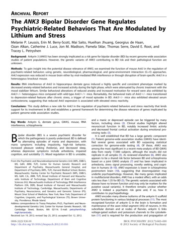
684 BIOL PSYCHIATRY 2013;73:683–690action potentials mediated by these channels (12). Ankyrin G isalso necessary for localization of inhibitory gamma-aminobutyricacid (GABA) synapses at excitatory neuron AIS (13), subventricularzone neurogenesis (14), and potentially synaptic transmission(15,16), among other functions.Increasingly, GWAS are identifying genetic loci associated withpsychiatric disorders, but determining the basis for these associations remains a formidable challenge. Although current diseaseknowledge may suggest a compelling hypothesis for the influence of a particular gene, such information is typically too sparseto support a specific mechanism. In this study, we explored anew role of ANK3 in neural circuits regulating mood using anintegrative approach encompassing genetic, neurobiological,pharmacologic, and environmental components. We used twocomplementary genetic approaches to suppress the mouse Ank3gene in brain, RNA interference targeting specific circuits andwhole brain transgenic knockout, that provide highly consistentand persuasive evidence for a novel function of Ank3 inmodulating psychiatric-related behaviors and stress reactivity.Our study provides compelling support for the human geneticdata implicating ANK3 in psychiatric disease. More broadly, thiswork establishes a general framework for validating newlyimplicated psychiatric GWAS risk genes and examining theirputative disease-relevant functions.Methods and MaterialsDetailed methods can be found in Supplement 1.AnimalsMale 8-week-old C57BL/6J mice were used for RNA interference studies. Male 8- to 11-week old Ank3 /– and Ank3 / knockout mice (12) were generated from male Ank3 /– andfemale C57BL/6J crosses.Lentiviral-Mediated RNA InterferenceTo minimize the possibility of off-target effects, two shorthairpin RNA sequences (shRNA1 or shRNA2 targeting Ank3 exons19 or 28) that suppress mRNA expression in vitro by 75% to 80%(Figure S1A in Supplement 1) were compared with a controlshRNA (shCON) that does not target any known mouse transcripts. Mouse hippocampal dentate gyrus was injected bilaterally with lentivirus expressing shRNA1, shRNA2, or shCON (n ¼10–11 mice/group).BehaviorMice were evaluated using conventional, well-validated assaysfor measuring behaviors mediated by neural circuits implicatedin psychiatric illness, as well as sensory and motor performance(17,18). These included locomotor activity in a novel open field,elevated plus maze (EPM), light-dark transition (LD), noveltysuppressed feeding (NSF), acoustic startle, prepulse inhibition,home cage activity, sucrose preference, forced swim test (FST),contextual and cued fear conditioning, and visual performance ina visible platform water maze.Drug TreatmentLithium chloride (85 mg/kg intraperitoneal; Sigma-Aldrich, St.Louis, Missouri) or vehicle (saline with .2% acetic acid) was administered once daily for 14 days before and throughout behavioraltesting 1 hour before testing.www.sobp.org/journalM.P. Leussis et al.ImmunohistochemistryCoronal sections from 4% paraformaldehyde fixed brains wereincubated with appropriate antibodies and processed usingstandard methods.Corticosterone MeasurementsPlasma corticosterone levels following acute restraint stresswere measured using an enzyme immunoassay (EIA).Statistical AnalysisAll data are presented as means and standard errors of themean (SEM). Where appropriate, one-way, two-way, and repeatedmeasures analysis of variance were used, and group differencesidentified using Fisher’s least significant difference post hoc tests.Statistical significance was accepted at the p .05 level.ResultsAnk3 RNA Interference in Dentate Gyrus Produces a HighlySpecific Phenotype Marked by Lower Anxiety-RelatedBehaviorAnk3 expression was reduced by viral-mediated RNA interference in hippocampal dentate gyrus (DG), given its role in BD,mood and stress regulation, and mood stabilizer response(19–23). Across nine behavioral assays, Ank3 suppression in theDG with either of two shRNA sequences targeting Ank3, shRNA1or shRNA2, produced a highly specific and sizable reduction inanxiety-related behavior in the EPM. Mice expressing shRNA1 orshRNA2 exhibited 60% to 75% shorter latencies (i.e., less time) toenter the EPM open arms, 50% more open arm entries, andthreefold more time in the open arms, compared with miceinjected with a control sequence, shCON (Figure S3 inSupplement 1). These changes were not due to hyperactivitybecause the number of total arm entries or rears in the EPM(Figure S3 in Supplement 1) and open field activity (Figure S4 inSupplement 1) did not differ from shCON. Ank3 knockdown micedid not differ from controls in conventional tasks assessingsensorimotor gating (prepulse inhibition), auditory and visualsensory performance (acoustic startle response and visible platform Morris water maze), associative learning (cued and contextual fear conditioning), or the FST (Figure S4 in Supplement 1).Overall, these data suggest that Ank3 suppression in DG resultsin a specific reduction in anxiety-related behavior.Following behavioral assessment and confirmation of correctvirus placement, we measured knockdown of ankyrin G expression in DG granule cells infected with shRNA lentivirus byidentifying cells expressing green fluorescent protein from thevirus. We specifically examined neuronal AIS because ankyrin Gis highly expressed and plays a critical role at this subcellularlocation. Ankyrin G AIS expression was significantly decreased by45% and 62% in shRNA1 and shRNA2 mice, respectively, relativeto shCON mice [n ¼ 5/group; F(2,8) ¼ 8.8, p .01; Figure 1].These results suggest that partial reduction of ankyrin G expression in DG granule cells is sufficient to cause marked behavioralchanges.Behavioral Changes Associated with Ank3 Knockdown AreReversed by Lithium TreatmentWe performed an independent experiment to evaluate theeffects of lithium in reversing the behavioral alterations inducedby Ank3 RNA interference. Given the consistent behavioral effectsnoted, we examined only shRNA2 because it targets more Ank3isoforms than shRNA1. Fourteen days after lentivirus injectioninto bilateral DG, mice were treated with lithium at a clinically
M.P. Leussis et al.relevant dose (85 mg/kg intraperitoneal; see Supplement 1) orwith vehicle for 14 days before and throughout behavioraltesting (n ¼ 8–12 mice/group). Lithium effects were assessedby comparing shRNA2 mice treated with lithium to shCON micetreated with vehicle that index the normal condition.Replicating the previous result, Ank3 RNA interference in DGsubstantially decreased anxiety-related behavior. Specifically,BIOL PSYCHIATRY 2013;73:683–690 685vehicle-treated shRNA2 mice exhibited 60% shorter latency and40% increased frequency to enter the EPM open arms [F(1,17) ¼5.54, p ¼ .03; F(1,17) ¼ 4.61, p ¼ .046; Figure 2A,B] comparedwith vehicle-treated shCON mice. Notably, lithium treatmentnormalized the behavior, in that lithium-treated shRNA2 micedid not significantly differ from vehicle-treated shCON mice inEPM open arm latency or entries (p .1 Figure 2A,B). Theseresults were confirmed using the LD task, where there was asignificant interaction between shRNA and lithium treatment inthe latency to leave the dark and enter the light side [F(1,33) ¼4.78, p ¼ .036; Figure 2C]. Vehicle-treated shRNA2 mice exhibited65% shorter latency to enter the light side compared withvehicle-treated shCON mice (post hoc p ¼ .02), and lithiumtreatment increased this latency such that shRNA2 mice did notdiffer from vehicle-treated shCON mice (post hoc p .1). We alsotested NSF, which assesses the latency to approach and eat foodin the center of a novel arena that mice innately avoid. There wasa significant shRNA by drug interaction [F(1,31) ¼ 5.44, p ¼ .026;Figure 2D] in the NSF approach latency. Vehicle-treated shRNA2mice exhibited 80% shorter latency to approach than vehicletreated shCON mice (post hoc p ¼ .005), whereas lithiumtreatment normalized the latency of shRNA2 mice such thatthere was no significant difference from vehicle-treated shCONmice (post hoc p ¼ .29). As in the behavioral screen describedearlier, mice expressing shRNA2 did not differ from shCON micein motor activity in the open field, EPM, or LD tasks (Figure S5 inSupplement 1). Thus, across three paradigms, Ank3 suppressionin DG was associated with substantially decreased anxiety-relatedbehaviors that were normalized by lithium treatment.Because BD patients report sleep disruptions during bothmanic and depressive episodes (24), we assessed changes inmotor activity across the light–dark cycle using an automatedhome-cage behavioral phenotyping system (25). Mice expressingshRNA2 showed no significant differences compared with shCONmice in the total time engaged in activity (walking, hanging,rearing, grooming, eating, drinking) during the dark phase [F(1,33)¼ 1.9, p .1; Figure 2F] when mice, being nocturnal, are mostactive. However, during the light phase, shRNA2 mice were 53%more active than shCON mice [F(1,33) ¼ 4.61, p ¼ .039; Figure 2E].Lithium treatment reversed the increased light phase activity ofshRNA2 mice to levels similar to vehicle-treated shCON mice (posthoc p ¼ .6; Figure 2E). These results indicate that in addition todecreased anxiety-related behaviors, Ank3 suppression in DG isassociated with elevated activity during the light phase of thelight–dark cycle, which is normalized by lithium treatment.Reduction of Ank3 Brain-Specific Isoforms Induces BehavioralChanges Consistent with Dentate Gyrus KnockdownTo gather further support and extend our behavioral findingsfrom Ank3 RNA interference mice, we performed aFigure 1. Ank3 RNA interference markedly decreases ankyrin G expression at neuronal axon initial segments (AIS). (A) In mouse brain, dentategyrus cells infected with lentivirus expressing shRNA (green, greenfluorescent protein (GFP)-positive infected cells; red, ankyrin G). Whitebox demarcates the region where ankyrin G expression was measured.(B) Representative images of neurons infected with lentiviral vectorsexpressing shCON, shRNA1, or shRNA2 targeting mouse Ank3. Ankyrin G(red) was measured at the AIS (demarcated by a white line) of neuronscontaining GFP (green) expressed from the lentiviral vector. (C) Quantification of ankyrin G expression showing decreases of 45% and 62% byshRNA1 and shRNA2, respectively, compared with shCON. Mean SEM;n ¼ 5 mice/group, 6 AIS/mouse; *p .05. GCL, granule cell layer; H, hilus;ML, molecular layer; shCON, control shRNA; shRNA, short hairpin RNA.www.sobp.org/journal
686 BIOL PSYCHIATRY 2013;73:683–690Figure 2. Ank3 RNA interference in dentate gyrus (DG) is associated withlower anxiety-related behavior and increased light-phase activity that isnormalized by lithium treatment. Vehicle-treated mice expressing shRNA2against Ank3 in the DG, when compared with shCON mice, exhibit (A)shorter latency to enter the open arms and (B) greater number of openarm entries in the elevated plus maze, (C) shorter latency to enter thelight–dark transition light side, (D) shorter latency to approach food in thenovelty-suppressed feeding task, and (E) increased activity during thelight phase but (F) no change in activity during the dark phase. (A–E)Lithium treatment (85 mg/kg intraperitoneal, 14 days) normalizes thebehavioral alterations of shRNA2 mice to similar levels as vehicle-treatedshCON mice. (F) Lithium has no impact on dark-phase activity in eithershCON or shRNA2 mice. Mean SEM; n ¼ 8–12 mice/group; *p .05,**p .01. shCON, control shRNA; shRNA, short hairpin RNA.complementary study using a knockout mouse in which the Ank3exon 1b locus is disrupted (12). This disruption results in theexclusive loss of Ank3 transcript variants containing exon 1b thatare specifically expressed in brain but has no effect on transcriptscontaining alternative leading exons, which shRNA1 and shRNA2target in addition to exon 1b transcripts. We confirmed in C57BL/6J mice that transcripts containing exon 1b are expressed infrontocortical and limbic regions relevant to mood-relatedbehaviors (Figure 3). Exon 1b expression was highest in cerebellum, as previously reported (12), and was also substantial in DGand all other regions examined (hippocampus, amygdala, orbitofrontal cortex, prefrontal cortex, and striatum). Exon 1b transcripts were more highly expressed in all brain regions comparedto transcripts containing an alternative leading exon 1e(Figure 3), which is found in brain and other tissues (12). Theseexpression data suggest that the brain-specific exon 1b isoformsfunction in many brain regions, including several implicated inmood regulation.Because Ank3–/– mutant mice exhibit early-onset ataxia (12)that interferes with general locomotion, only Ank3 /– andwww.sobp.org/journalM.P. Leussis et al.Ank3 / mice were examined (n ¼ 7–18/group). Comparedwith wild-type Ank3 / mice, heterozygous Ank3 /– mice arereported to have reduced forebrain and cerebellar expression ofthe 270- and 480-kD ankyrin G isoforms that localize to the AIS(12,26). We verified that Ank3 /– mice have a significant 39%reduction in ankyrin G expression at neuronal AIS in the DGgranule cell layer compared with Ank3 / littermates [t(6 df) 4.6, p .01; Figure 4A], similar to the RNA interference mice(Figure 1). Furthermore, ankyrin G expression at the AIS of corticalneurons was decreased by 41% in Ank3 /– mice compared withAnk3 / mice [t(8 df) ¼ 6.2, p .001; Figure 4B]. Together withthe published data, these results suggest that Ank3 /– mice witha single functional copy of brain-specific isoforms have reducedankyrin G expression at neuronal AIS throughout the brain.We found that male Ank3 /– mice exhibited behavioral alterations that were highly consistent with the decreased anxiety-relatedphenotype of RNA interference mice (Table S3 in Supplement 1) andalso displayed other relevant behaviors. Compared to Ank3 / littermates, Ank3 /– mice exhibited substantially shorter latencies toenter the EPM open arms (57% reduced, post hoc p .05, Figure 5A)and LD light side (59% reduced, post hoc p ¼ .07, Figure 5B), and toapproach food in the NSF task (83% reduced, post hoc p .05,Figure 5C). To consider other attributes relevant to mood, wemeasured preference to drink a 1% sucrose solution over water.Ank3 /– mice exhibited 53% greater sucrose preference comparedto Ank3 / littermates (post hoc p .05, Figure 5D), suggestingheightened motivation to obtain a reward. As with Ank3 RNAinterference mice, Ank3 /– mice were normal in all other behavioraltests including open field activity, acoustic startle, prepulse inhibition,contextual and cued fear conditioning, and motor activity in the EPMand LD tasks (Figure 5E; Tables S1 and S3 in Supplement 1). Thehighly consistent behavioral changes in Ank3 /– mice furtherdemonstrate a previously unknown function of Ank3 brain-specificisoforms in regulating psychiatric-related behaviors.Chronic Stress Induces a Shift to Depressive-like Features inAnk31/2 MiceBecause stress is a major risk factor for BD episode recurrence (2)and the hypothalamic-pituitary-adrenal (HPA) axis is implicated inBD (27), we examined the effect of chronic stress on Ank3 /– mice.Figure 3. Ank3 exon 1b transcripts are expressed in multiple brainregions. Normalized expression of Ank3 transcripts containing the brainspecific exon 1b or widely expressed exon 1e across brain regions, asmeasured by Nanostring gene expression analysis. For each brain region,mRNA from eight C57BL/6J mice was pooled before measurement. Exon1b transcripts are more highly expressed than exon 1e transcripts acrossall brain regions examined. DG, dentate gyrus; dHipp, dorsal hippocampus; mPFC, medial prefrontal cortex; OFC, orbitofrontal cortex; vHipp,ventral hippocampus.
M.P. Leussis et al.BIOL PSYCHIATRY 2013;73:683–690 687Figure 4. Ank3 /– mice have substantially reducedankyrin G expression at the axon initial segments (AIS)of dentate gyrus and cortical neurons. Compared withAnk3 / littermates, heterozygous Ank3 /– mice exhibit an approximately 40% reduction in ankyrin G at theAIS of neurons in the (A) dentate gyrus and (B) cortex,as measured by immunohistochemistry. Mean SEM;n ¼ 5–7 mice/group, 6–7 AIS/mouse; **p .01.Compared with Ank3 / littermates, Ank3 /– mice singly housedfor 6 weeks did not exhibit increased motivation for reward ordecreased anxiety-related behaviors (Figure 5 isolated condition),as found under standard group-housed conditions describedabove (Figure 5, group condition). Specifically, isolated Ank3 /–mice did not display significantly shorter latencies to enter theEPM open arms or LD light side or to approach food in the NSFtask when compared with either group-housed or isolatedAnk3 / mice (all post hoc p values .09, Figure 5A–C). Therewas also a change in motivation (Figure 5D): isolated Ank3 /–mice did not display increased sucrose preference compared withisolated Ank3 / mice (p .05), as was observed under grouphoused conditions, and had substantially lower sucrose preference, relative to group-housed Ank3 /– mice (post hoc p .05).Chronic isolation also resulted in 50% greater FST immobility inAnk3 /– mice versus Ank3 / mice (p .05, Figure 5E),whereas group-housed Ank3 /– and Ank3 / mice did notdiffer (p .05, Figure 5E). The latter results are suggestive of adepression-related phenotype in Ank3 /– mice exposed tostress, because the increase in FST immobility is opposite tothe reduced immobility induced by antidepressant treatment(28). Thus, chronic stress induced a transition in Ank3 /– micefrom decreased anxiety-related behaviors and increased motivation to depression-related features, highlighting anenvironmental component in mood regulation. Interestingly,the isolation stress had no effect on Ank3 / mice (all pvalues .09; Figure 5, group vs. isolation housed), suggestinga putative gene-environment (G E) interaction in which diminished Ank3 is associated with elevated stress sensitivity.Evidence for Altered Stress Hormone Reactivity in Ank3 /–MiceTo investigate the mechanism through which chronic stressmodifies the phenotype of Ank3 /– mice, we measured plasmalevels of corticosterone, the predominant stress hormone inrodents, and weight of the adrenal gland that produces corticosterone, as a measure of chronic stress load (29). Under grouphousing, the ratio of adrenal weight to body weight ratio did notdiffer between Ank3 / and Ank3 /– mice, nor did isolationhave any effect on the adrenal:body weight ratio of wild-typeAnk3 / mice (all post hoc p values .1; Figure 6A). In contrast,isolated Ank3 /– mice exhibited a 27% increase in adrenal:bodyweight ratio compared to group-housed Ank3 /– mice (p .01;Figure 6A), suggesting that Ank3 /– mice are more reactiveto chronic stress than wild-type mice. Ank3 /– mice exhibitedhigher basal corticosterone levels than Ank3 / littermates,regardless of group or isolation housing [F(1,10) ¼ 9.03 andF(1,11) ¼ 9.04, both p values .05; Figure 6C,D; Table S2 inFigure 5. Ank3 /– mice exhibit distinctive behavioral alterations that are modified by chronic stress. Under standard group-housed conditionscompared with Ank3 / littermates, Ank3 /– mice display shorter latencies (A) to enter the elevated plus maze (EPM) open arms, (B) to enter the light–dark (LD) transition light side, and (C) to approach food in the novelty-suppressed feeding (NSF) task; (D) they also exhibit greater preference for sucroseover water. In contrast, after prolonged isolation housing, Ank3 /– mice no longer exhibit shorter latencies to enter (A) the EPM open arms or (B) the LDlight side, (C) or to approach food in the NSF task, (D) nor do they display greater sucrose preference, compared with isolation-housed Ank3 / mice.(E) Group-housed Ank3 /– mice do not differ from Ank3 / mice in FST immobility time, whereas isolation housing significantly increased Ank3 /–immobility time. Mean SEM; n ¼ 7–18 mice/genotype; **p .01, *p .05, #p .07, unpaired t test.www.sobp.org/journal
688 BIOL PSYCHIATRY 2013;73:683–690Figure 6. Ank3 /– mice exhibit heightened physiologic stress reactivity.Following chronic social isolation, (A) adrenal gland weight (milligrams)relative to body weight (grams) is higher in Ank3 /– mice compared withisolation-housed Ank3 / mice or to group-housed mice, suggesting ahigher sustained response to chronic stress in Ank3 /– mice. (B)Isolation-housed Ank3 / and Ank3 /– mice exhibit a smaller increasein body weight than group-housed mice. Corticosterone levels (ng/mL)are elevated in Ank3 /– mice compared to Ank3 / mice whether (C)group-housed or (D) isolated for 6 weeks. In particular, Ank3 /– miceexhibit a higher corticosterone peak 30 min after acute restraint stress aswell as a slower return to baseline after 180 min. Mean SEM; n ¼ 6–8mice/genotype; **p .01, *p .05.Supplement 1). Following acute stress (20-min restraint), corticosterone levels were more elevated and returned to baselinemore slowly in Ank3 /– mice compared with Ank3 / mice(Figure 6C,D). These results demonstrate a persistent elevation inbasal corticosterone levels in Ank3 /– mice that is furtherexaggerated by acute stress, suggesting impaired HPA axisregulation.DiscussionThis study uncovered a novel function of Ank3 in theregulation of psychiatric-related traits that implicates a centralrole in neural circuits underlying psychopathology. Ank3 suppression in mouse brain, either by viral delivery of shRNAs to the DGor disruption of brain-specific Ank3 in a heterozygous knockoutmouse, resulted in lower anxiety-related behavior denoted byrapid entry into aversive novel environments in three tasks.Notably, these changes were reversed by chronic lithium, a firstline treatment for BD mania. Decreased anxiety-like behaviors inthe absence of anxiolytic drugs can be interpreted as increasedrisk taking (30,31), a classic feature of BD mania (1). Risk takingcan be considered on the same spectrum but at the opposite endfrom anxiety, thus the rapid entry into aversive environments ofmice with diminished Ank3 could reflect increased risk taking. Wealso detected increased activity during the light phase in theAnk3 RNA interference mice, and characterization of Ank3 /–mice identified heightened motivation to obtain a reward, whichare both features of BD mania (1). Thus, converging evidencefrom two genetic models and several behavioral paradigmswww.sobp.org/journalM.P. Leussis et al.(Table S3 in Supplement 1) suggests that ankyrin G functions inneural circuits relevant to BD, given that the behavioral changeswere reversed by lithium, a clinically effective prophylactic formanic episodes.Another striking finding of this study was the observation thatchronic isolation stress reversed the lower anxiety-related behavior and elevated motivation of Ank3 /– mice and induceddepression-related behavior (increased FST immobility). Thestress-induced transition is particularly interesting because stresspredicts relapse of both depressive and manic episodes in BD (2).The elevated stress reactivity of Ank3 /- mice compared toAnk3 / mice suggests a G E interaction in which the level ofAnk3 expression modulates the impact of stress on brainfunction. One putative mechanism through which Ank3 maymediate stress reactivity is through dysregulated HPA axisactivity, as suggested by elevated corticosterone levels andadrenal gland weight following chronic isolation stress inAnk3 /– mice compared with Ank3 / mice. Notably, micewith reduced neural expression of the glucocorticoid receptorexhibit a phenotype remarkably similar to Ank3 /– mice, including decreased anxiety-related behavior, impaired behavioralresponse to stress, and altered HPA axis regulation (32), supporting a link between HPA axis function and the behavioral changesobserved in Ank3 /– mice. Future experiments to examine HPAaxis integrity in Ank3 /– mice, as well as ankyrin G functionfollowing chronic stress, will be important to elucidate the roleof ankyrin G in stress reactivity.Our data suggest that Ank3 may influence a broader repertoire of emotional behaviors beyond those associated mainlywith BD. Further insight into its nuanced role could be obtainedthrough human genetic association studies of other psych
support for its involvement in BD and establishes a general framework for determining the disease relevance of genes implicated by patient genome-wide association studies. Key Words: Ankyrin G, dentate gyrus, GWAS, mouse, RNA . Department of Psychiatry, Massachusetts General Hospital, and Department of Psychiatry (MPL, TLP), Harvard Medical .