Transcription
Bauer et al. BMC Medical Education(2021) CHNICAL ADVANCEOpen AccessDevelopment, production and evaluation of2-dimensional transfer tattoos to simulateskin conditions in health professionseducationDaniel Bauer1*† , Andrea C. Lörwald1†, Sandra Wüst1, Helmut Beltraminelli2, Miria Germano1, Adrian Michel1 andKai P. Schnabel1AbstractBackground: Moulages can greatly extend the possibilities of simulation in teaching and assessment. Sincemoulages that fit an educator’s exact needs are often unavailable commercially, this paper explains how 2dimensional transfer tattoos can be independently developed, produced, and evaluated.Methods: From representative photographs of the specific skin condition an analogue copy of the pathologicalfinding is drawn. Once validated by the medical expert, it can be digitized by scanning and processed using graphicssoftware. The final digital image file is printed onto transfer paper. Once applied and fixed onto the intended wearer,usually a simulated patient, its authenticity can be confirmed, and further transfer tattoos can be produced.Results: Using this moulage technique we produced 10 different 2-dimensional transfer tattoos to date, includinghematoma, Janeway lesions and splinter nails. These moulages are used in clinical skills training, formative and highstakes summative assessment in undergraduate medical and nursing programs.Conclusions: By sharing our development process for 2-dimensional transfer tattoos, health profession educators canproduce their own that best fit their local educational needs. Due to their high authenticity and standardization, 2dimensional transfer tattoos are ideal for use in high-stakes assessment.Keywords: Medical moulage, Simulation engagement, Clinical skills, OSCE, Standardized patient* Correspondence: daniel.bauer@iml.unibe.ch†Daniel Bauer and Andrea C. Lörwald contributed equally to this work.1Institute for Medical Education; Faculty of Medicine, University of Bern,Mittelstrasse 43, 3012 Bern, SwitzerlandFull list of author information is available at the end of the article The Author(s). 2021 Open Access This article is licensed under a Creative Commons Attribution 4.0 International License,which permits use, sharing, adaptation, distribution and reproduction in any medium or format, as long as you giveappropriate credit to the original author(s) and the source, provide a link to the Creative Commons licence, and indicate ifchanges were made. The images or other third party material in this article are included in the article's Creative Commonslicence, unless indicated otherwise in a credit line to the material. If material is not included in the article's Creative Commonslicence and your intended use is not permitted by statutory regulation or exceeds the permitted use, you will need to obtainpermission directly from the copyright holder. To view a copy of this licence, visit http://creativecommons.org/licenses/by/4.0/.The Creative Commons Public Domain Dedication waiver ) applies to thedata made available in this article, unless otherwise stated in a credit line to the data.
Bauer et al. BMC Medical Education(2021) 21:350BackgroundSince its inception in the 1960s, simulated patient (SP)methodology has spread around the world and is nowwidely accepted in simulation-based teaching and assessment in health professions education. SPs can portraymany pathological conditions [1]. For those nonportrayable, workarounds include the use of simulators(e.g., delivery), part-task trainers (e.g., rectal exam), ormaterial presented as video (e.g., seizures), audio (e.g.,heart murmurs) or photographs (e.g., rashes). However,these all constitute media breaks which can negativelyinfluence the learners’ or examinees’ simulation engagement [2] and threaten the simulation’s validity argument.One method to remedy this is the use of modern medical moulage. Moulages allow the depiction of visualfindings directly on the SP, rendering the delivery ofphotographs by the simulation facilitator or clinicalexaminer redundant. The range of findings that can bedisplayed with moulages encompasses traumatological[3–6], infectious diseases and medical conditions [7–10],and dermatological findings [11–16]. While there are examples where moulages were mounted onto a mannequin [17–19], they are most commonly applied directlyonto SPs [20–22]. Educational scenarios in which moulage are used are diverse. Garg et al. describe the use ofmoulage in a dermatology course and report a positiveeffect on long-term learning outcomes [12]. Other usesinclude the use of moulage in the assessment of clinicalskills, e.g., detecting [21] and managing skin lesions [23].Beyond examination skills, moulage have also beenemployed to stimulate empathy in learners [16].While modern medical moulage employs technologiesused in the movie industry, its use in health professionseducation demands an additional set of criteria. Whilemost moviegoers will not be put off by minor medicalinconsistencies, the intended users of moulage in medical simulation usually have at least some expertise, alsoconsidering inconspicuous details during the simulation,while building their diagnostic and therapeutic plan.Correct tactile information adds another criterionrelevant in simulations but not movies. Beyond theseauthenticity issues, the use of moulages in medical assessment requires the best possible produce quality andhigh levels of standardization ensuring a level playingfield for examinees and as not to compromise the justiciability of the exam. In the following we provide a shortoverview of different approaches to medical moulages insimulation.2-dimensional moulage techniques Special effects makeup, based on grease, water oralcohol soluble colours, allows for relatively quickand cheap application of 2-dimensional findingsPage 2 of 8onto an SP. They can be used to cover large areas ofthe SP’s body including hairy areas and near the eyes(water and grease-based colours) and can be editedeasily if necessary. Their degree of standardizationdepends primarily on the makeup artist’s expertise,the photo template used and desired granularity ofdetail. The indication for special effects makeupincludes scenarios where the findings depicted servefor quick diagnoses and decisions, e.g. in masscasualty triage, or as visual cues. They can usuallylast for at least a day but do not convey spatial ortactile information. 2-dimensional transfer tattoos are printed on decalpaper and have a higher potential forstandardization. Development requires a goodphotographic template and some artistic and digitalediting skills. Production and application are quickand easy [13]. They can be used to cover large,preferably flat areas of the SP’s body includingfinger- and toenails, but usually excluding overlyhairy areas. The quality depends on the base imageand printing process. Once mounted, additionalediting is limited. Indications include medicalconditions of a 2-dimensional nature (e.g., somerashes). Can last for several days but convey neitherspatial nor tactile information.3-dimensional moulage techniques3-dimensional moulages open a new range of findingsthat can be simulated, as they include spatial and sometimes correct tactile information. 3-dimensional moulage can be hand-modelleddirectly onto the SP or simulator, often using wax,silicone, or gelatine materials (gelatine-basedmoulage can be modelled into hairy areas includingthe scalp). The development phase includes obtaininggood photographs as templates. The application takestime and is highly dependent on the makeup artist’sskill and experience. Once applied, these moulagescan be further edited as required. Standardizationdepends heavily on the desired granularity of detailsand again on the makeup artist. Hand-modelledmoulages are a relevant choice for scenarios withlower requirements for standardization and detail,e.g., triage exercises, for budget-constrained settingsand for moulage requested at short notice. Thesemoulages can hold for several hours, limited mostlyby their adherence to the SP. 3-dimensional silicone appliances are quite sturdyand allow reutilization. Development and productionare quite elaborate, but application is quick and easy.Once mounted, the degree of further editing islimited. Their sturdiness often comes at the price of
Bauer et al. BMC Medical Education(2021) 21:350increased thickness, so the transition into the SP’sown skin often remains clearly visible [24]. Theirsize can be considerable but tend to stick out asartificial with increasing size. This is of lessimportance in scenarios where the edges of themoulage would be covered anyway (e.g., in somewound care tasks) or when the moulage is notcrucial for reaching a diagnosis but serves todemonstrate a clinical procedure. Within theselimitations, their high standardization andcommercial availability makes them suitable forcertain assessment settings. Tactile authenticity isseldomly achieved. A variation of 3-dimensional moulages, prepared inadvance, makes use of movie makeup artistry toproduce thin-edged Probondo transfer tattoos, at thecost of being single-use. They have the sameadvantages as the thicker versions described. Likeprinted tattoos, they offer high levels ofstandardization, however at the cost of an elaboratedevelopment and production process. Theirauthenticity depends largely on the makeup artist’sexpertise. A specialist’s suggestions are crucialregarding peculiar morphological parameters to finetune colour nuances, distribution, size, and shape ofthe lesion(s). Since these moulages blend well intothe SP’s own skin, they can be used in scenarioswhere they should not be too obvious to spot andrequire learners or examinees to uncover andinterpret them, before building their diagnostic andtherapeutic plan. Can be applied quickly and easilyand last for a whole day, especially if attached withsealer. Once mounted, the degree of further editingis limited. Can convey tactile information.Page 3 of 8MethodsPreparing an analogue templateThe process begins with the provision of representative,high-resolution photographs of the specific skin condition by a medical expert. From this, the artist draws ananalogue copy of the pathological finding, i.e., everythingbut the healthy skin. This is done with alcohol or watersoluble colours on a semi-transparent parchment-likepaper. A normalized colour fan acts as inexpensive andhandy common standard between artist and medical expert when discussing colour nuances. Using a commonindustrial standard allows for reliable identification ofcolour values, conversion into other colour systems, anddirect comparison with the physical product. Minorretouching and corrections can usually be realized digitally later.Digitizing the dataOnce the medical expert confirms the correctness of thedrawing, it is fixed onto a blank sheet of regular whiteoffice paper using adhesive tape for scanning. The scanengine of a regular office printer delivers useable results(full colour text/photo scan, 600 dpi, pdf file format).Once digitized, the image is processed using AdobePhotoshop. First, the yellowish tint of the parchmentlike paper is colour-corrected using the graduation curveadjustment layer. Colours are enhanced using the vibrance adjustment layer. Regular proofs are printedwhile keeping print settings unchanged to show if further colour adjustment is necessary. The original photographs and the expert-validated drawing act as opticalreferences.1Manipulating the templateMoulages offer the possibility to integrate a myriad ofpathologies into simulation visually. However, the rangeof commercially available moulages is narrow and theirauthenticity not necessarily up to every educator’s individual needs. In order to independently prepare 2-dimensionaltransfer tattoos for their own simulation needs, educatorshave to overcome some obstacles. These include achievinga medically correct visualisation of the pathology and thenaffixing that visualisation onto the wearer (SP or mannequin), guaranteeing a smooth visual transition into the SP’sown skin. For this, relevant competencies beyond medicalexpertise are needed and include painting skills, digitalimage editing, and knowledge of transfer materials andtechniques. Since these are skills not all medical educatorshave, our aim is show how to approach the preparation ofmedical transfer tattoos with little prior knowledge andhow to how to integrate these skills. By doing so we willenable people with little experience to prepare their own 2dimensional moulages using easily available materials.In some cases (e.g., when designing a rash) one mightdecide to prepare just a section of the rash and digitallyincrease its size. To achieve this, the digital templateproduced to begin with can be flipped, inverted, andotherwise transformed multiple times, with the partsthen spliced together to increase the size of the findingwhich will be printed later, avoiding a tiled appearance.1Colour fidelity is influenced by several factors. Full colourmanagement can prove quite resource-consuming: Each parameterwould need its own ICC profile, which can only be achieved by graphics professionals carrying out the calibration. Further, printing/scanning equipment at a given institution cannot be changed easily. Lastbut not least, the final output medium, i.e. the SP’s skin, cannot becovered technically (visible edges), or standardized at that, why fullcolour management remains unfeasibly after all. Considering this, wesuggest for an iterative opportune colour management approach withpartially calibrated devices, visual image editing using standard profiles,and multiple proofs to ensure flexibility with limited resources. In thecourse of an opportune colour management, we have also renouncedthe ICC profiling of the printer. Instead, the printer should be locatedin close proximity to image processing.
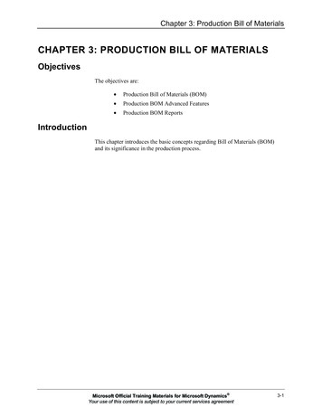
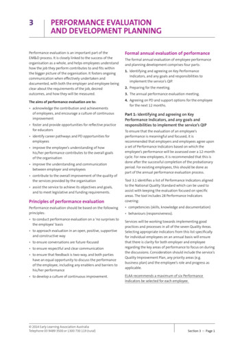
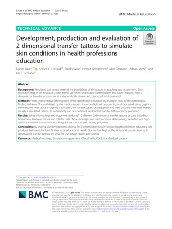
Bauer et al. BMC Medical Education(2021) 21:350Prominent parts of the condition that could give awaythis manipulation can be excluded from this process andlater re-set into the picture once its desired size isachieved.With skin conditions where the orientation of thefinding is of importance, the print needs to be mirrorinverted, as the transfer paper is flipped over when applying the tattoo to the SP’s skin.If applied to the SP’s skin, every colour point printedon it will, as a rule, subtly darken the underlying skin.Only yellow colour tones can have a brightening effect.The SP’s skin colour thus defines the brightest possiblehue of the presented skin condition (subtractive colourmixing), therefore depicting, e.g., dried pus or dandruffis unfeasible.Printing and finishing the transfer tattooPage 4 of 8Fig. 1 A 2D tattoo prototype of splinter nails on a healthy 40-yearold SP’s middle finger, intended for a case of bacterial endocarditis.The haemorrhages should be less sequential but continuous, parallelto each other as well as the nail’s growth lines. The colouringsuggests an old subungual haemorrhageThe final digital image file is then printed on transferpaper, using the previous print settings. The prints canbe stored in this form until used.Removing the moulageApplication and EvaluationAfter use, soak cotton pads with makeup remover andgently rub the moulage starting at the edges. Use additional rehydrating skin care as warranted.To determine if the 2D transfer tattoo produced in thismanner is authentic, it needs to be fixed onto theintended wearer. For this, the front of the print issprayed with adhesive spray and left until completelydry. Scissors are used to cut off superfluous sections. Itis then placed onto the intended place on the wearer’sbody, the paper facing upwards, away from the body(i.e., the print facing the SP’s skin). The paper is thensoaked with water until it can be moved sideways, carefully, and easily. Once any remaining water has evaporated, 96% isopropyl-soaked cotton swabs can be used toremove any unwanted residual paste on the edges. Thensealer2 is applied gently with a fresh cotton swab, airbrush, or sponge, covering the whole moulage and immediately adjacent areas and left until completely dry.3The sealer fixes the moulage, so it does not stick, collectlint or tear when manipulated.4 The applied transfer tattoo can now be judged against prior established criteriaor other tools [25]. If unsatisfactory, earlier steps mustbe revisited. As soon as the product is satisfactory, production can start. Figure 1 depicts an example of a printin need of further editing.2When using sealer that has to be mixed beforehand, prepare in timeand according to manufacturer’s specifications. We recommendBluebird FX sealer (matt or gloss), mixed with 96% isopropyl at a 1:1ratio.3In cases where some strain on the tattoo is foreseeable, apply thetattoo, let it dry, apply first layer of sealer, let it dry, then apply secondlayer of sealer and again leave to dry.4Although the application can be done by lay people, we stillrecommend training in the application and post-production process.MaterialsTable 1 Additional material: paintbrushes, 96% isopropyl, colour fan deck, office quality white paper, officequality adhesive tape, office scissors, cotton swabs, cotton pads. Throughout the production process, maintainsafety standards as per manufacturer’s specifications andinstitutional and national guidelines.ResultsUsing this technique, we were able to produce 10 different 2-dimensional (e.g., hematoma, Janeway lesions,splinter nails) moulages. They are currently used in formative and high-stakes summative assessment (includingthe Swiss Federal Licensing Examination [26]) as well asclinical skills training in undergraduate medical andnursing programs. Figure 2 depicts an example of a printused in an exam as print template, Fig. 3 shows the sametattoo affixed onto an SP.To ensure highest quality, we integrated a sequence ofcontrol steps. When planning an exam, a meeting withexam stakeholders and the makeup artist is scheduled todetermine which cases should and can be realized withmoulages, considering the overall exam and available resources and, if possible, showcasing prototypes. Afterthis meeting development and production take place asdescribed. On the morning of the exam day, examinerscovering any moulages case are invited to assess themoulages affixed to the SP. Based on the examiners’feedback, moulages are either approved, or can still get
Bauer et al. BMC Medical Education(2021) 21:350Page 5 of 8Table 1 Components used in the development and production of 2D Transfer TattoosComponentProduct nameProducerTracing paperHigh-quality tracing paper 80 g/m2, smooth front andmatt back surfaceAMI Art Material International Warenhandelsgesellschaft mbH,Kaltenkirchen, GermanyAlcohol solublecoloursPPI Skin Illustrator, e.g., FX PalettePremiere Products Inc., Pacoima, CA, USAPrinter/ScannerAficio Infotec MP C2800Ricoh Company Ltd., Tokio, JapanColour CalibrationSpyder 4 EliteDatacolor AG, Lucerne, SwitzerlandImage processinghardwareiMac (27-in., Late 2013 configuration)Apple Inc., Cupertino, CA, USAImage processingsoftwarePhotoshop CS5.5Adobe Systems, San Jose, CA, USATransfer paperTattoo 2.1 Transfer PaperTheMagicTouch GmbH, Dieburg, GermanyAdhesive SprayGolden Phoenix Super Spray AdhesiveBaoding OUPU Electronic Science and Technology Co. Ltd, Baoding,China gpbodyart.comMakeup removerBEN NYE Bond Off!!Ben Nye Company, Inc., Los Angeles, CA, USAThe process of developing and producing 2D transfer tattoo moulages described above is generic and should work with materials of different producers. Forreproducibility reasons, this table declares the precise materials used by the authorstouched up, exchanged with a backup moulage or in extreme cases taken off, reverting to backup photographs.SPs are instructed to reach out to examination supportstaff should they notice any problem with their moulage(well-prepared moulage usually hold for at least half aday, often longer). In this case, a (moulaged) backup SPtakes their place while makeup staff tend to the moulage.SPs are also directed to report to makeup staff duringbreaks for check-up. Photographic documentation of themoulages further supplements quality control.Once the exam is over, we collect systematic and casual feedback from the examiners, examinees, and observations from makeup staff regarding the moulages, e.g.,durability. SP feedback regarding moulages is collectedduring the debriefing session. Relevant information regarding the moulages is discussed in a post-examdebriefing meeting and should the need arise, actedupon accordingly.DiscussionMoulages can greatly extend the possibilities of simulation but since their dissemination is low and since moulages that fit an educator’s exact needs are often unlikelyto be available commercially. This paper describes howwe developed, produced, and evaluated 2-dimensionalmoulages ourselves.We used our moulages mainly for high-stakes assessment in which authenticity and standardization weredeemed of importance. During assessment, examiners,SPs, and makeup staff were key for quality control. Afterthe exams, we obtained additional feedback from examinees and more elaborate feedback from examiners andSPs as basis for future development.Moulages can and should be evaluated in a more systematic manner, e.g., using the MARS instrument [25].This tool takes into account not only the anatomical orvisual correctness of a moulage (e.g., regarding colour,size, position, shape, likeness to real world) but alsowhether it fits the scenario (e.g., logical fit) and its effecton the participant (does not distract/confuse theparticipant).Development and production of a moulage as described requires some resources (time, materials, amakeup artist, and access to clinical experts). Beforeeven embarking the development of a moulage, the effort and assumed benefit of using moulages should bedetermined. This requires some knowledge of the educational scenario and the specific case, both in teachingscenarios and assessment. Is the affliction that is to berealized central to the case, relevant for the learner orexaminee to correctly reach the diagnosis and build theirmanagement plan; would the affliction be missed if itwere not visible on the SP; is the affliction scripted inthe case as near the eyes, on mucous membranes, onjoints that get flexed a lot or hairy parts of the body,which count as relative contraindications for moulages,or could the case be adapted to better fit the simulationwith a moulage; will the resources be available on theday required to competently apply the moulages; oncedeveloped, will there be chances to re-use the moulagetemplates with other learners? Could cooperation withother health professions institutions reduce the cost foran individual site?The relatively high (initial) effort required in the development of moulages calls for cost-benefit analysis [27].The evidence available so far seems limited. In the wakeof a systematic review investigating the impact of moulages on learner engagement, Stokes-Parish and colleagues [28] found only a small body of literature onmoulages in simulation, with most only investigating the
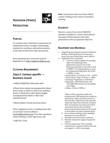
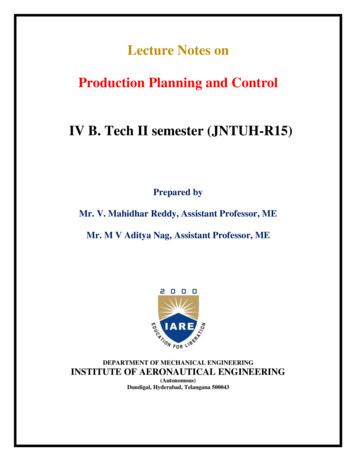
Bauer et al. BMC Medical Education(2021) 21:350Page 6 of 8Fig. 2 The print template to a case of petechial bleeding in a patient with thrombocytopenia. The image is shrunk in size from 21 by 29.7 cm inthe originalfield of dermatology. Several studies were poorly reported and yielded only low-level evidence, so that thesum of generalizable findings on moulages in simulationis still undersized.Two studies made use of moulages to have studentsexperience the impact that dermatological diagnoseshave on patients (psoriasis [16] and melanoma [29]).While the effects on students were remarkable, this alsoraises the question if SPs as wearers of such moulagesneed special debriefing and support.Strengths and limitationsIn our article, we describe methods for developing andproducing 2-dimensional moulages. This descriptionshould be accurate enough for others to copy theprocess and adapt it to their needs locally. We believethat it is important to share this knowledge in stark contrast, for example, to the historical moulage artists, whousually kept their recipes secret throughout history [30].For development, we worked very closely with clinicalexperts. This not only benefits moulage authenticity but
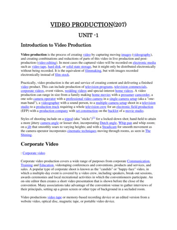
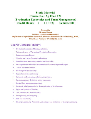
Bauer et al. BMC Medical Education(2021) 21:350Page 7 of 8the top colour layer that depicts the pathology, but bothapproaches warrant further experiments. From an integrative standpoint on the other side, this calls for acloser look how the preparation and use of moulagemight differ for SP with different skins tones. The factthat moulage have been successfully used e.g. in Brazil,South Africa, Pakistan, or Spain, where darker skin tonesare more common [11, 33–35], as well as settings withmore diverse SP [14] shows that the concept of moulagein education is as such not restricted by skin tones.also increases their acceptance, since the experts wereinvolved right from the start. To date our team developed and set up a manufacturing process. In the future,more exchange and cooperation with other simulationeducators would be desirable. Our approach to evaluation of the moulages has so far been predominantlyqualitative. In the future, this could also be done in aquantitative manner, e.g. using the MARS instrument[25] and include their effect in simulations [2].ConclusionsThe use of modern medical moulage can enhancesimulation-based education and assessment. Moulagesoffer the possibility to advance simulation of a widerange of pathologies and conditions. Due to their highauthenticity and standardization, 2-dimensional transfertattoos are ideal for use in high-stakes assessment. Sincethe range of commercially available moulages is narrowand their authenticity not always guaranteed we decidedto share our experiences in this report. By doing so, wehope health profession educators can overcome theobstacles in moulage preparation and successfully andindependently produce their own that best fit their localeducational needs.Using 2D transfer tattoos on different skin tonesSupplementary InformationThere seems to be a bias in dermatology education (but notrestricted to that discipline) that is attracting increasing attention. It has been shown that educational materialsunderrepresent patients with darker skin tones [31] e.g., indermatology textbooks [32]. Along this line, it appears thatmost academic work on modern medical moulage alsostems from North America and Europe. This might well beanother aspect of that bias that needs to be addressed.The authors’ experiences stem from a Central Europeansimulation and SP program that, while actively workingon its diversity, so far has most experience with moulageon light-skinned SP.The production of printed transfer tattoos as describedin this report utilizes a colour pigment on a quasitransparent film that is transferred to the SP’s own skinsurface. From a technological standpoint, the challengelies in a basic conception of inkjet printers: commonprinters mix five colours to achieve the desired print,CMYK and white, with the latter not added from a cartridge but contributed by the white printer paper. However, when printing moulage, the fifth colour in the mixis the SP’s own skin colour, which now limits the rangeof printable findings. This effect is less prominent inlighter skin tones that have a higher white fraction thanin darker skin tones. One approach to counter this limitation would be the use either of white printer toner, or,adding a second, partially opaque white base film belowThe online version contains supplementary material available at https://doi.org/10.1186/s12909-021-02763-z.Fig. 3 The 2D Tattoo affixed onto an SP. The knuckles and back of thehand were left out, as regular flexing would have broken the printAdditional file 1. “MakeUp DB hohe.Quali.mp4”. The video file showsthe transfer of a 2-dimensional print onto an SP.AcknowledgmentsThe authors would like to extend their gratitude to all SP, SP trainers, andclinician experts who contributed to the development of our moulages.Authors’ contributionsAll authors collectively conceived the technical innovation described. DB,ACL, and SW wrote the initial draft of the manuscript. Further expertisecontributed includes the medical and dermatological domains (HB), make-upartistry (MG), digital image editing (AM), and simulations in healthcare (KPS).All authors read and approved the final manuscript.FundingThe work described took place as part of the regular curriculum andreceived no external funding.Availability of data and materialsNot applicable.DeclarationsEthics approval and consent to participateNot applicable.Consent for publicationNot applicable.Competing interestsThe authors declare that they have no competing interests.
Bauer et al. BMC Medical Education(2021) 21:350Author details1Institute for Medical Education; Faculty of Medicine, University of Bern,Mittelstrasse 43, 3012 Bern, Switzerland. 2Department of Dermatology,Inselspital, Bern, Switzerland.Received: 6 August 2020 Accepted: 26 May 2021References1. Barrows HS. An overview of the uses of standardized patients for teachingand evaluating clinical skills. Acad Med. 1993;68:443.2. Hagiwara MA, Backlund P, Söderholm HM, Lundberg L, Lebram M,Engström H. Measuring participants’ immersion in healthcare simulation: thedevelopment of an instrument. Adv Simul. 2016;1(1):1.3. Saiboon IM, Jaafar MJ, Harunarashid H, Jamal SM. The effectiveness ofsimulation based medical education in teaching concepts of major incidentresponse. Procedia Soc Behav Sci. 2011;18:372–8. https://doi.org/10.1016/j.sbspro.2011.05.053.4. Pywell M, Evgeniou E, Highway K, Pitt E, Estela C. High fidelity, low costmoulage as a valid simulation tool to improve bu
usually a simulated patient, its authenticity can be confirmed, and further transfer tattoos can be produced. Results: Using this moulage technique we produced 10 different 2-dimensional transfer tattoos to date, including hematoma, Janeway lesions and splinter nails. These moulages are used in clinical skills training, formative and high-