Transcription
Cancer Education ProjectCancer and the Cell CycleOverview:This series of 6 learning experiences is designed to give students a basic understanding of thecell cycle in the context of skin cancer. As students move through the activities, theirunderstanding shifts from a simplistic definition towards an understanding of regulation of thecell cycle and how lack of regulation can lead to cancer. Learning experiences include: Part 1: Information gathering in the context of a young female who has been diagnosedwith skin cancer. Part 2: Exploration of a basic understanding of cancer from a historical perspective.This experience utilizes a series of four short video clips from the NIH Supplement: CellBiology and Cancer. Part 3: Class Lecture Notes. Part 4: Mitosis Lab that includes a mitosis simulation using red and yellow poppingbeads, and online and hands-on experiences identifying cells in various stages of mitoticdivision. Part 5: Cell animations from the NIH Supplement, Cell Biology and Cancer that bridgecell cycle and cancer information. Part 6: Modeling the cell cycle in a normal cell Part 7: Modeling the cell cycle in a cancer cellLiving Environment Major Understandings: Gene mutations in a cell can result in uncontrolled cell division, called cancer. Exposureof cells to certain chemicals and radiation increases mutations and thus increases thechance of cancer. Feedback mechanisms have evolved that maintain homeostasis.Life Sciences Learning Center – Cancer Education ProjectCopyright 2007, University of RochesterMay be copied for classroom use1
Lesson Setup:TimePart #: TitleMaterialsHomework20 minutes forreview ofanswers20 minutesPart 1: Catching SomeKiller Rays Part 1: Catching Some Killer Rays handout Student text or other resource to gatherinformation Cell Cycle Chart Handout for Activity 2 from NIH: Cell Biology andCancer. Master 2.1a Section 1 available on CDor online 1/cancer/default.htm To print, select “Teachers Guide” then “PDFFiles for Printing” then “Activity 2 Masters”,download and print “Understanding Cancer”worksheet. To view videos s/nih1/cancer/ Click on “Web Portion of StudentActivities”, then click on “Cancer and the CellCycle” Part 3: Class Notes on Mitosis handout. Studentversion and key for teachers provided. Part 4: Mitosis in Plant and Animal Cellslaboratory handout. Popping bead kits Internet access to: www.biology.arizona.eduClick on “Onion Root Tips” on left side column. Slides of onion root tip or other sample cells inmitosis Microscope Worksheet for Activity 2 from NIH: Cell Biologyand Cancer. Master 2.1a Section 2 available onCD or online 1/cancer/default.htm To print, select “TeachersGuide” then “PDF Files for Printing” then “Activity2 Masters”, download and print “UnderstandingCancer” worksheet. To view animations s/nih1/cancer/ Click on “Web Portion of StudentActivities”, then click on “Cancer and the CellCycle”, then click on “Cell Cycle Animations” Cell Cycle Wheel Cell Cycle Modeling Kit (one per group of 4students) Part 6: Modeling the Normal Cell Cycle20 minutes80 minutesPart 2: Cancer aHistorical PerspectivePart 3: Class Notes onMitosisPart 4: Mitosis in Plantand Animal Cells40 minutesPart 5: Causes ofCancer Animations40 minutesPart 6: Modeling theNormal Cell CycleLife Sciences Learning Center – Cancer Education ProjectCopyright 2007, University of RochesterMay be copied for classroom use2
40 minutesPart 7: Modeling theCell Cycle and CancerLife Sciences Learning Center – Cancer Education ProjectCopyright 2007, University of RochesterMay be copied for classroom use Bingo chips Cell Cycle Wheel Cell Cycle Modeling Kit (one per student group of4 students) Part 7: Modeling the Cell Cycle and Cancer3
Teacher Instructions - Part 1Catching Some Killer Rays1. Distribute copies of Part 1: Catching Some Killer Rays.2. Read through scenario in class.3. Assign questions #1-6 to be completed for homework using textbooks or other resources.Alternate: May be assigned in class as group work.4. Review answers in class the following day round-robin style.Life Sciences Learning Center – Cancer Education ProjectCopyright 2007, University of RochesterMay be copied for classroom use4
Part 1: Catching Some Killer RaysSheena had always liked to lie out in the sun. She just wasn’t happy unless her skin was agolden brown color. Even in the winter, she insisted upon removing the cold whiteappearance of her skin. “I’ve got to get to the tanning bed!” would be her weekly slogan.Towards the end of her senior year in college, Sheena began to notice a strange black spoton her back. It had not been there a few years ago, and it seemed to look a littledifferent everyday.Sheena decided to show this strange black mark to her doctor. He diagnosed her withmalignant melanoma, a serious form of skin cancer. Her doctor described “Melanoma” as adisease of the skin in which cancer (malignant) cells are found in the cells that color theskin (melanocytes).” He further explained that the first step in treatment is the removalof the melanoma, and the standard method of doing this is by surgical excision - cutting itout. If the cancer has spread, then chemotherapy will be necessary.Sheena’s head was spinning. She knew she needed a minute to digest the information justgiven to her. Before Sheena made any decisions, she decided to do a little research.Much of the information that Sheena found centered upon the cell cycle. Sheremembered taking biology in high school, but couldn’t recall anything about the cell cycle.She decided to investigate a little further.1. Define cancer.2. What is the cell cycle?3. What is the process of cell division called?Life Sciences Learning Center – Cancer Education ProjectCopyright 2007, University of RochesterMay be copied for classroom use5
4. What is the period of growth between cell divisions called?5. Using the following terms (mitosis, G1, G2, S, and cytokinesis), complete the Cell CycleDiagram that shows the phases of the cell cycle.Life Sciences Learning Center – Cancer Education ProjectCopyright 2007, University of RochesterMay be copied for classroom use6
5. Complete the following chart by explaining what happens to the cell and thechromosomes during each phase:Cell DivisionMitosisCytokinesisInterphaseG1SG2Life Sciences Learning Center – Cancer Education ProjectCopyright 2007, University of RochesterMay be copied for classroom use7
Teacher Instructions - Part 2Cancer a Historical Perspective1.Explain to students that the cancer and cell cycle information Sheena found was not theresult of one person’s research. Rather, our knowledge of cancer has developed slowlyover a period of time. The four video clips students will watch highlight some historicalknowledge of the causes of cancer.2.Distribute one copy of the worksheet from Activity 2 Section1 to each student. Thishandout for Part 2 is available from NIH: Cell Biology and Cancer. Master 2.1a, Section 1available on CD or online 1/cancer/default.htm To print, select“Teachers Guide” then “PDF Files for Printing,” then “Activity 2 Masters”, download andprint “Understanding Cancer” worksheet.3.Using a computer projector, distribute one copy of CD to each student group, open “CellBiology and Cancer”. Click on “Cancer and the Cell Cycle”, then click on “news AlertVideos”. Or, to view videos online go h1/cancer/ Click on “Web Portion ofStudent Activities”, then click on “Cancer and the Cell Cycle”.4.Watch News Alert Video #1, “Cancer and Chemical Poisons”.5.Have students individually fill in the chart on worksheet that corresponds to this video.6.Watch News Alert Video # 2, “Cancer and Your Family History”.7.Have students individually fill in the chart on worksheet that corresponds to this video.8.Watch News Alert Video #3, “Cancer and Radiation Exposure”.9.Have students individually fill in the chart on worksheet that corresponds to this video.10. Watch News Alert Video #4, “Cancer and UV Light”.11. Have students individually fill in the chart on worksheet that corresponds to this video.12. Give students 5-7 minutes to discuss their answers with their group.13. Create a classroom “Understanding Cancer” chart by discussing answers with whole group.Life Sciences Learning Center – Cancer Education ProjectCopyright 2007, University of RochesterMay be copied for classroom use8
Teacher Instructions - Part 3Class Notes on Mitosis1. Explain to students that much of our knowledge of cancer focuses on how cells divide. Thisactivity allows students to follow the major changes in the nucleus during cell division.Distribute one copy of Part 3: Class Notes on Mitosis to each student2. Complete as lecture notes or as a group study guide.Life Sciences Learning Center – Cancer Education ProjectCopyright 2007, University of RochesterMay be copied for classroom use9
Part 3: Class Notes on Mitosis1. The process of mitosis is used as a method of forsingle-celled organisms and some multicellular organisms. Mitosis is also used for forand in multicellular organisms.2. When mitosis is used for reproduction, it is considered .What does this mean?3. Examples of asexual reproduction include:a.b.c.d.4. Mitosis occurs during early embryonic development. Remember that the one-celledzygote must divide and become the millions of cells that make a human body!!.Life Sciences Learning Center – Cancer Education ProjectCopyright 2007, University of RochesterMay be copied for classroom use10
5. Process of mitosis:Step 1: Chromosomes .What does this mean?Step 2: Chromosomes become double stranded (label)Step 3: Chromosomes line up in the of the cell.Step 4: Double stranded chromosomesStep 5: CytokinesisLife Sciences Learning Center – Cancer Education ProjectCopyright 2007, University of RochesterMay be copied for classroom use11
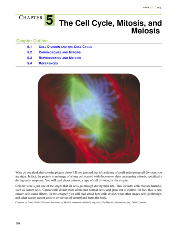
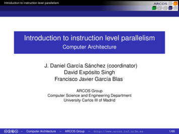
6.Differences between plant and animal cell cytokinesis:Cleavage of an animal cellCleavagefurrowContracting ring ofmicrofilamentsDaughter cellsCell plate formation in a plant cellCell wallNew cell wallCell plateVesiclescontaining cellwall material7.Daughter cells2n2n2nLife Sciences Learning Center – Cancer Education ProjectCopyright 2007, University of RochesterMay be copied for classroom use12
Part 3: Class Notes on Mitosis Teacher Key1. The process of mitosis is used as a method of reproduction forsingle-celled organisms and some multicellular organisms. Mitosis is also used forgrowth and repair in multicellular organisms.2. When mitosis is used for reproduction, it is considered asexual .What does this mean?One parent, offspring are genetically identical toparent and to each other.3. Examples of asexual reproduction include:a. binary fissionb. buddingc. tubers, bulbs, runners, rhizomesd. vegetative propagation4. Mitosis occurs during early embryonic development. Remember that the one-celledzygote must divide and become the millions of cells that make a human body!!. (Draw onboard and have students copy) draw gastrula/labelEGG Sperm ZygoteLife Sciences Learning Center – Cancer Education ProjectCopyright 2007, University of RochesterMay be copied for classroom usedraw blastula and labelCleavage13
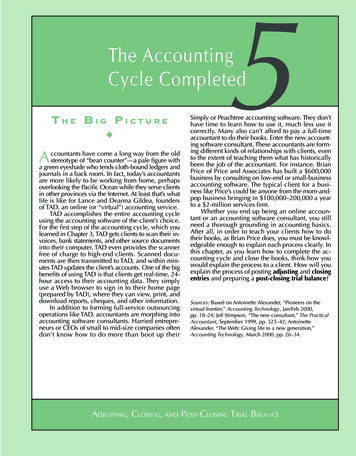
5. Process of mitosisStep 1: Chromosomes replicate .What does this mean?Make exact copies of themselves, Make reference to DNAreplication.Step 2: Chromosomes become double stranded: (Label)chromosomecentromerechromatidsLife Sciences Learning Center – Cancer Education ProjectCopyright 2007, University of RochesterMay be copied for classroom use14
Step 3: Chromosomes line up in the middle of the cell.Step 4: Double stranded chromosomes begin to separate to the poles of the cell.Step 5: Cytokinesis- After division of the chromosomes, the cytoplasm dividesforming two new cells.6. Differences between plant and animal cell cytokinesis:Cleavage of an animal cellCleavagefurrowContracting ring ofmicrofilamentsDaughter cellsCell plate formation in a plant cellCell wallVesiclescontaining cellwall materialLife Sciences Learning Center – Cancer Education ProjectCopyright 2007, University of RochesterMay be copied for classroom useNew cell wallCell plateDaughter cells15
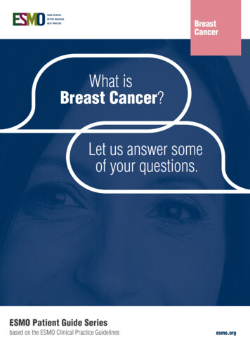
7.2n2nDaughtercell (diploid)Life Sciences Learning Center – Cancer Education ProjectCopyright 2007, University of RochesterMay be copied for classroom useParent cell(diploid)2nDaughter cell(Diploid)16
Teacher Instructions - Part 4Mitosis in Plant and Animal Cells1.Distribute one copy of Mitosis in Plant and Animal Cells lab instructions.2.Distribute one supply bag of popping bead models to each pair of students. These beadsare available from Wards # 36W1602, Chromosome Simulation Lab activity. Each bagshould contain: two 10-bead long red chromosomes two 10-bead long yellow chromosomes two 4-bead long red chromosomes two 4-bead long yellow chromosomes3.Instruct students to divide a blank sheet of paper into 6 sections (6 “boxes”).4.Label box 1 “original cell, 2n 4”. Instruct students to pull out four chromosomes from thesupply bag: 1 long red (10 beads), 1 long yellow (10 beads), 1 short red (4 beads), 1 shortyellow (4 beads). Lay these chromosomes on the table in front of them. Draw what theyhave in box 1. It helps to use colored pencils. Use this opportunity to discusschromosome packing, structure, homology, etc. NOTE: This simulation is imperfectbecause the DNA in the original cell would be unpacked as chromatin and would be similarto a piece of thread.5.Label box 2 “chromosomes replicate”. Have students pull out the remaining chromosomesfrom their supply bag and “replicate” the original ones on their desk, making sure thatcopies are held together by the centromere magnet. This is a good time to explain thatreplication means exact copies. (the long red will make a long red copy, etc.) Alsodiscuss chromosome structure, chromatid, centromere, etc. Students should draw whatthey have on their desk in box 2 and label their drawings.6.Label box 3 “Metaphase, chromosomes line up in the middle”. Have students orient theirred and yellow chromosomes so that the chromosomes are lined up along the equator ofthe cell. Students draw their results in box 3.7.Label box 4 “Anaphase, chromosomes begin to separate.” Students should separatechromatids and move them about 2” away from each other towards the poles of the cell.Students draw what they see in box 4.8.Label box 5 “Telophase, nuclear division complete.” Students should move chromatids topoles. Students draw what they see in box 5.9.Label box 6 “Cytokinesis, division of the cytoplasm.” Students should then draw thechromosomes as they see them, but this time a cell membrane should be drawn aroundeach set of chromosomes, creating two new cells. Teacher should spend time discussingchromosome number, two daughter cells, identical to each other and to original parent cell.10. Using the materials, each lab partner should explain the sequence of events in mitosis withthe other partner. Switch roles.11. Gain student access to internet website: www.biology.arizona.edu Click on “Onion RootTips” on left side column.12. Students may work individually, in pairs, or as a whole class if access to multiplecomputers is not available.Life Sciences Learning Center – Cancer Education ProjectCopyright 2007, University of RochesterMay be copied for classroom use17
13. Teacher should explain that this website will give students a chance to gain confidence inlocating cells in the different phases of mitosis. Each time a new cell is shown, the studentshould select the phase that is best demonstrated by that cell. If the student is incorrect, ahint is given and the student must choose again.14. Students should create a data table logging the number of cells in each phase as theymove through the internet activity.15. After completing, the internet lesson, students should then continue with step 6 in their labinstructions. Students should obtain a microscope, and a slide of onion root tip mitosis.16. Draw five circles on a sheet of paper.17. Students should be instructed to locate one cell for each phase of mitosis (Interphase,Prophase, Metaphase, Anaphase, Telophase). These cells should be drawn in the circlesin their data section and correctly labeled with the appropriate mitotic phase.18. Conclusion questions should be assigned for homework.Life Sciences Learning Center – Cancer Education ProjectCopyright 2007, University of RochesterMay be copied for classroom use18
Part 4: Lab - Mitosis in Plant and Animal cellsIntroduction:Mitosis, as you remember from seventh grade life science, is the process by whichcells reproduce themselves. Mitosis is only part of a bigger process called the cell cycle.Although mitosis is usually studied in depth, the most important phase of the cell cycle isthe first stage (interphase) in which the chromosomes are replicated. After this, thechromosomes line up in the middle and then separate to the poles. Following chromosomalseparation, cytokinesis occurs , dividing the cytoplasm into two new cells. This method ofreproduction is asexual.Pre lab:1. Give several characteristics of asexual reproduction.2. Why is mitosis important?3. If our original cell has fourteen chromosomes, how many should each of our finalcells have? How do you know?Procedure:1. Draw five circles in your data section. (About the size of a beaker bottom)2. Label the circles using the following phrasesa. Original cell 2n b. Chromosomes replicatec. Chromosomes line up in the middled. Chromosomes separate to polese. Chromosomes at poles, cytoplasm begins to divide3. Following your teacher’s instructions, and using your popping bead chromosomes,perform the process of mitosis. Draw each phase in the appropriate circle.4. Have each lab member orally perform the process again.5. Log on to a computer and navigate towww.biology.arizona.edu/cell bio/activities/cell cycle/cell cycle.html6. Complete the online activity to practice identifying stages of mitosis in actual cells.Copy the data table into the data section of your lab report.7. When finished, Obtain a microscope, and slide of onion skin mitosis. Scan in low(10X) power. Focus in high (40X) power. Locate, draw and label each of the fivephases of mitosis.Life Sciences Learning Center – Cancer Education ProjectCopyright 2007, University of RochesterMay be copied for classroom use19
Conclusions:1. There are many signals that control cell division. What do you think would happen if thesignal to “shut down” mitosis was never given? What do you think this is called?2. Some cells can have several nuclei in a single cell. Considering the events in a typical cellcycle, which phase of the cell cycle is not working?3. The nerve cells in humans seldom undergo mitosis. Based on this information, explainwhy complete recovery from injuries to the nervous system may not occur.Life Sciences Learning Center – Cancer Education ProjectCopyright 2007, University of RochesterMay be copied for classroom use20
Teacher Instructions - Part 5Causes of Cancer Animations1.Give each student has a copy of the “Understanding Cancer” worksheet from the NIH: CellBiology and Cancer curriculum guide. This is available online h1/cancer/guide/pdfs/ACT2M.PDF(To see the entire Cell Biology and Cancer curriculum guide, go 1/cancer/default.htm)2.Use a computer and projector to show students the 5 animations available online h1/cancer/activities/activity2 animations.htm3.Watch each animation and then complete Section 2 of the worksheet. Have students workindividually to write a one-sentence summary on the worksheet that corresponds to eachanimation.4.Give students 5-7 minutes to discuss their answers with their group.5.Discuss answers with whole group.6.Student groups should complete Section 3, “Explaining Factors Associated with Cancer”.7.Discuss answers as a group.Life Sciences Learning Center – Cancer Education ProjectCopyright 2007, University of RochesterMay be copied for classroom use21
Teacher Instructions - Part 6Modeling the Normal Cell Cycle1.2.Prepare Cell Cycle wheels before class. Purchase enough materials so that each team offour students will have one wheel. 10” cardboard cake bases (available at most craft stores) Color copy of the labeled cell cycle wheel (see document entitled cell “cyclewheel.doc”) One brad to hold the paper to the wheel One copy of the arrow (found in graphics folder) Optional: The color copy of the cell cycle can be laminated or covered in clearcontact paper to add to its durability. Optional: Teachers may choose to use “business card magnets” cut into smallpieces to use as a centomere. With this option, chromatids will “stick” togetherand will be harder to pull apart.Prepare a 1 gallon Ziplock bag of materials for each team of 4 students and label this bag“Materials”. The bag contains the following items: 1 strip of 4 hole reinforcements 8 pieces of black or white plastic lanyard (boondoggle) approximately 10” long.Take four pieces and tie into a knot at one end. Do the same with the other four.o Tie a paper clip to each untied end of the lanyard. ½ sheet of blank pink paper, ½ sheet blank blue paper black markers dried beans A sandwich size Ziploc bag, labeled “cell” with the following inside:o 2 chromosomes graphics copied in pink (see document entitled “pinkchromosome master.doc”), cut out and a small white ring reinforcementattached to the black dot.o 2 chromosomes graphics copied in blue (see document entitled “bluechromosome master.doc”), cut out and a small white ring reinforcementattached to the black dot.3.Prepare the following Teacher Signs: Ras Cyclin, p53, Mad1, ATM/Nibrin. (see documententitled “teacher signs.doc”)4.Students should be divided into groups of four. Each student in the group is responsiblefor one chromosome in the cell model activity.5.Distribute one copy of Part 6: Modeling the Normal Cell Cycle worksheet to each student.6.Distribute one large Ziploc bag of materials, one balance, 2-4 pairs of scissors, tape andan assembled Cell Cycle Wheel to each group.Life Sciences Learning Center – Cancer Education ProjectCopyright 2007, University of RochesterMay be copied for classroom use22
7.Beginning at step 1 on the worksheet, read the instructions together as a class. Studentsshould locate the two pairs of chromosomes. Teacher should circulate to check that eachgroup has correctly located the two chromosome pairs. Discuss DNA packaging and theaccompanying diagram.8.Continue reading steps 2-3.9.Read step 4. Students should add beans to their small cell bag one at a time to simulatecell growth. Give students a minute or two to add beans.10. Read step 5. Teacher should now wear the “Ras Cyclin” sign. Students should completeprocedure by checking the mass of their cell. Teacher circulates and checks the mass ofthe “cells”. Cells that are large enough ( grams) are given a bingo chip as a signal fromRas Cyclin that they may proceed.11. Read step 6 together. Teacher should now wear “p53”sign. Teacher should circulate andcheck for DNA damage (there is none). Make a big point about how nice their DNA is!12. Read step 7. Teacher should monitor progress of student groups. Each student is incharge of copying one chromosome.13. Read step 8. Teacher should now wear the “ATM/Nibrin” sign. Teacher should circulateamong groups checking their replicated chromosomes. Student groups who accuratelycopy their chromosomes should be given a bingo chip as their “go ahead” signal.14. Read step 9.15. Read step 10. Students should follow directions to perform mitosis. Chromatids shouldNOT be separated at this point!16. Read step 11. Teacher should now wear the “Mad1” sign. The teacher should circulateamong groups and check their mitosis setup. If it is OK, give them a bingo chip as a signalto continue. If their setup is not OK, discuss changes to be made. Note: the mostcommon error is to attach non-chromatids together at the small white rings.17. Read step 12.18. Read step 13.19. Complete step 14.20. A closing discussion can revolve around the make up the two new cells. Students shouldrecognize that the chromosomes are the same as when we began, however, the size ofthe cell is much smaller because of the division of cytoplasm.Life Sciences Learning Center – Cancer Education ProjectCopyright 2007, University of RochesterMay be copied for classroom use23
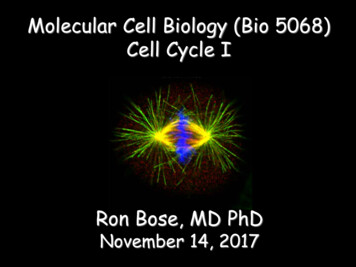
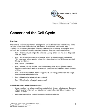
Part 6: Modeling the Normal Cell CycleYour Task: Model the events and the controls of the cell cycle. Work in groups of 4students. At your station you should have: A cell cycle wheel A large Ziploc bag with materials Scissors Tape Dried lima beans Balance1.First, become familiar with the chromosomes that will be used in your cellmodel. Your cell has 2 pairs of chromosomes. These are made from blue andpink paper. Each pair is the same length. Find the 2 pairs. Throughout this simulation,each person in the group will be responsible for one of the chromosomes. Each pair of chromosomes is made up of 2 partners, a maternal chromosomeand a paternal someDNA coiled around histone proteinsDuring this simulation, how would you tell the difference between the maternal andpaternal chromosomes?Life Sciences Learning Center – Cancer Education ProjectCopyright 2007, University of RochesterMay be copied for classroom use24
2.The chromosomes are found inside your cell that is represented by the smallZiploc bag labeled “CELL”. Place your 2 pairs of chromosomes into the smallZiploc bag. The remainder of the materials will be used throughout theactivity and should be kept in the large bag labeled “MATERIALS”, as asupply/synthesis area.3.Let’s Begin Mitosis! A cell next to your cell has died. This cell’s death causesan external growth signal and tells your cell to begin the cell cycle.Cell death acts as an external growthsignal that stimulates cell division.4.Turn your wheel to the G1 phase. This is the growth phase. According to the cellcycle chart (from Part 1), what should happen during G1 phase?5.To simulate growth of your cell, beans will be added to your small bag torepresent an increase in the mass of your cell. Please add beans one at a timeto your cell until the mass of your cell is grams. Leave your cell withincreased mass on the balance6.Turn your wheel until you reach the first checkpoint (). At this checkpoint,a growth regulator protein, called Ras cyclin, checks that cells are big enoughto enter the next part of the cell cycle. Ras cyclin is a protein that functions during this checkpoint. As the cellgrows, the amount of Ras cyclin increases. When the amount of Rascyclin reaches a certain level, it signals the cell to proceed to the nextpart of the cycle. Raise your hand so that Ras cyclin (your teacher) maycheck the mass of your cell.Life Sciences Learning Center – Cancer Education ProjectCopyright 2007, University of RochesterMay be copied for classroom use25
If Ras cyclin does not give cells the signal to continue, the cells remain inthis part of the cycle and continue to grow. Why is it important to check that the cell is big enough to continue withthe cell cycle? Once you have been given the signal to continue, proceed to step 7.7. Before your cell is allowed to enter the next part of the cell cycle, it must passa second checkpoint. Turn your wheel to the next checkpoint ().8. DNA in these chromosomes can be damaged by a number of agentsincluding radiation, toxic chemicals, and free radicals. At thischeckpoint, another protein known as p53 will inspect the chromosomes’DNA for damage. If there is DNA damage, p53 will stop the cell cycleuntil DNA repair enzymes can fix the damage. If the DNA damage can’tbe repaired, p53 may signal programmed cell death, called apoptosis. Raise your hand to have p53 (your teacher) check your DNA for damage. What do you think would happen if this p53 surveillance system did notwork properly?If your cell has cleared the two checkpoints (that is, it is large enough and hassuffered no DNA damage), turn your wheel to the S phase of the cell cycle.During this part of the cell cycle, DNA must be replicated. You must completethe following steps in order to replicate your DNA: Using the materials from the supply bag, (black markers, pink and bluepaper strips, scissors, small white rings) make exact copies of each ofyour DNA strands. These two copies (chromatids) are held together at the centromereregion. Place the centromere regions of the chromatids on top of eachother to simulate how the chromatids are held together. Summarize the events that occur during the S phase of the cell cycleLife Sciences Learning Center – Cancer Education ProjectCopyright 2007, University of RochesterMay be copied for classroom use26
9.Turn your wheel to the next checkpoint (). At this checkpoint, anotherprotein complex called ATM/Nibrin inspects to make sure that the DNA wascopied without mistakes. Raise your hand to have ATM/Nibrin (your teacher) check yourreplicated DNA for damage. Why is it important to check the replicate DNA?10. Once you have been given the ‘go ahead’ to continue, turn your wheel and enterthe G2 part of the cell cycle. During this part of the cell cycle, the cellprepares for mitosis by synthesizing the materials it will need. Please prepareyour cell for mitosis by assembling the following materials: Spindle apparatus
Life Sciences Learning Center - Cancer Education Project . To print, select "Teachers Guide" then "PDF Files for Printing" then "Activity 2 Masters", . Cell Cycle Wheel Cell Cycle Modeling Kit (one per student group of 4 students)