Transcription
Placenta2267PReferences and ReadingsPlaceboFred R. VolkmarDirector – Child Study Center, Irving B. HarrisProfessor of Child Psychiatry, Pediatrics andPsychology, School of Medicine, YaleUniversity, New Haven, CT, USASynonymsPlacebo effect; Placebo responseMcCracken, J. T., McGough, J., Shah, B., Cronin, P.,Hong, D., Aman, M. G., et al. (2002). Risperidone inchildren with autism and serious behavioral problems.New England Journal of Medicine, 347(5), 314–321.Sandler, A. D., & Bodfish, J. W. (2000). Placebo effects inautism: Lessons from secretin. Journal of Developmental & Behavioral Pediatrics, 21(5), 347–350.Shapiro, A., & Shapiro, E. (2000). The powerful placebo.Baltimore, MD: Johns Hopkins University Press.Volkmar, F. (2001). What is a “placebo controlled” study?Journal of Autism & Developmental Disorders, 31(2),251–252.Volkmar, F., & Wiesner, L. (2009). A practical guide toautism. Hoboken, NJ: John Wiley.DefinitionA simulated intervention (often a drug but potentially other interventions) used as a control/comparison in research studies. As suggested in thetitle of a book on the subject (Shapiro & Shapiro,2000), the effects of such treatment can be powerful (what is termed to placebo response oreffect). In drug studies, for example, the use ofa placebo (inert agent that is packaged so as to beindistinguishable from the drug being studied)control provides one of the most stringent testsin demonstrating drug efficacy.As Volkmar and Wiesner (2009) note that thenature of the placebo response in studies involving children with autism reflects several factors:high levels of attention from parents, teachers,and clinicians; provision of high-quality care during a study; variation in symptoms over time; andthe effects of expecting a change with a newtreatment (see also Volkmar, 2001). The effectsof placebo treatments can be quite strong, asdemonstrated in studies of secretin which didnot significantly outperform secretin (Sandler &Bodfish 2000); on the other hand, studies ofagents like risperidone (McCracken et al.,2002), the active drug, proved significantly betterthan placebo even after only a few weeks ofadministration.Placebo Effect PlaceboPlacebo Response PlaceboPlacement-Pending Requirement Stay-Put RequirementPlacentaHarvey J. KlimanReproductive and Placental Research Unit,Department of Obstetrics, Gynecology andReproductive Sciences, Yale University Schoolof Medicine, New Haven, CT, USADefinitionSee Also Risperidone SecretinIf we could noninvasively analyze the detailedneuroanatomy of all newborns at a cellular, synapse by synapse level, we might be able toP
P2268identify those children who will go on to have anautistic spectrum disorder (ASD). As that isunlikely to be possible anytime soon, doctorsseek surrogates that can make the same diagnoses. Luckily, the placenta, an organ that is oftenroutinely discarded at birth, may be able to identify those babies who will exhibit overt signs ofautism months or years later.The human placenta is not only an integralpart of the fetus during all of pregnancy, but italso shares in the vast majority of cases the exactgenetic makeup of the fetus. As such, it offersthe potential to reveal abnormal morphologic patterns that may be at the basis of ASD and othergenetic and developmental abnormalities.Historical BackgroundFormation of the PlacentaHumans start off as a symmetrical ball of cells.Even as our first few dozen cells begin to separateinto an inner cell mass (which will become theembryo, fetus, and eventually baby) and the trophoblasts (which will become the placenta), genesare regulating the creation of the developmentalaxes that will form the basis of the entire organism(Kliman, 1999). Defects in the genes that regulatethese processes lead to a wide range of embryonic,fetal, and neonatal defects from minor cosmeticabnormalities to disasters that terminate pregnancywithin a few days to weeks after fertilization.By 21 days after fertilization, the trophoblastshave begun to sort themselves into what willbecome the treelike structures that make up theplacenta: the chorionic trees, branches, and villi(Fig. 1). The terminal villi (from the Latin forshaggy hair) are fingerlike structures that are covered with a double cell layer. This trophoblastbilayer is made up of an inner cytotrophoblastlayer made of single nucleated cells and an outersyncytiotrophoblast layer made of giantmultinucleated sheets (Fig. 2). Starting with purified cytotrophoblasts, we demonstrated usingin vitro time lapse cinematography thatcytotrophoblasts fuse to form syncytiotrophoblast(Kliman, Nestler, Sermasi, Sanger, & Strauss,1986) (Fig. 3), an observation that has beenPlacentaconfirmed in situ (Huppertz, Tews, & Kaufmann,2001). The critical conclusion from these studiesis that only the cytotrophoblasts proliferate,making the growth of the syncytiotrophoblastlayer completely dependent on the absorption offusing cytotrophoblasts (Fig. 4).Current KnowledgeTrophoblast InclusionsThe relative rates of cytotrophoblast proliferationand incorporation into the outer syncytiotrophoblast layer appear to determine the morphology of the fingerlike chorionic villi (Huppertzet al., 2001; Kliman & Segel, 2003; Rejniak,Kliman, & Fauci, 2004). In the normal placenta,new villus branches are formed by outward bending bulges of the trophoblast bilayer (Fig. 5).However, when these critical processes go awry,the bilayer can inappropriately bulge inward intothe villi, creating invaginations (Fig. 6) and trophoblast inclusions (Fig. 7) that can be readilydetected upon histological examination of sectioned placental tissue.The basis of trophoblast invaginations andinclusions, therefore, appears to be an imbalancebetween the rate of cytotrophoblast proliferationand fusion (Rejniak et al., 2004). This could bethe result of either an increased rate of proliferation – due to either endogenous genetic factorswithin these cells or exogenous factors such asincreased exposure to paracrine or endocrinegrowth factors – or the result of a decreased rateof fusion – due to decreased production of thefactors that facilitate cell fusion. Endogenous cellproliferation rates may be affected by genes thatregulate the mitotic cycle, such as the cyclins(Gillett & Barnes, 1998). Equally potent are hormones that regulate cellular proliferation, such asgrowth hormone and insulin. Abnormalities ofcytotrophoblast fusion have been shown in theplacentas of Down syndrome (trisomy 21)children, showing a direct relationship betweenthis particular genetic disorder and placentalmorphogenesis (Malassine, Frendo, & EvainBrion, 2010).
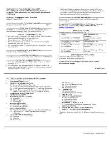
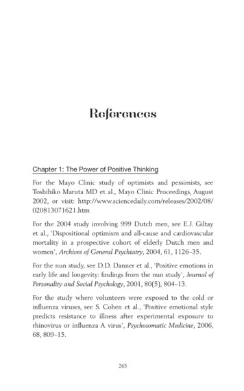
Placenta2269PPlacenta, Fig. 1 Diagram of the human placenta. Theplacenta, which is part of the fetus, attaches to the maternal decidua via the anchoring villi. The maternal blood isinjected into the intervillous space where it circulatesaround the chorionic villi and then returns to the maternalcirculation via the endometrial veins. The fetus pumps itsblood into the placenta via the umbilical arteries, whichbranch in the chorionic plate and eventually dive down toform the villus trees. The fetal circulation terminates in thefingerlike chorionic villi, which are covered by a layer oftrophoblast cells (see Fig. 2). (From Moore KL, TheDeveloping Human, 4th edition, WB Saunders, 1988,used with permission)Understanding how trophoblast invaginationsand inclusions are formed helps to identify themin placental tissues and leads to criteria for diagnosing these dysmorphic features. In almost allcases of invagination, there is an increased number of cytotrophoblasts at the point of infoldingcompared to the density of cytotrophoblasts awayfrom the invagination (Fig. 8). Likewise, carefulexamination of a trophoblast inclusion revealsa central region of syncytiotrophoblasts,surrounded by cytotrophoblasts (Fig. 8). Like anepidermal inclusion cyst, which continues to getlarger and larger over time, the cytotrophoblastscontinue to proliferate and fuse with the adjacentsyncytiotrophoblasts, which in some cases leadsto very large trophoblast inclusions (see Fig. 7d).Adherence to the criteria of identifying increasednumbers of cytotrophoblasts along the invagination and around the syncytiotrophoblast core ofan inclusion helps to distinguish trophoblastinvaginations and inclusions from tangential sections of curved chorionic villus surfaces.Although trophoblast inclusions were firstdescribed as a marker of triploid gestations(a complete extra one set of chromosomes)(Szulman, Philippe, Boue, & Boue, 1981), it isnow appreciated that the presence of trophoblastinclusions in placentas is associated with a longlist of genetically abnormal gestations, includingtetraploidy (a complete extra two sets of chromosomes), trisomies (an extra individual chromosome, such as trisomy 21 or Down syndrome,trisomy 18, and trisomy 13), Turner’s syndrome(female with one X chromosome missing), andeven genetic diseases without obvious chromosome abnormalities (Honore, Dill, & Poland,1976; Novak et al., 1988; Silvestre, Cusi, Borras,& Antich, 1996; Szulman, 1984; van Lijnschoten,P
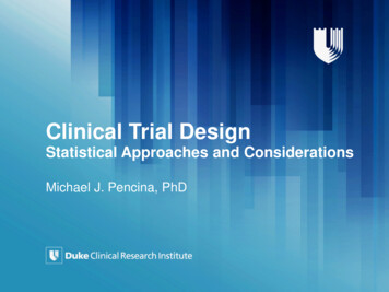
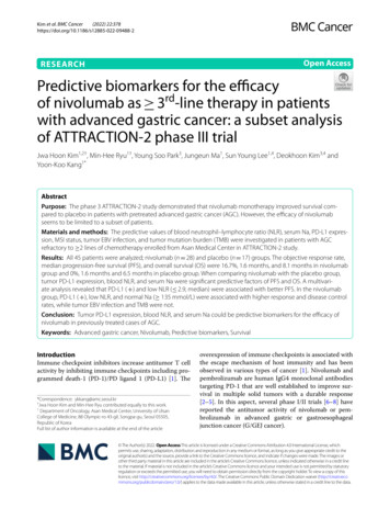
P2270PlacentaFetal capillaryMesenchymal coreHofbauer cellSyncytiotrophoblastCytotrophoblastPlacenta, Fig. 2 Diagrammatic cross section of firsttrimester chorionic villus. The villus core of villi containsfetal capillaries embedded in a loose matrix which contains fibroblasts and macrophages (also called Hofbauercells). In the first trimester, a villus cross section revealstwo distinct trophoblast layers, the outer syncytiotrophoblast layer which is in direct contact with maternalblood and the inner cytotrophoblast layer, the stem cellof the placenta and the source of new trophoblasts. (Modified from Moore KL, The Developing Human, 4th edition, WB Saunders, 1988, used with permission)Arends, De La Fuente, Schouten, & Geraedts,1993). Thus, many different genetic defects manifest themselves in the placenta as trophoblastinclusions. Since cytotrophoblast proliferationand absorption is no doubt regulated by manygenes, it appears that abnormalities in any part ofthese multigene processes may result in thesevillus dysmorphic features.Frequency of Trophoblast InclusionsFewer than 3% of placentas from uncomplicated,normal gestations manifest trophoblast inclusions. But 70% of placentas of fetuses withknown chromosomal abnormalities exhibit inclusions (Kliman et al., 2003). Since the presence ofa normal karyotype does not exclude the possibility of a genetic defect (e.g., cystic fibrosis,Tay-Sachs, sickle cell disease), it should not besurprising that trophoblast inclusions are alsoseen in cases with a normal karyotype. Themost common example of the later situation isa spontaneous pregnancy loss where the karyotype is normal, but clearly, there is somethingabnormal about the gestation. The genetic basisof such losses is reinforced by the finding of botha high recurrence risk for these families and thefact that the losses almost always occur at thesame gestational age for each particular family,suggesting a specific programming error.The more severe the genetic abnormality, orthe earlier the pregnancy loss, the more inclusions are found (Fig. 9). This is consistent withstudies that have concluded that over 90% of firsttrimester losses are secondary to genetic causes(Zhang et al., 2009). As pregnancies progressfrom the first to the third trimester, the likelihoodof a genetic basis for a pregnancy loss decreasesbut does not go to zero even at term. Not surprisingly, the number of trophoblast inclusions seenin cases that reach term are fewer than those thatterminate early in pregnancy. This is most likelyrelated to the severity of the genetic abnormality,with the most severe terminating before 13 weeksof gestation.The only documented nongenetic cause fortrophoblast inclusions appears to be gestationaldiabetes. In such cases, it is believed thatincreased glucose and insulin levels lead toincreased cytotrophoblast proliferation, whichas described above, would lead to invaginationof the trophoblast bilayer. This is consistent withthe observation that cultured cytotrophoblastsgrow better in high glucose media compared tonormal media (Kliman et al., 1986). It is also ofinterest that diabetics have a much higher fetalanomaly rate (Correa et al., 2008), suggestingthat the high glucose insulin environment leadsnot only to abnormalities in the placenta but alsoin the fetus. This has been confirmed in rat gestations where it has been shown that high glucoselevels alone can lead to both deformed fetusesand pregnancy terminations (Reece, Pinter,Homko, Wu, & Naftolin, 1994).Trophoblast Inclusions and AutismIt was in the context of trophoblast inclusions asa general marker of genetic and developmentalabnormalities that the association of trophoblastinclusions and autism was first suggested. Thelinkage was first observed anecdotally in twocases of Asperger’s syndrome, which was thenfollowed by a retrospective study. Trophoblast
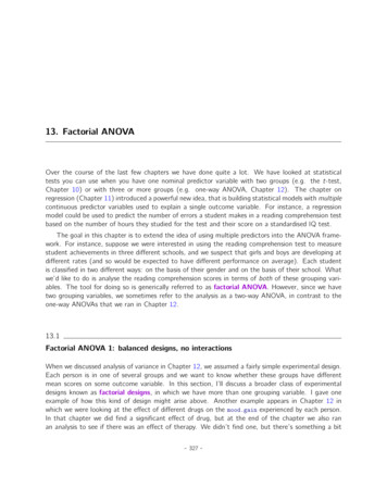
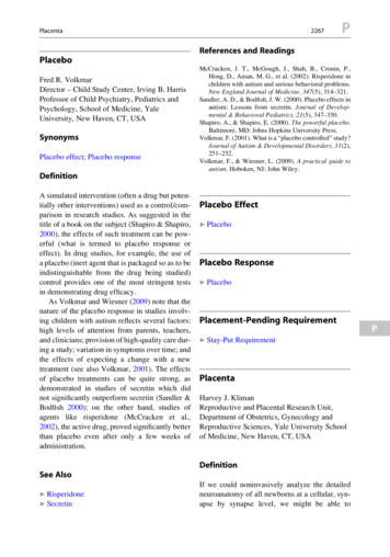
PlacentaPlacenta, Fig. 3 In vitro conversion of cytotrophoblastsinto syncytiotrophoblasts. Individual cytotrophoblasts(left) migrate like amoeba, eventually making contactwith each other. Once in contact, the cytotrophoblastsform aggregates (middle), and in time, the cells fuse toform syncytiotrophoblasts. Eventually, all thecytotrophoblasts have merged and fused to make a largePlacenta, Fig. 4 Cytotrophoblast proliferation andfusion. Cytotrophoblasts (blue) either proliferate toincrease the number of trophoblastic stem cells (bluearrows) or occasionally fuse upward (gold arrows) intothe multinucleated syncytiotrophoblast layer (red). Thebalance of these two processes – proliferation and fusion– determines the overall morphology of the placenta’schorionic villi (see also Fig. 5)inclusions were found in significantly morecases of ASD than would be predicted from thenormal population (Anderson, Jacobs-Stannard,Chawarska, Volkmar, & Kliman, 2007).This result fits into the consensus view thatASD is largely genetically based. It also suggeststhat this seemingly polygenetic, heterogeneouscondition may ultimately be caused by subtleabnormalities in common morphogenetic processes, such as bilayer folding.A Problem of FoldingThere are only a few developmental processesin the embryology tool box. These include cellproliferation, cell death, cell migration, cellhypertrophy, differentiation, fusion, and cellular2271Psyncytiotrophoblast (right). (From Kliman HJ, Nestler JE,Sermasi E, Sanger JM, and Strauss JF III. (1986) Purification, characterization and in vitro differentiation ofcytotrophoblasts from human term placentae. Endocrinology 118: 1567–1582, used with permission. Copyright1986, The Endocrine Society)Placenta, Fig. 5 Proliferation fusion model. A modelillustrating the different ratios of proliferation (P) andfusion (F). Cytotrophoblasts (pink circles) proliferateand intermittently fuse into the upper syncytiotrophoblastlayer (blue bars). Stability (relative flatness) of the bilayeris maintained at, or near, an ideal ratio: P ¼ 2F. Normaloutward budding (evagination) is observed from the ratio:P 2F, while an abnormal trophoblast inclusion (invagination) results from the ratio: P 2F. (Kliman HJ, SegelL. (2003) The placenta may predict the baby. J Ther Biol,225: 143–145, used with permission)dissociation. With these tools, all the variousorgans and body parts are made. One of the common problems in development is how to increasesurface area. There are two basic solutions to thisproblem: increased branching or increased folding. In both cases, these are most often achievedby differential growth of layers of cells, whichdue to increased tension being built up in onelayer or another, results in bending. This isexactly how the placenta forms new buds andvilli to make the villus tree. It is also how theheart forms, the bronchi branch in the lung, thekidney tubules form, the gut surface areaP
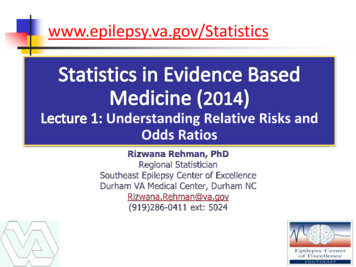
P2272PlacentaPlacenta, Fig. 6 Trophoblast invaginations. (a, b) Trophoblast invaginations forming cleft-like structures. Notethe many cytotrophoblasts lining the invaginations(arrowheads) and the fewer cytotrophoblasts underlyingthe normal bilayers (*). (c) Invagination ending in an areaof increased syncytiotrophoblasts (arrow). Even thoughthe bilayer is invaginated, syncytiotrophoblasts still form.(d) Bulb-like prominence of syncytiotrophoblasts at baseof an invagination (arrow). (Kliman HJ, Segel L.(2003) The placenta may predict the baby. J Ther Biol,225: 143–145, used with permission)Placenta, Fig. 7 Trophoblast inclusions. (a) Trophoblast inclusion within the villous core. Note how thecytotrophoblasts of the bilayer (arrowheads) and thecytotrophoblasts of the inclusion (*) both are adjacent tothe villous core. (b) Trophoblast inclusion witha prominent syncytiotrophoblast layer and a lonecytotrophoblast (arrowhead). Fetal vessels (V). (c) Chorionic villous with four prominent trophoblast inclusions(arrows). (d) Trophoblast inclusion with very expandedsyncytiotrophoblast component (arrow). (Kliman HJ,Segel L. (2003) The placenta may predict the baby.J Ther Biol, 225: 143–145, used with permission)
Placenta2273PPlacenta, Fig. 8 Formation of trophoblast invaginationsand inclusions. (a) Histologic section of a placental villuswhich exhibits both a trophoblast invagination (I) andinclusion (TI). Note the increased numbers ofcytotrophoblasts (arrow heads) beneath the syncytiotrophoblast layer in the region of the invagination and theirpaucity away from the invagination (*). When an invagination is sectioned perpendicular to its long axis (S-S), itappears as an inclusion (TI), with dark syncytiotrophoblastnuclei in its center surrounded by cytotrophoblasts (arrowheads). IVS Intervillus space. (b) Diagram of a villus crosssection showing the outer syncytiotrophoblast layer (blue)and inner cytotrophoblast layer (pink cells) witha trophoblast invagination (I) and inclusion (TI) illustrating the relevant morphology and disposition ofcytotrophoblasts in the region of the invagination. (FromAnderson GM, Jacobs-Stannard A, Chawarska K,Volkmar FR, Kliman HJ. (2007) Placental TrophoblastInclusions in Autism Spectrum Disorder, Biological Psychiatry, 61:487–491, used with permission)Placenta, Fig. 9 Trophoblast inclusions as a function ofgenetic abnormality severity. Normal placentas rarelyexhibit trophoblast inclusions. As the degree of geneticabnormality increases, the more trophoblast inclusionscan be identified per unit area of placenta examined. Theearlier the pregnancy loss, the more frequent the inclusions. Many pregnancy losses reveal normal karyotypes;however, a significant number of trophoblast inclusionscan be seen in many of these cases. The trisomies (e.g.,trisomy 21, 13, 18) may or may not lead to an earlypregnancy loss, depending on which chromosome isaffected. In very abnormal gestations, such as triploidyand tetraploidy, as many as 50 or more inclusions per slidecan be seen. ASD, a subtle condition that does not lead topregnancy loss, usually exhibits only 1–3 trophoblastinclusions per placenta slide examinedincreases, and most dramatically how the brain,especially the human brain, fits into the skull.Since the upper limit of human skull diameteris directly related to the size of the female pelvis,increases in brain surface area could only beachieved by folding. And the human brain isone of the most folded brains in the animal kingdom (Hilgetag & Barbas, 2006). Behind thisfolded structure are the cellular processes thatlead to differential growth in the many neurallayers that make up the brain. Could the developmental abnormalities that lead to abnormal folding and trophoblast inclusions in the placenta alsobe at work in the brains of ASD children?Researchers have demonstrated significantlyabnormal brain folding in children on the autismspectrum using surface mapping and magneticresonance imaging (MRI) (Awate, Win,Yushkevich, Schultz, & Gee, 2008; Kates,Ikuta, & Burnette, 2009; Nordahl et al., 2007).These abnormalities may in part explain theobservation of increased head size in ASDchildren (Awate et al., 2008; McCaffery &Deutsch, 2005). Basically, with less folding, thebrain tissue has no other option but to expandthe skull to make up for the less compact natureP
P2274of the neural tissue. Translating this gross macroscopic observation to cellular processes andultimately to abnormal behaviors is much harder.However, neuropathologists have describedabnormalities in ASD children at the tissue levelof the brain that may be associated with thesemacroscopic changes (Bauman & Kemper,2003, 2005; Kemper & Bauman, 1998; Whitney,Kemper, Rosene, Bauman, & Blatt, 2009).Future DirectionsIf one of the basic pathologies in ASD is relatedto problems in tissue folding, then we should seeevidence for this in any tissue where folding isa critical part of the anatomy or function of thattissue. Some organs may be impervious to lowfrequencies of misfolding, such as the liver,which does not have a critical requirement formultilayer cellular organization. Other organs,such as the gut, may not function as well if itsfolded structure is disrupted. If commonalitiescan be ascertained in cases of ASD, then candidate genes could be sought that control and regulate these processes. Further, in the future, onemight anticipate that genetic or medical interventions might be forthcoming that can amelioratethe abnormalities that may exist in ASD children.In the meantime, knowing that trophoblast inclusions are related to ASD could lead to earlyidentification and intervention before overtsymptoms arise.See Also Developmental ChangeReferences and ReadingsAnderson, G. M., Jacobs-Stannard, A., Chawarska, K.,Volkmar, F. R., & Kliman, H. J. (2007). Placentaltrophoblast inclusions in autism spectrum disorder.Biological Psychiatry, 61, 487–491.Awate, S. P., Win, L., Yushkevich, P., Schultz, R. T., &Gee, J. C. (2008). 3D cerebral cortical morphometry inautism: Increased folding in children and adolescentsin frontal, parietal, and temporal lobes. InternationalPlacentaConference on Medical Image Computing andComputer-Assisted Intervention, 11, 559–567.Bauman, M. L., & Kemper, T. L. (2003). The neuropathology of the autism spectrum disorders: What havewe learned? Novartis Foundation Symposium, 251,112–22; discussion 122–128, 281–297.Bauman, M. L., & Kemper, T. L. (2005). Neuroanatomicobservations of the brain in autism: A review andfuture directions. International Journal of Developmental Neuroscience, 23, 183–187.Correa, A., Gilboa, S. M., Besser, L. M., Botto, L. D.,Moore, C. A., Hobbs, C. A., et al. (2008). Diabetesmellitus and birth defects. American Journal of Obstetrics & Gynecology, 199, 237.e1-9.Gillett, C. E., & Barnes, D. M. (1998). Demystified . . . cellcycle. Molecular Pathology, 51, 310–316.Hilgetag, C. C., & Barbas, H. (2006). Role of mechanicalfactors in the morphology of the primate cerebral cortex. PLoS Computational Biology, 2, e22.Honore, L., Dill, F. J., & Poland, B. J. (1976). Placentalmorphology in spontaneous human abortuses with normal and abnormal karyotypes. Teratology, 14,151–166.Huppertz, B., Tews, D. S., & Kaufmann, P. (2001). Apoptosis and syncytial fusion in human placental trophoblast and skeletal muscle. International Review ofCytology, 205, 215–253.Kates, W. R., Ikuta, I., & Burnette, C. P. (2009).Gyrification patterns in monozygotic twin pairs varying in discordance for autism. Autism Research, 2,267–278.Kemper, T. L., & Bauman, M. (1998). Neuropathology ofinfantile autism. Journal of Neuropathology & Experimental Neurology, 57, 645–652.Kliman, H. J. (1999). Trophoblast to human placenta. In E.Knobil & J. D. Neill (Eds.), Encyclopedia of reproduction, 4, 834–846. San Diego: Academic Press.Kliman, H. J., McSweet, J. C., Franco, A., Ying, X.,Zhao, Y., & Stetten, G. (2003). Trophoblast inclusionsare rare in elective terminations and normal deliveries,but common in cases with karyotypic abnormalities.Fertility and Sterility, 80, 88–88.Kliman, H. J., Nestler, J. E., Sermasi, E., Sanger, J. M., &Strauss, J. F., 3rd. (1986). Purification, characterization,and in vitro differentiation of cytotrophoblasts fromhuman term placentae. Endocrinology, 118, 1567–1582.Kliman, H. J., & Segel, L. (2003). The placenta maypredict the baby. Journal of Theoretical Biology,225, 143–145.Malassine, A., Frendo, J.-L., & Evain-Brion, D. (2010).Trisomy 21- affected placentas highlight prerequisitefactors for human trophoblast fusion and differentiation. International Journal of Developmental Biology,54, 475–482.McCaffery, P., & Deutsch, C. K. (2005). Macrocephalyand the control of brain growth in autistic disorders.Progress in Neurobiology, 77, 38–56.Nordahl, C. W., Dierker, D., Mostafavi, I., Schumann, C. M.,Rivera, S. M., Amaral, D. G., et al. (2007). Cortical
Plasticity, Neuralfolding abnormalities in autism revealed by surfacebased morphometry. The Journal of Neuroscience, 27,11725–11735.Novak, R., Agamanolis, D., Dasu, S., Igel, H., Platt, M.,Robinson, H., et al. (1988). Histologic analysis ofplacental tissue in first trimester abortions. PediatricPathology, 8, 477–482.Reece, E. A., Pinter, E., Homko, C., Wu, Y. K., &Naftolin, F. (1994). The yolk sac theory: Closing thecircle on why diabetes-associated malformationsoccur. Journal of the Society for Gynecologic Investigation, 1, 3–13.Rejniak, K. A., Kliman, H. J., & Fauci, L. J. (2004).A computational model of the mechanics of growthof the villous trophoblast bilayer. Bulletin of Mathematical Biology, 66, 199–232.Silvestre, E., Cusi, V., Borras, M., & Antich, J. (1996).Cytogenetic and morphologic findings in chorionicvilli from spontaneous abortions. Birth Defects Original Article Series, 30, 353–357.Szulman, A. E. (1984). Syndromes of hydatidiform moles.Partial vs. complete. The Journal of ReproductiveMedicine, 29, 788–791.Szulman, A. E., Philippe, E., Boue, J. G., & Boue, A.(1981). Human triploidy: Association with partialhydatidiform moles and nonmolar conceptuses.Human Pathology, 12, 1016–1021.van Lijnschoten, G., Arends, J. W., De La Fuente, A. A.,Schouten, H. J., & Geraedts, J. P. (1993). Intra- andinter-observer variation in the interpretation of histological features suggesting chromosomal abnormalityin early abortion specimens. Histopathology, 22,25–29.Whitney, E. R., Kemper, T. L., Rosene, D. L.,Bauman, M. L., & Blatt, G. J. (2009). Density ofcerebellar basket and stellate cells in autism: Evidencefor a late developmental loss of Purkinje cells. Journalof Neuroscience Research, 87, 2245–2254.Zhang, Y. X., Zhang, Y. P., Gu, Y., Guan, F. J., Li, S. L.,Xie, J. S., et al. (2009). Genetic analysis of firsttrimester miscarriages with a combination of cytogenetic karyotyping, microsatellite genotyping andarrayCGH. Clinical Genetics, 75, 133–140.PL-ADOS Prelinguistic Autism Diagnostic ObservationSchedulePlanning and Placement Team (PPT) Interdisciplinary Team2275PPlasticity, NeuralJoshua Trachtenberg and Peyman GolshaniDavid Geffen School of Medicine at UCLA,Los Angeles, CA, USASynonymsCortical remapping; Cortical rewiring; NeuralreorganizationStructureAutism spectrum disorders are characterized byprofound impairments in language acquisitionand social interaction that are often comorbidwith repetitive behaviors and sensory and motorabnormalities. Communicative and social skillsemerge in early childhood and grow exponentially in complexity. The establishment of appropriate neural circuitry in the cerebral cortexsubserving language acquisition, social interaction, and indeed, all aspects of cortical function,including vision and motor control, relies on theproper execution of genetic programs that regulate neural proliferation, migration, differentiation, axon guidance, and recognition of synaptictargets. A characteristic conserved between thesedisparate systems is that this circuitry is ineffective at birth. Sensory experience within specifiedsensitive or so-called “critical” periods duringdevelopment sculpts the fine structure and function of cortical circuitry. However, the timing andinfluence of these critical periods varies dramatically among the functional domains of the cerebral cortex. This entry examines recent advancesin our understanding of how autism-candidategenes regulate neural growth, synaptic plasticity,and the closure of critical periods.Anatomical Plasticity of Neural StructureThe establishment of precise neural circuitry inthe cortex hinges on five major events. First,neurons must be born in correct numbers. Second, these neurons must migrate to appropriateP
Placebo effect; Placebo response Definition A simulated intervention (often a drug but poten-tially other interventions) used as a control/com-parison in research studies. As suggested in the title of a book on the subject (Shapiro & Shapiro, 2000), the effects of such treatment can be pow-erful (what is termed to placebo response or effect).