Transcription
clinical articleJ Neurosurg 123:897–905, 2015Neuroophthalmological outcomes associated with use ofthe Pipeline Embolization Device: analysis of the PUFStrial resultsDaniel H. Sahlein, MD,1,2 Mohammad Fouladvand, MD,1,3 Tibor Becske, MD,1,2 Isil Saatci, MD,4Cameron G. McDougall, MD,5 István Szikora, MD, PhD,6 Giuseppe Lanzino, MD,7Christopher J. Moran, MD,8 Henry H. Woo, MD,9 Demetrius K. Lopes, MD,10 Aaron L. Berez, MD,12Daniel J. Cher, MD,11 Adnan H. Siddiqui, MD, PhD,13 Elad I. Levy, MD,13 Felipe C. Albuquerque, MD,5David J. Fiorella, MD, PhD,9 Zsolt Berentei, MD,6 Miklos Marosfoi, MD,6 Saruhan H. Cekirge, MD,14David F. Kallmes, MD,7 and Peter K. Nelson, MD2,15Departments of 1Neurology, 2Radiology, 3Ophthalmology, and 15Neurosurgery, New York University Langone Medical Center,New York; 9Department of Neurological Surgery, Stony Brook University, Stony Brook; 13Department of Neurosurgery,University at Buffalo, Buffalo, New York; 4Department of Interventional Neuroradiology, Koru Hospitals, Ankara; 14Departmentof Interventional Neuroradiology, Bayindir Hospitals, Ankara/Istanbul, Turkey; 5Division of Neurological Surgery, BarrowNeurological Institute, Phoenix, Arizona; 6National Institute of Neurosciences, Budapest, Hungary; 7Department of Neurosurgery,Mayo Clinic, Rochester, Minnesota; 8Division of Interventional Neuroradiology, Mallinckrodt Institute of Radiology, WashingtonUniversity School of Medicine, St. Louis, Missouri; 10Department of Neurological Surgery, Rush University, Chicago, Illinois;11Wild Iris Consulting LLC, Palo Alto; and 12Alembic LLC, Mountain View, CaliforniaAbbreviations APD afferent pupillary defect; CN cranial nerve; ICA internal carotid artery; PED Pipeline Embolization Device; PUFS Pipeline for Uncoilable orFailed Aneurysms; VA visual acuity; VF visual field.submitted August 1, 2014. accepted December 18, 2014.include when citing Published online July 10, 2015; DOI: 10.3171/2014.12.JNS141777.Disclosure Dr. Becske is a consultant/proctor in the use of the Pipeline Embolization Device for Covidien. Dr. Saatci’s institution received fees for participation in reviewactivities from Chestnut Medical, and she received consultancy fees from ev3/Covidien. Dr. McDougall received consultancy fees from the Covidien Medical AdvisoryBoard. Dr. Szikora and his institution received consulting fees or honoraria from Chestnut Medical, and he received consultancy fees from Stryker Neurovascular and ev3/Covidien and speaker’s and service fees for being on speakers’ bureaus of Stryker Neurovascular and ev3/Covidien. Dr. Lanzino’s institution received consulting fees orhonoraria from ev3/Covidien and support for travel to meetings for the study or other purposes from ev3/Covidien for presentation to a FDA panel; his institution receivedan unrestricted educational grant from ev3/Covidien. Dr. Moran is a chief medical officer for Covidien, and he received fees from ev3/Covidien for lectures and for development of educational presentations. Dr. Woo’s institution received funds for grants or for pending grants from Siemens and from MicroVention; he received royalties paidby Codman and Shurtleff for an acute stroke device after acquisition of Revasc and travel/accommodations/meeting expenses unrelated to this study from Siemens; andhe is the President/CFO of Vascular Simulations. Dr. Berez was an employee of Chestnut Medical and ev3/Covidien and received patent royalties from Covidien; he hasstock/stock options in Chestnut Medical and Covidien, and is a founder and employee of Chestnut; he received travel/accommodations/meeting expenses unrelated to thisstudy from Chestnut Medical and ev3/Covidien; and he is the founder and CEO of Chestnut Medical, the company that developed the PED, and is the inventor named onthe relevant patents. Dr. Cher was an employee of Chestnut Medical and ev3/Covidien during the data collection phase of this study, is currently a consultant for Covidien/Medtronic, and he had stock/stock options in Chestnut Medical and Covidien. Dr. Siddiqui received financial compensation from Codman & Shurtleff for serving on its advisory board and from Codman & Shurtleff, Concentric Medical, ev3/Covidien, Guide Point Global Consulting, and Penumbra for consultancy; he has grants or grants pendingfrom the National Institutes of Health and the University at Buffalo (SUNY); he received fees for lectures and for serving on speakers bureaus from Codman & Shurtleff andGenetech; he received funds for stock/stock options from Hotspur, Intratech Medical, Stim-Sox, and Valor Medical; his institution received funds for travel/accommodations/meeting expenses unrelated to this study from the 44th Neuroendovascular Complication Meeting and Asia Pacific Stroke Conference; he received honoraria from AbbottVascular, American Association of Neurological Surgeons’ courses, and Genetech; and he has a nonfinancial relationship with Penumbra and serves on the National Steering Committee for 3D Separator Trial. Dr. Levy is a board member of Codman & Shurtleff; received consultancy fees from Codman & Shurtleff, ev3/Covidien, and TheraSynSensors; provided expert testimony for Medical Legal Review; has grants or grants pending from SARIS, Codman & Shurtleff, ev3/Covidien, and Boston Scientific; receivedhonoraria and lecture fees from Boston Scientific; received funds from stocks or stock options from Intratech Medical, Mynx; and received fees for carotid stent training fromAbbott Vascular. Dr. Fiorella received grant support from SAMMPRIS-NIH and consultancy fees from Codman & Shurtleff, ev3/Covidien, NFocus, and Cordis; has grants orgrants pending from Siemens, MicroVention; and holds stocks or stock options from Vascular Simulators. Dr. Cekirge’s institution received financial compensation for reviewactivities from Chestnut Medical, and he received consultancy fees from ev3/Covidien and MicroVention. Dr. Kallmes’s institution received a grant from Chestnut Medicaland ev3/Covidien for funding of a clinical trial; support for travel to meetings for the study or other purposes from MictroVention; consultancy fees not related to this studyfrom Medtronic and ev3/ Covidien; financial support for grants or pending grants from ev3/Covidien, MicroVention, Sequent, Micrus, and Benvenue Medical for preclinicalstudies and clinical trials; development funds for educational presentations from ev3/Covidien and CareFusion; and travel/accommodations/meeting expenses unrelatedto this study from MicroVention and ev3/Covidien; and he received royalties from the UVA Patent Foundation. Dr. Nelson is a consultant/proctor in the use of the PipelineEmbolization Device for Covidien. The PUFS trial was supported by Chestnut Medical and ev3 (Covidien). AANS, 2015J Neurosurg Volume 123 October 2015897Unauthenticated Downloaded 06/22/22 12:51 PM UTC
D. H. Sahlein et al.Object Neuroophthalmological morbidity is commonly associated with large and giant cavernous and supraclinoidinternal carotid artery (ICA) aneurysms. The authors sought to evaluate the neuroophthalmological outcomes after treatment of these aneurysms with the Pipeline Embolization Device (PED).Methods The Pipeline for Uncoilable or Failed Aneurysms (PUFS) trial was an international, multicenter prospectivetrial evaluating the safety and efficacy of the PED. All patients underwent complete neuroophthalmological examinationsboth before the PED procedure and at a 6-month follow-up. All examinations were performed for the purpose of thisstudy and according to study criteria.Results In total, 108 patients were treated in the PUFS trial, 98 of whom had complete neuroophthalmologicalfollow-up. Of the patients with complete follow-up, 39 (40%) presented with a neuroophthalmological baseline deficitthat was presumed to be attributable to the aneurysm, and patients with these baseline deficits had significantly largeraneurysms. In 25 of these patients (64%), the baseline deficit showed at least some improvement 6 months after PEDtreatment, whereas in 1 patient (2.6%), the deficits only worsened. In 5 patients (5%), new deficits had developed at the6-month follow-up, while in another 6 patients (6%), deficits that were not originally assumed to be related to the aneurysm had improved by that time. A history of diabetes was associated with failure of the baseline deficits to improve afterthe treatment. The aneurysm maximum diameter was significantly larger in patients with a new deficit or a worse baseline deficit at 6 months postprocedure.Conclusions Patients treated with the PED for large and giant ICA aneurysms had excellent neuroophthalmologicaloutcomes 6 months after the procedure, with deficits improving in most of the patients, very few deficits worsening, andfew new deficits developing.Clinical trial registration no.: NCT00777088 171/2014.12.JNS141777Key Words neuroophthalmology; aneurysm; flow diversion; Pipeline Embolization Device; aneurysm embolization;vascular disordersAlthough the risks of mortality or severe neurologi-cal morbidity from large and giant cavernous andsupraclinoid internal carotid artery (ICA) aneurysms are low,6,7,13 they often are associated with substantial neuroophthalmological disorders and facial pain.4,8,13Mass effects from large and giant aneurysms of the ICAinvolving the petrous through supraclinoid segments maycause cranial neuropathies that affect cranial nerves (CNs)III, IV, V, and VI. In addition, optic neuropathy may develop from direct compression of the optic nerve, resultingin visual field (VF) deficits, visual acuity (VA) problems,and/or afferent pupillary defects (APDs). Although the effects of both endovascular and surgical aneurysm treatments on neuroophthalmological morbidity rates havebeen studied,1,3–5,8,9,11,13,15,16 the literature on this matter asa whole is diminished by the lack of prospective studies,the variable length of reported follow-up, and the lack ofconsensus on fundamental neuroophthalmological endpoints; for example, there is a lack of agreement whetherobjective findings such as “ophthalmoparesis” should begrouped with subjective findings such as “diplopia.”In April 2011, the Pipeline Embolization Device (PED,Covidien) received FDA clearance for the endovascularreconstruction of large and giant wide-necked aneurysmsof the ICA. The safety and efficacy of the PED has beenunequivocally demonstrated for these aneurysms of thecavernous and supraclinoid segments of the ICA.2,10 Although the treatment of large and giant ICA aneurysmswith mass-occupying coils is very frequently associatedwith negative neuroophthalmological outcomes17—observed with both hydrogel-coated coils12,14 and platinumcoils5—the absence of an intrasaccular element in PEDbased treatment theoretically permits a reduction in mass898effect accompanying aneurysmal involution with the potential to facilitate resolution of compression-related CNdeficits and optic neuropathy. The Pipeline for Uncoilableor Failed Aneurysms (PUFS) is a prospective, multicenter,and single-arm clinical trial of the PED for the treatment ofunruptured large and giant aneurysms of the ICA. Herein,we present an analysis of the baseline and 6-month neuroophthalmological findings from the trial to determinewhether PED treatment improves neuroophthalmologicalsymptoms associated with the target aneurysm.MethodsDesignThe details of the PUFS trial, which was approved bythe internal review boards of all institutions involved, havebeen published previously.2 Briefly, PUFS is an international, multicenter clinical trial conducted at 10 centers: 8in the United States, 1 in Hungary, and 1 in Turkey.PatientsPatients were included in this study if they had an aneurysm arising from the ICA (petrous through the superior hypophysial segments and proximal to the posteriorcommunicating segment) that measured at least 10 mmin maximum diameter and also had an aneurysm neck ofat least 4 mm in one dimension. Patients were excluded ifthey had had a subarachnoid hemorrhage in the previous60 days, any intracranial hemorrhage, or a major operationwithin the last 42 days; a history of bleeding disorders orlow platelet count; undergone placement of an indwellingstent at the target aneurysm site; a contraindication to CTJ Neurosurg Volume 123 October 2015Unauthenticated Downloaded 06/22/22 12:51 PM UTC
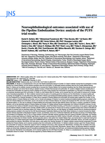
Neuroophthalmological outcomes in the PUFS trialand MRI; an allergy to platinum or cobalt/chromium alloys; evidence of an active infection; or major stenosis ofthe ipsilateral carotid artery. Patients with recurrent intracranial aneurysms after surgical or endovascular therapywere included, except if an indwelling stent was present inthe parent artery. All study participants provided writteninformed consent before study participation.AssessmentsBefore a PED placement, all patients underwent a baseline neurological examination, which included a detailedneuroophthalmological assessment to identify any oculomotor, or VA, and/or VF deficits. The neuroophthalmological examination was performed by neuroophthalmologistsat each study site and included a fundus photograph, VAexamination by Snellen with correction, VF examinationby static perimetry, and a detailed CN examination witha prism, if required. Patients underwent a repeat examination at 180 days after PED placement. All neuroophthalmological data were independently reanalyzed by a singleneuroophthalmologist (M.F.) to ensure that baseline andfollow-up examinations adhered to the study protocol. Thebaseline findings of the neuroophthalmological examination were recorded as normal, abnormal and probably related to the aneurysm, abnormal and probably unrelated tothe aneurysm, or abnormal with unknown relationship tothe aneurysm. Only those neuroophthalmological findingsthat were presumed to be related to the aneurysm were included in the analysis as aneurysm related. At the followup, elements of the neuroophthalmological examinationwere graded as completely resolved (100% improvement),highly improved ( 50% improvement), moderately improved (20%–50% improvement), stable ( 20% improvement), moderately worse (20%–50% worsened), markedlyworse ( 50% worsened), or cannot tell because of equivocal or otherwise unclear examination findings.The procedural efficacy (degree of occlusion) for allaneurysms was assessed at 6 months postprocedure by acore laboratory using a previously published scale (complete occlusion, residual neck, or residual aneurysm).2,13Neuroophthalmological outcome was analyzed as a function of procedural efficacy.Statistical AnalysisCategorical variables were analyzed with chi-square orFisher’s exact tests as appropriate. Continuous variableswere analyzed with Student’s t-test for 2-group comparisons, and 1-way ANOVA with a post hoc Tukey’s test wasused for 3 or more group comparisons.ResultsIn total, 108 patients were treated in the PUFS trial.For several reasons, 10 of these patients were excludedfrom the 180-day neuroophthalmological follow-up (Table1). Therefore, the analysis included 98 patients (91%) forwhom complete follow-up data were available. The demographics and baseline aneurysm characteristics of theincluded patients are shown in Table 2. The average age ofthe patients was 56.4 11.6 years, and 88 of the patients(90%) were female.TABLE 1. Reasons for exclusion of patients from this studyReason for Elimination From AnalysisNo. of Patients (%)DeathDeclined 180-day examNo 180-day examNonparticipatoryNot treated w/ PEDWithdrawal from studyLoss to follow-up3 (3)2 (2)1 (1)1 (1)1 (1)1 (1)1 (1)Thirty-nine patients (40%) had baseline neuroophthalmological deficits at the time of the PED treatment,and several of these patients had multiple deficits. Alsoincluded in Table 2 are the results of a univariate analysisof baseline clinical and aneurysm characteristics that wereassociated with a presentation of a baseline deficit. Thosewith a deficit tended to be older (age 58.0 10.8 years)than those with no baseline deficits (55.3 12.0 years), butthis age difference did not reach statistical significance (p 0.26). We also noted a trend toward statistical significance of the association between aneurysm location andpresentation of symptoms (p 0.058), with a higher proportion of patients with cavernous segment aneurysms exhibiting neuroophthalmological deficits (57%) than thosewith aneurysms of the petrous (0%), superior hypophysial(22%), paraophthalmic (27%), or supraclinoid (25%) segments. The size of the aneurysm (its maximum dimension) was most statistically significantly associated with abaseline deficit (22.0 5.9 mm for patients with deficits vs15.7 5.9 mm for those with no deficits, p 0.001). Domesize (16.8 5.2 mm for patients with deficits vs 13.3 5.5mm for those with no deficits, p 0.003) and, to a lesserextent, neck size (10.0 4.6 mm for those with deficits vs8.1 4.3 mm for those with no deficit, p 0.032) were alsosignificantly associated with baseline deficits.Figure 1 shows the results of PED treatment of a giant, wide-neck paraophthalmic segment aneurysm of theleft ICA, with both angiographically defined vascularexclusion of the aneurysm 6 months after treatment andinvolution of the excluded aneurysm sack. Three sequential transaxial CT images immediately after PED placement (Fig. 1, panels D–F) show contrast layering withinthe giant aneurysm and mass effect on the left optic nerveand inferomedial frontal lobe. Six months after treatment(panels G–I), the aneurysm sac had completely resolved(disappeared on CT). Three frontal oblique angiographicimages show the aneurysm (panel A), the stent construct(immediately after placement in the left ICA) (panel B),and complete angiographically defined occlusion of theaneurysm at the 6-month follow-up (panel C).The relationship between individual baseline deficitsand aneurysm location is shown in Table 3. A statisticallysignificant predominance of cavernous segment aneurysms was detected in patients with symptoms of CN IIIor CN VI palsies.The 6-month outcomes for all patients with neuroophthalmological findings during the course of this study arepictorially summarized in Fig. 2. One patient with a centralJ Neurosurg Volume 123 October 2015899Unauthenticated Downloaded 06/22/22 12:51 PM UTC
D. H. Sahlein et al.TABLE 2. Patient demographics and aneurysm characteristics ordered by baseline neuroophthalmological findingsPatientsVariableAllw/ Baseline Deficit§w/o Baseline Deficit§p ValueNo. of patients in group (%)Female sex (%)Mean age in yrs (SD)Mean BMI (SD)Medical history, no. of pts (%)SAHHypertensionCocaine useSmokingPrevious smokerCurrent smokerCoronary artery diseaseMyocardial infarctionDiabetes mellitusAneurysm side (rt)Aneurysm location, no. of pts (%)PetrousCavernousCarotid caveSup hypophysialLat clinoidalParaophthalmicSupraclinoidPCoAMean aneurysm size in mm (SD)NeckDomeMax dimensionNeck-to-dome ratio98 (100.0)88 (89.8)56.4 (11.6)27.1 (5.3)39 (39.8)35 (89.7)58.0 (10.8)27.0 (5.3)59 (60.2)53 (89.8)55.3 (12.0)27.1 (5.3)NA1.0*0.26†0.94†6 (6.1)52 (53.1)1 (1.0)4 (10.3)24 (61.5)1 (2.6)2 (3.4)28 (47.5)0 (0.0)0.21*0.31‡0.40*0.94‡27 (27.6)29 (29.6)6 (6.1)3 (3.1)7 (7.1)49 (50.0)10 (25.6)12 (30.8)1 (2.6)0 (0.0)4 (10.3)17 (43.6)17 (28.8)17 (28.8)5 (8.5)3 (5.1)3 (5.1)32 (54.2)4 (4.1)42 (42.9)2 (2.0)9 (9.2)2 (2.0)30 (30.6)8 (8.2)1 (1.0)0 (0.0)24 (61.5)1 (2.6)2 (5.1)1 (2.6)8 (20.5)2 (5.1)0 (0.0)4 (6.8)18 (30.5)1 (1.7)7 (11.9)1 (1.7)22 (37.3)6 (10.2)1 (1.7)8.8 (4.5)14.7 (5.6)18.2 (6.6)1.8 (0.7)10.0 (4.6)16.8 (5.2)22.0 (5.9)1.80 (0.56)8.1 (4.3)13.3 (5.5)15.7 (5.9)1.77 3† 0.001†0.79†BMI body mass index; PCoA posterior communicating artery; Pts patients; SAH subarachnoid hemorrhage; Sup superior.* The p value was determined with a 2-sided Fisher exact test.† The p value was determined with an independent samples t-test.‡ The p value was determined with a chi-square test.§ Presumed attributable to the target aneurysm.retinal artery occlusion and resulting VF and VA deficitsand an APD is not included on this chart because that outcome was adjudicated by an independent clinical eventscommittee as being a thromboembolic event. Each row inFig. 2 represents findings from an individual patient. Baseline deficits adjudicated as probably related to the aneurysm are shown in solid colors. Deficits that were not present at baseline but that appeared at 6 months postprocedureare indicated by colored rectangles with horizontal hatching. Also included (and depicted by colored rectangles withvertical hatching) are those baseline deficits that were initially presumed not to be likely related to the aneurysm buthad improved at the 6-month follow-up, thereby suggestingthat the original adjudication was incorrect. Among the 39patients with a baseline deficit, 25 (64%) had a deficit thatshowed at least some improvement. Only 1 patient (2.6%)had a VF deficit that exhibited worsening from baseline at9006 months, which was accompanied by a deterioration inVA. The same patient had improvement of a baseline CNVI palsy.Of the 43 distinct baseline deficits (observed in 25patients) that had improved by the 6-month follow-up,26 (61%) had moderately improved, 8 (19%) had highlyimproved, and 9 (21%) had completely resolved. We observed single new deficits in 5 patients (5%) at 6 monthspostprocedure: 3 moderate CN VI palsies, 1 moderate CNIV palsy, and 1 moderate VA deficit. Three of these newdeficits (2 CN VI palsies and the CN IV palsy) developedin patients who did not have deficits at the baseline presentation (represented in Fig. 2 by a colored rectangle withhorizontal hatching). A new CN VI palsy appeared in apatient who had a preexisting CN III palsy that had moderately improved by the 6-month follow-up. The VA deficit appeared in the patient with the worsening baseline VFJ Neurosurg Volume 123 October 2015Unauthenticated Downloaded 06/22/22 12:51 PM UTC
Neuroophthalmological outcomes in the PUFS trialFIG. 1. Preembolization and postembolization imaging results of PED treatment for a large paraophthalmic segment aneurysm ofthe left ICA. A: Arterial-phase digital subtraction angiogram (frontal projection) showing a large, complex-neck cerebral aneurysmarising from the ophthalmic segment of the left ICA. B: Unsubtracted frontal radiographic image showing the PED deployed withinthe left ICA. C: Arterial-phase digital subtraction angiogram (the same projection as in B) taken at the 6-month follow-up, confirming occlusion of the aneurysm. D–F: Serial transaxial CT scans of the head immediately after treatment, showing contrast-bloodlevel within the fundus of the ophthalmic segment aneurysm (arrows). G–I: Serial transaxial CT scans 6 months after treatment,showing involution of the aneurysm.deficit, but moderately improved CN VI palsy. Six patients(6%) with baseline deficits that were considered not likelyto be related to the aneurysm (1 with a deficit of unclearrelationship to the aneurysm and 5 with deficits that werepresumed unlikely to be related to the aneurysm) showedimprovement in their deficits. Two of these patients hadmoderate improvement in VFs, both ipsilateral and contralateral to the aneurysm; 1 had moderate improvementin ipsilateral VF; 2 showed completely resolved VF deficit—1 with resolution in both ipsilateral and contralateralfields; and 1 exhibited complete resolution of a CN VIpalsy.Overall, 17 patients showed symptoms of a compressive optic neuropathy (that is, findings of VF or VA deficitsor APD), which were presumed to be related to the aneurysm at baseline, with 9 of these patients (53%) showingany improvement in these symptoms. In total, 27 patientspresented with any combination of CN III, IV, and VIpalsies, 17 of whom (63%) showed any improvement insymptoms. Three patients showed symptoms of trigeminal neuralgia, 1 of whom (33%) showed any improvementin these symptoms.Univariate analysis of factors associated with neuroophthalmological improvement after PED treatment indicatedonly 1 characteristic that reached statistical significance.None of the 4 patients with diabetes who presented with abaseline deficit showed an improvement in their deficits (p 0.012 by 2-sided Fisher’s exact test). The maximum dimension of an aneurysm was significantly larger in patientsin whom examinations revealed a new or worse finding at6 months postprocedure than in those in whom findingswere stable or improved: 25.5 4.1 mm for the patientswith worse outcomes versus 17.9 6.5 mm in those withstable or improved outcomes (p 0.013, 2-tailed independent samples t-test). Four of the 5 patients whose examination indicated worsening of their neuroophthalmologicalJ Neurosurg Volume 123 October 2015901Unauthenticated Downloaded 06/22/22 12:51 PM UTC
D. H. Sahlein et al.TABLE 3. Neuroophthalmological findings attributable to the aneurysm stratified by aneurysm neck location*Aneurysm Neck LocationDeficit/CN AffectedPetrousVF—CN III—CN IV—CN V—CN VI—APD—VA—No symptoms attribut4 (100.0)able to the aneurysmNo. of pts w/ aneurysm 4neck at locationCavernous Carotid Cave Sup Hypophysial Lat Clinoidal Paraophthalmic SupraclinoidPCoAp Value0.3100.0010.1200.7600.0040.2400.2100.0586 (14.3)13 (31.0)—3 (7.1)17 (40.5)5 (11.9)1 (2.4)18 (42.9)—1 (50.0)——1 (50.0)——1 (50.0)2 (22.2)————2 (22.2)2 (22.2)7 (77.8)1 (50.0)————1 (50.0)1 (50.0)1 (50.0)6 (20.0)———2 (6.7)6 (20.0)4 (13.3)22 (73)1 (12.5)—1 (12.5)——1 (12.5)1 (12.5)6 (75.0)1 (100.0)1 (100.0)———1 (100.0)—0 (0.0)422923081NANA not applicable; — none.* Values represent the number of patients (%; the percentages indicate proportion of patients with a symptom for a given aneurysm location); note that a given aneurysm can result in 1 deficit, and therefore the number of patients in a given column may exceed the column total.findings had cavernous segment aneurysms. In these patients, the smallest aneurysm was 19.4 mm in maximumdimension and the largest was 30.0 mm.In aggregate, 80 (82%) of the aneurysms treated in thePUFS trial and evaluated at the 6-month follow-up showedangiographic occlusion, 8 (8%) had a neck remnant, 8 (8%)resulted in a residual aneurysm, and 2 (2%) had indeterminate changes with respect to procedure-related efficacy. Ofthe 25 patients with symptomatic aneurysms at the time oftreatment and who clinically improved, 21 (84%) had complete occlusion of their aneurysm at 6 months post–PEDimplantation. In contrast, only 9 aneurysms (64%) in the14 patients whose baseline deficits did not improve wereoccluded at 6 months, but this difference failed to reachstatistical significance (p 0.16, chi-square test).DiscussionTo the best of our knowledge, this study is the first toreport the neuroophthalmological findings from endovascular stent-only reconstruction of large and giant ICAaneurysms. For the purpose of this study, the data in theanalyses reported here were collected from thorough neuroophthalmological examinations both before and 6 monthsafter PED treatment. Even given the relatively brief followup period of 6 months, the results are striking, with 64%of patients experiencing an objective improvement fromtheir baseline deficit after treatment of their aneurysmswith PED.Drawing meaningful comparisons between the results presented here and those reported in the literatureis limited by differences in design, end point definition,and patient cohorts between our study and those of otherauthors. Many of these previous studies have no definedthreshold for reporting that a symptom has changed frombaseline,1,13,15,16 while only 2 define a clear threshold.4,11Likewise, most of the studies examining changes in extraocular motor function nonspecifically include changesin both the objective extraocular motor examination anda patient’s perception of double vision as a broad single902outcome measure,1,4,13,15,16 as opposed to relying purely onthe objective extraocular motor examination.The inclusion of a patient’s subjective experience ofdouble vision in the broad metric of extraocular motorfunction does not account for circumstances in which theperception of double vision may change in the absence ofimproved extraocular motor function. Two of the aforementioned studies provide evidence that patients with noimprovement in oculomotor paresis, or who even haveworsening paresis, may have decreased perception of double vision. Stiebel-Kalish et al.13 note that 2 of their patientshad worsening of misalignment, but their diplopia was unnoticed on a follow-up examination, using a primary endpoint of “resolved, improved, or became unnoticed” forfollowing up patients who initially presented with oculomotor palsies. What the authors did not address explicitlywas the number of patients whose misalignment remainedunchanged on physical examination, but who reported thattheir diplopia had improved or became unnoticed. Anotherstudy reported that 19 patients had baseline diplopia and23 had ophthalmoparesis, clearly suggesting that a reasonable proportion of patients with ophthalmoparesis do notreport double vision.4 The change in the perception of double vision is an interesting subject and is likely dependenton patient age and duration of symptoms, but does little toaddress the question of restoration of CN function following CN decompression.2The aneurysm treatment series most similar to that reported here come from a retrospective study of 17 patientswith large ophthalmic segment aneurysms and symptomsof anterior optic pathway compression treated with endovascular therapy (16 patients were initially treated withendosaccular coils and 1 with arterial occlusion).5 The aneurysms of patients included in this series each had a necksize of at least 4 mm and a dome size of at least 10 mm,which are anatomical thresholds shared by aneurysms inour study. That retrospective study reported that after theprimary procedure, vision improved in 38% of the patients,remained unchange
d. h. sahlein et al. A lthough the risks of mortality or severe neurologi- cal morbidity from large and giant cavernous and supraclinoid internal carotid artery (ICA) aneu-rysms are low,6,7,13 they often are associated with substan- tial neuroophthalmological disorders and facial pain.4,8,13 Mass effects from large and giant aneurysms of the ICA