Transcription
Human Physiology/The gastrointestinal system1Human Physiology/The gastrointestinal system The respiratory system — Human Physiology — Nutrition Homeostasis — Cells — Integumentary — Nervous — Senses — Muscular — Blood — Cardiovascular — Immune — Urinary — Respiratory— Gastrointestinal — Nutrition — Endocrine — Reproduction (male) — Reproduction (female) — Pregnancy — Genetics — Development —AnswersIntroductionWhich organ is the most important organ in the body? Most people would say the heart or the brain, completelyoverlooking the gastrointestinal tract (GI tract). Though definitely not the most attractive organs in the body, theyare certainly among the most important. The 30 foot long tube that goes from the mouth to the anus is responsiblefor the many different body functions which will be reviewed in this chapter. The GI tract is imperative for our wellbeing and our life-long health. A non-functioning or poorly functioning GI tract can be the source of many chronichealth problems that can interfere with your quality of life. In many instances the death of a person begins in theintestines.The old saying "you are what you eat" perhaps would be more accurate if worded "you are what you absorb anddigest". Here we will be looking at the importance of these two functions of the digestive system: digestion andabsorption.The Gastrointestinal System is responsible for the breakdown and absorption of various foods and liquids neededto sustain life. Many different organs have essential roles in the digestion of food, from the mechanical disrupting bythe teeth to the creation of bile (an emulsifier) by the liver. Bile production of the liver plays a important role indigestion: from being stored and concentrated in the gallbladder during fasting stages to being discharged to thesmall intestine.In order to understand the interactions of the different components we shall follow the food on its journey throughthe human body. During digestion, two main processes occur at the same time; Mechanical Digestion: larger pieces of food get broken down into smaller pieces while being prepared forchemical digestion. Mechanical digestion starts in the mouth and continues into the stomach. Chemical Digestion: starts in the mouth and continues into the intestines. Several different enzymes break downmacromolecules into smaller molecules that can be absorbed.The GI tract starts with the mouth and proceeds to the esophagus, stomach, small intestine (duodenum, jejunum,ileum), and then to the large intestine (colon), rectum, and terminates at the anus. You could probably say the humanbody is just like a big donut. The GI tract is the donut hole. We will also be discussing the pancreas and liver, andaccessory organs of the gastrointestinal system that contribute materials to the small intestine.Layers of the GI TractThe GI tract is composed of four layers or also know as Tunics. Each layer has different tissues and functions. Fromthe inside out they are called: mucosa, submucosa, muscularis, and serosa.Mucosa: The mucosa is the absorptive and secretory layer. It is composed of simple epithelium cells and a thinconnective tissue. There are specialized goblet cells that secrete mucus throughout the GI tract located within themucosa. On the mucosa layer there are Villi and Micro Villi.Submucosa: The submucosa is relatively thick, highly vascular, and serves the mucosa. The absorbed elements thatpass through the mucosa are picked up from the blood vessels of the submucosa. The submucosa also has glands andnerve plexuses.
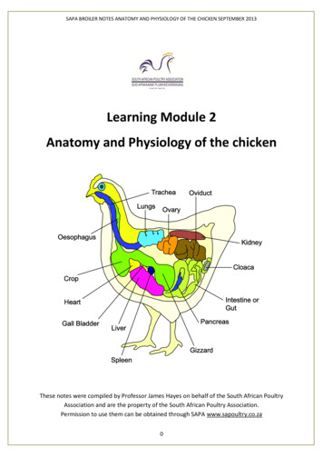
Human Physiology/The gastrointestinal system2Muscularis: The muscularis is responsible for segmental contractions and peristaltic movement in the GI tract. Themuscularis is composed of two layers of muscle: an inner circular and outer longitudinal layer of smooth muscle.These muscles cause food to move and churn with digestive enzymes down the GI tract.Serosa: The last layer is a protective layer. It is composed of avascular connective tissue and simple squamousepithelium. It secretes lubricating serous fluid. This is the visible layer on the outside of the organs.Accessory Organs1.Salivary glands Parotid gland, submandibular gland, sublingual gland Exocrine gland that produces saliva which begins the process ofdigestion with amylase2. Tongue Manipulates food for chewing/swallowing Main taste organ, covered in taste buds3. Teeth For chewing food up4. LiverTeeth, Tongue, and Salivary Glands Produces and excretes bile required for emulsifying fats. Some of the bile drains directly into the duodenum andsome is stored in the gall bladder. Helps metabolize proteins, lipids, and carbohydrates. Urea, chief end product of mammalian metabolism, is formed in liver from amino acids and compounds ofammonia. Breaks down insulin and other hormones. Produces coagulation factors.5. Gallbladder Bile storage.6. Pancreas Exocrine functions: Digestive enzyme secretion. Stores zymogens (inactive enzymes) that will be activated by the brush boarder membrane in the smallintestine when a person eats protein (amino acids). Trypsinogen – Trypsin: digests protein. Chymotypsinogen – Chymotrypsin: digests proteins. Carboxypeptidases: digests proteins. Lipase-lipid: digests fats. Amylase: digests carbohydrates. Endocrine functions: Hormone secretion. Somatostatin: inhibits the function of insulin. Produced if the body is getting too much glucose. Glucagon: stimulates the stored glycogen in the liver to convert to glucose. Produced if the body does not haveenough glucose. Insulin: made in the beta cells of the Islets of Langerhans of the pancreas. Insulin is a hormone that regulatesblood glucose.7. Vermiform appendix
Human Physiology/The gastrointestinal system3 There are a few theories on what the appendix does. Vestigal organ Immune function Helps maintain gut floraThe Digestive SystemThe first step in the digestive system can actuallybegin before the food is even in your mouth. Whenyou smell or see something that you just have to eat,you start to salivate in anticipation of eating, thusbeginning the digestive process.Food is the body's source of fuel. Nutrients in foodgive the body's cells the energy they need to operate.Before food can be used it has to be broken downinto tiny little pieces so it can be absorbed and usedby the body. In humans, proteins need to be brokendown into amino acids, starches into sugars, and fatsinto fatty acids and glycerol.During digestion two main processes occur at thesame time: Mechanical Digestion: larger pieces of food getbroken down into smaller pieces while beingprepared for chemical digestion. Mechanicaldigestion starts in the mouth and continues in tothe stomach. Chemical Digestion: several different enzymesbreak down macromolecules into smallermolecules that can be more efficiently absorbed.Chemical digestion starts with saliva andcontinues into the intestines.The digestive system is made up by the alimentary canal, or the digestive tract, and other abdominal organs that playa part in digestion such as the liver and the pancreas. The alimentary canal is the long tube of organs that runs fromthe mouth (where the food enters) to the anus (where indigestible waste leaves). The organs in the alimentary canalinclude the esophagus, stomach and the intestines. The average adult digestive tract is about thirty feet (30') long.While in the digestive tract the food is really passing through the body rather than being in the body. The smoothmuscles of the tubular digestive organs move the food efficiently along as it is broken down into absorbable atomsand molecules. During absorption, the nutrients that come from food (such as proteins, fats, carbohydrates, vitamins,and minerals) pass through the wall of the small intestine and into the bloodstream and lymph. In this way nutrientscan be distributed throughout the rest of the body. In the large intestine there is reabsorption of water and absorptionof some minerals as feces are formed. The parts of the food that the body passes out through the anus is known asfeces.MasticationDigestion begins in the mouth. A brain reflex triggers the flow of saliva when we see or even think about food.Saliva moistens the food while the teeth chew it up and make it easier to swallow. Amylase, which is the digestiveenzyme found in saliva, starts to break down starch into simpler sugars before the food even leaves the mouth. The
Human Physiology/The gastrointestinal system4nervous pathway involved in salivary excretion requires stimulation of receptors in the mouth, sensory impulses tothe brain stem, and parasympathetic impulses to salivary glands.Swallowing your food happens when the muscles in your tongue and mouth move the food into your pharynx. Thepharynx, which is the passageway for food and air, is about five inches (5") long. A small flap of skin called theepiglottis closes over the pharynx to prevent food from entering the trachea and thus choking. For swallowing tohappen correctly a combination of 25 muscles must all work together at the same time. Salivary glands also producean estimated three liters of saliva per day.EnzymeProduced InSite of ReleasepH LevelCarbohydrate Digestion:Salivary amylaseSalivary glands MouthNeutralPancreatic amylasePancreasSmall intestineBasicMaltaseSmall intestine Small intestineBasicPepsinGastric glandsStomachAcidicTrypsinPancreasSmall intestineBasicPeptidasesSmall intestine Small intestineBasicNucleasePancreasSmall intestineBasicNucleosidasesPancreasSmall intestineBasicPancreasSmall intestineBasicProtein Digestion:Nucleic Acid Digestion:Fat Digestion:Lipase
Human Physiology/The gastrointestinal esophagus) or gullet is themuscular tube in vertebrates through whichingested food passes from the throat to thestomach. The esophagus is continuous withthe laryngeal part of the pharynx at the levelof the C6 vertebra. It connects the pharynx,which is the body cavity that is common toboth the digestive and respiratory systemsbehind the mouth, with the stomach, wherethe second stage of digestion is initiated (thefirst stage is in the mouth with teeth andtongue masticating food and mixing it withsaliva).After passing through the throat, the foodmoves into the esophagus and is pusheddown into the stomach by the process ofperistalsis (involuntary wavelike musclecontractions along the G.I. tract). At the endof the esophagus there is a sphincter thatallows food into the stomach then closesback up so the food cannot travel back up into the esophagus.HistologyThe esophagus is lined with mucus membranes, and uses peristaltic action to move swallowed food down to thestomach.The esophagus is lined by a stratified squamous epithelium, which is rapidly turned over, and serves a protectiveeffect due to the high volume transit of food, saliva, and mucus into the stomach. The lamina propria of theesophagus is sparse. The mucus secreting glands are located in the submucosa, and are connective structures calledpapillae.The muscularis propria of the esophagus consists of striated muscle in the upper third (superior) part of theesophagus. The middle third consists of a combination of smooth muscle and striated muscle, and the bottom(inferior) third is only smooth muscle. The distal end of the esophagus is slightly narrowed because of the thickenedcircular muscles. This part of the esophagus is called the lower esophageal sphincter. This aids in keeping food downand not being regurgitated.The esophagus has a rich lymphatic drainage as well.5
Human Physiology/The gastrointestinal systemStomachThe stomach a thick walled organ that lies between the esophagus and the first part of the small intestine (theduodenum). It is on the left side of the abdominal cavity; the fundus of the stomach lying against the diaphragm.Lying beneath the stomach is the pancreas. The greater omentum hangs from the greater curvature.A mucous membrane lines the stomach which contains glands (with chief cells) that secrete gastric juices, up to threequarts of this digestive fluid is produced daily. The gastric glands begin secreting before food enters the stomach dueto the parasympathetic impulses of the vagus nerve, making the stomach also a storage vat for that acid.The secretion of gastric juices occurs in three phases: cephalic, gastric, and intestinal. The cephalic phase is activatedby the smell and taste of food and swallowing. The gastric phase is activated by the chemical effects of food and thedistension of the stomach. The intestinal phase blocks the effect of the cephalic and gastric phases. Gastric juice alsocontains an enzyme named pepsin, which digests proteins, hydrochloric acid and mucus. Hydrochloric acid causesthe stomach to maintain a pH of about 2, which helps kill off bacteria that comes into the digestive system via food.The gastric juice is highly acidic with a pH of 1-3. It may cause or compound damage to the stomach wall or its layerof mucus, causing a peptic ulcer. On the inside of the stomach there are folds of skin call the gastric rugae. Gastricrugae make the stomach very extendable, especially after a very big meal.The stomach is divided into foursections, each of which has differentcells and functions. The sections are:1) Cardiac region, where the contentsof the esophagus empty into thestomach, 2) Fundus, formed by theupper curvature of the organ, 3) Body,the main central region, and 4) Pylorusor atrium, the lower section of theorgan that facilitates emptying thecontents into the small intestine. Twosmooth muscle valves, or sphincters,keep the contents of the stomachcontained. They are the: 1) Cardiac oresophageal sphincter, dividing the tractabove, and 2) Pyloric sphincter, dividing the stomach from the small intestine.After receiving the bolus(chewed food) the process of peristalsis is started; mixed and churned with gastric juices thebolus is transformed into a semi-liquid substance called chyme. Stomach muscles mix up the food with enzymes andacids to make smaller digestible pieces. The pyloric sphincter, a walnut shaped muscular tube at the stomach outlet,keeps chyme in the stomach until it reaches the right consistency to pass into the small intestine. The food leaves thestomach in small squirts rather than all at once.Water, alcohol, salt, and simple sugars can be absorbed directly through the stomach wall. However, most substancesin our food need a little more digestion and must travel into the intestines before they can be absorbed. When thestomach is empty it is about the size of one fifth of a cup of fluid. When stretched and expanded, it can hold up toeight cups of food after a big meal.Gastric GlandsThere are many different gastric glands and they secret many different chemicals. Parietal cells secrete hydrochloricacid; chief cells secrete pepsinogen; goblet cells secrete mucus; argentaffin cells secrete serotonin and histamine; andG cells secrete the hormone gastrin.Vessels and nerves6
Human Physiology/The gastrointestinal system7Arteries: The arteries supplying the stomach are the left gastric,the right gastric and right gastroepiploic branches of the hepatic,and the left gastroepiploic and short gastric branches of thelineal. They supply the muscular coat, ramify in the submucouscoat, and are finally distributed to the mucous membrane.Capillaries: The arteries break up at the base of the gastrictubules into a plexus of fine capillaries, which run upwardbetween the tubules, anatomizing with each other, and ending ina plexus of larger capillaries, which surround the mouths of thetubes, and also form hexagonal meshes around the ducts.Nerves in the lower abdomen.Veins: From these the veins arise, and pursue a straight coursedownward, between the tubules, to the submucous tissue; theyend either in the lineal and superior mesenteric veins, or directlyin the portal vein.Lymphatics: The lymphatics are numerous: They consist of a superficial and a deep set, and pass to the lymphglands found along the two curvatures of the organ.Nerves: The nerves are the terminal branches of the right and left urethra and other parts, the former beingdistributed upon the back, and the latter upon the front part of the organ. A great number of branches from theceliac plexus of the sympathetic are also distributed to it. Nerve plexuses are found in the submucous coat andbetween the layers of the muscular coat as in the intestine. From these plexuses fibrils are distributed to themuscular tissue and the mucous membrane.Disorders of the StomachDisorders of the stomach are common. There can be a lot of different causes with a variety of symptoms. Thestrength of the inner lining of the stomach needs a careful balance of acid and mucus. If there is not enough mucus inthe stomach, ulcers, abdominal pain, indigestion, heartburn, nausea and vomiting could all be caused by the extraacid.Erosions, ulcers, and tumors can cause bleeding. When blood is in the stomach it starts the digestive process andturns black. When this happens, the person can have black stool or vomit. Some ulcers can bleed very slowly so theperson won't recognize the loss of blood. Over time, the iron in your body will run out, which in turn, will causeanemia.There isn't a known diet to prevent against getting ulcers. A balanced, healthy diet is always recommended. Smokingcan also be a cause of problems in the stomach. Tobacco increases acid production and damages the lining of thestomach. It is not a proven fact that stress alone can cause an ulcer.Histology of the human stomachLike the other parts of the gastrointestinal tract, the stomach walls are made of a number of layers.From the inside to the outside, the first main layer is the mucosa. This consists of an epithelium, the lamina propriaunderneath, and a thin bit of smooth muscle called the muscularis mucosa.The submucosa lies under this and consists of fibrous connective tissue, separating the mucosa from the next layer,the muscularis externa. The muscularis in the stomach differs from that of other GI organs in that it has three layersof muscle instead of two. Under these muscle layers is the adventitia, layers of connective tissue continuous with theomenta.The epithelium of the stomach forms deep pits, called fundic or oxyntic glands. Different types of cells are atdifferent locations down the pits. The cells at the base of these pits are chief cells, responsible for production ofpepsinogen, an inactive precursor of pepsin, which degrades proteins. The secretion of pepsinogen prevents
Human Physiology/The gastrointestinal system8self-digestion of the stomach cells.Further up the pits, parietal cells produce gastric acid and a vital substance, intrinsic factor. The function of gastricacid is two fold 1) it kills most of the bacteria in food, stimulates hunger, and activates pepsinogen into pepsin, and2) denatures the complex protein molecule as a precursor to protein digestion through enzyme action in the stomachand small intestines. Near the top of the pits, closest to the contents of the stomach, there are mucous-producing cellscalled goblet cells that help protect the stomach from self-digestion.The muscularis externa is made up of three layers of smooth muscle. The innermost layer is obliquely-oriented: thisis not seen in other parts of the digestive system: this layer is responsible for creating the motion that churns andphysically breaks down the food. The next layers are the square and then the longitudinal, which are present as inother parts of the GI tract. The pyloric antrum which has thicker skin cells in its walls and performs more forcefulcontractions than the fundus. The pylorus is surrounded by a thick circular muscular wall which is normally tonicallyconstricted forming a functional (if not anatomically discrete) pyloric sphincter, which controls the movement ofchyme.Control of secretion and motilityThe movement and the flow of chemicals into the stomach are controlled by both the nervous system and by thevarious digestive system hormones.The hormone gastrin causes an increase in the secretion of HCL, pepsinogen and intrinsic factor from parietal cellsin the stomach. It also causes increased motility in the stomach. Gastrin is released by G-cells into the stomach. It isinhibited by pH normally less than 4 (high acid), as well as the hormone somatostatin.Cholecystokinin (CCK) has most effect on the gall bladder, but it also decreases gastric emptying. In a different andrare manner, secretin, produced in the small intestine, has most effects on the pancreas, but will also diminish acidsecretion in the stomach.Gastric inhibitory peptide (GIP) and enteroglucagon decrease both gastric motility and secretion of pepsin. Otherthan gastrin, these hormones act to turn off the stomach action. This is in response to food products in the liver andgall bladder, which have not yet been absorbed. The stomach needs only to push food into the small intestine whenthe intestine is not busy. While the intestine is full and still digesting food, the stomach acts as a storage for food.Small IntestineThe small intestine is the site where most of the chemical andmechanical digestion is carried out. Tiny projections called villi linethe small intestine which absorbs digested food into the capillaries.Most of the food absorption takes place in the jejunum and the ileum.The functions of a small intestine is, the digestion of proteins intopeptides and amino acids principally occurs in the stomach but somealso occurs in the small intestine. Peptides are degraded into aminoacids; lipids (fats) are degraded into fatty acids and glycerol; andcarbohydrates are degraded into simple sugars.The three main sections of the small intestine is The Duodenum, TheJejunum, The Ileum.The DuodenumDiagram showing the small intestineIn anatomy of the digestive system, the duodenum is a hollow jointedtube connecting the stomach to the jejunum. It is the first and shortest part of the small intestine. It begins with theduodenal bulb and ends at the ligament of Treitz. The duodenum is almost entirely retro peritoneal. The duodenum isalso where the bile and pancreatic juices enter the intestine.
Human Physiology/The gastrointestinal system9The JejunumThe Jejunum is a part of the small bowel, located between the distal end of duodenum and the proximal part ofileum. The jejunum and the ileum are suspended by an extensive mesentery giving the bowel great mobility withinthe abdomen. The inner surface of the jejunum, its mucous membrane, is covered in projections called villi, whichincrease the surface area of tissue available to absorb nutrients from the gut contents. It is different from the ileumdue to fewer goblet cells and generally lacks Preyer's patches.The IleumIts function is to absorb vitamin B12 and bile salts. The wall itself is made up of folds, each of which has many tinyfinger-like projections known as villi, on its surface. In turn, the epithelial cells which line these villi possess evenlarger numbers of micro villi. The cells that line the ileum contain the protease and carbohydrate enzymesresponsible for the final stages of protein and carbohydrate digestion. These enzymes are present in the cytoplasm ofthe epithelial cells. The villi contain large numbers of capillaries which take the amino acids and glucose producedby digestion to the hepatic portal vein and the liver.The terminal ileum continues to absorb bile salts, and is also crucial in the absorption of fat-soluble vitamins(Vitamin A, D, E and K). For fat-soluble vitamin absorption to occur, bile acids must be present.Large IntestineThe large intestine (colon) extends from the end of the ileum to theanus. It is about 5 feet long, being one-fifth of the whole extent of theintestinal canal. It's caliber is largest at the commencement at thececum, and gradually diminishes as far as the rectum, where there is adilatation of considerable size just above the anal canal. It differs fromthe small intestine in by the greater caliber, more fixed position,sacculated form, and in possessing certain appendages to its externalcoat, the appendices epiploicæ. Further, its longitudinal muscular fibersdo not form a continuous layer around the gut, but are arranged in threelongitudinal bands or tæniæ.The large intestine is divided into the cecum, colon, rectum, and anal canal. In its course, describes an arch whichsurrounds the convolutions of the small intestine. It commences in the right iliac region, in a dilated part, the cecum.It ascends through the right lumbar and hypochondriac regions to the under surface of the liver; here it takes a bend,the right colic flexure, to the left and passes transversely across the abdomen on the confines of the epigastric andumbilical regions, to the left hypochondriac region; it then bends again, the left colic flexure, and descends throughthe left lumbar and iliac regions to the pelvis, where it forms a bend called the sigmoid flexure; from this it iscontinued along the posterior wall of the pelvis to the anus.There are trillions of bacteria, yeasts, and parasites living in our intestines, mostly in the colon. Over 400 species oforganisms live in the colon. Most of these are very helpful to our health, while the minority are harmful. Helpfulorganisms synthesize vitamins, like B12, biotin, and vitamin K. They breakdown toxins and stop proliferation ofharmful organisms. They stimulate the immune system and produce short chain fatty acids (SCFAs) that are requiredfor the health of colon cells and help prevent colon cancer. There are many beneficial bacteria but some of the mostcommon and important are Lactobacillus Acidophilus and various species of Bifidobacterium. These are available as"probiotics" from many sources.
Human Physiology/The gastrointestinal systemPancreas, Liver, and GallbladderThe pancreas, liver, and gallbladder are essential for digestion. The pancreas produces enzymes that help digestproteins, fats, and carbohydrates, the liver produces bile that helps the body absorb fat, and the gallbladder stores thebile until it is needed. The enzymes and bile travel through special channels called ducts and into the small intestinewhere they help break down the food.PancreasThe pancreas is located posterior to the stomach and in close association with the duodenum.In humans, the pancreas is a 6-10 inch elongated organ in the abdomen located retro peritoneal. It is often describedas having three regions: a head, body and tail. The pancreatic head abuts the second part of the duodenum while thetail extends towards the spleen. The pancreatic duct runs the length of the pancreas and empties into the second partof the duodenum at the ampulla of Vater. The common bile duct commonly joins the pancreatic duct at or near thispoint.The pancreas is supplied arterially by the pancreaticoduodenal arteries, themselves branches of the superiormesenteric artery of the hepatic artery (branch of celiac trunk from the abdominal aorta). The superior mesentericartery provides the inferior pancreaticoduodenal arteries while the gastroduodenal artery (one of the terminalbranches of the hepatic artery) provides the superior pancreaticoduodenal artery. Venous drainage is via thepancreatic duodenal veins which end up in the portal vein. The splenic vein passed posterior to the pancreas but issaid to not drain the pancreas itself. The portal vein is formed by the union of the superior mesenteric vein andsplenic vein posterior to the body of the pancreas. In some people (as many as 40%) the inferior mesenteric vein alsojoins with the splenic vein behind the pancreas, in others it simply joins with the superior mesenteric vein instead.The function of the pancreas is to produce enzymes that break down all categories of digestible foods (exocrinepancreas) and secrete hormones that affect carbohydrates metabolism (endocrine pancreas). ExocrineThe pancreas is composed of pancreatic exocrine cells, whose ducts are arranged in clusters called acini (singularacinus). The cells are filled with secretory granules containing the precursor digestive enzymes (mainly trypsinogen,chymotrypsinogen, pancreatic lipase, and amylase) that are secreted into the lumen of the acinus. These granules aretermed zymogen granules (zymogen referring to the inactive precursor enzymes.) It is important to synthesizeinactive enzymes in the pancreas to avoid auto degradation, which can lead to pancreatitis.The pancreas is near the liver, and is the main source of enzymes for digesting fats (lipids) and proteins - theintestinal walls have enzymes that will digest polysaccharides. Pancreatic secretions from ductal cells containbicarbonate ions and are alkaline in order to neutralize the acidic chyme that the stomach churns out. Control of theexocrine function of the pancreas are via the hormone gastrin, cholecystokinin and secretin, which are hormonessecreted by cells in the stomach and duodenum, in response to distension and/or food and which causes secretion ofpancreatic juices.The two major proteases which the pancreas are trypsinogen and chymotrypsinogen. These zymogens are inactivatedforms of trypsin and chymotrypsin. Once released in the intestine, the enzyme enterokinase present in the intestinalmucosa activates trypsinogen by cleaving it to form trypsin. The free trypsin then cleaves the rest of the trypsinogenand chymotrypsinogen to their active forms.Pancreatic secretions accumulate in intralobular ducts that drain the main pancreatic duct, which drains directly intothe duodenum.Due to the importance of its enzyme contents, injuring the pancreas is a very dangerous situation. A puncture of thepancreas tends to require careful medical intervention. EndocrineScattered among the acini are the endocrine cells of the pancreas, in groups called the islets of Langerhans. They are:10
Human Physiology/The gastrointestinal systemInsulin-producing beta cell
Main taste organ, covered in taste buds 3. Teeth For chewing food up 4. Liver Produces and excretes bile required for emulsifying fats. Some of the bile drains directly into the duodenum and some is stored in the gall bladder. Helps metabolize proteins, lipids, and carbohydrates.