Transcription
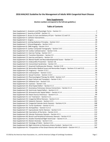
2018 AHA/ACC Guideline for the Management of Adults With Congenital Heart DiseaseData Supplements(Section numbers correspond to the full-text guideline.)Table of ContentsData Supplement 1. Anatomic and Physiologic Terms – Section 2.1 . 5Data Supplement 2. Severity of ACHD – Section 2.2 . 7Data Supplement 3. Access to Care and Delivery of Care – Sections 3.2 and 3.3 . 16Data Supplement 4. Catheter Interventions. 17Data Supplement 5. Surgery . 18Data Supplement 6. Ionizing Radiation Principles – Section 3.4.2 . 20Data Supplement 7. Echocardiography – Section 3.4.3. 21Data Supplement 8. CMR Imaging – Section 3.4.4 . 24Data Supplement 9. Cardiac Computed Tomography – Section 3.4.5 . 28Data Supplement 10. Cardiac Catheterization – Section 3.4.6 . 30Data Supplement 11. Exercise Testing – Section 3.4.7 . 34Data Supplement 12. Transition Education – Section 3.5 . 35Data Supplement 13. Exercise and Sports – Section 3.6 . 36Data Supplement 14. Mental Health and Neurodevelopmental Issues – Section 3.7. 39Data Supplement 15. Endocarditis Prevention – Section 3.8 . 41Data Supplement 16. Concomitant Syndromes – Section 3.9 . 43Data Supplement 17. Acquired Cardiovascular Disease – Section 3.10 . 45Data Supplement 18. Noncardiac Medical Issues and Noncardiac Surgery – Sections 3.11 and 3.12 . 54Data Supplement 19. Pregnancy – Section 3.13.1 . 58Data Supplement 20. Contraception – Section 3.13.2 . 69Data Supplement 21. Sexual Function – Section 3.13.4 . 70Data Supplement 22. Pharmacological Therapy for ACHD – Section 3.17 . 71Data Supplement 23. Heart Failure and Transplant – Section 3.14.2. 76Data Supplement 24. Palliative Care – Section 3.15 . 78Data Supplement 25. Cyanosis – Section 3.16. 78Data Supplement 26. Atrial Septal Defect – Section 4.1.1 . 80Data Supplement 27. Anomalous Pulmonary Venous Connections – Section 4.1.2 . 92Data Supplement 28. Ventricular Septal Defect – Section 4.1.3 . 96Data Supplement 29. Atrioventricular Septal Defect – Section 4.1.4 . 98Data Supplement 30. Patent Ductus Arteriosus – Section 4.1.5 . 99Data Supplement 31. Cor Triatriatum – Section 4.2.1 . 101Data Supplement 32. Congenital Mitral Stenosis – Section 4.2.2 . 101Data Supplement 33. Subaortic Stenosis – Section 4.2.3 . 102Data Supplement 34. Congenital Valvular Aortic Stenosis – Sections 4.2.4 . 108Data Supplement 35. Turner Syndrome – Section 4.2.4.1 . 109Data Supplement 36. Aortopathies – Section 4.2.4.2 . 110Data Supplement 37. Supravalvular Aortic Stenosis – Section 4.2.5 . 112Data Supplement 38. Coarctation of the Aorta – Section 4.2.6 . 116Data Supplement 39. Valvular Pulmonary Stenosis – Section 4.3.1 . 123Data Supplement 40. Branch and Peripheral Pulmonary Stenosis – Section 4.3.2 . 125Data Supplement 41. Double-Chambered Right Ventricular – Section 4.3.3 . 128Data Supplement 42. Ebstein Anomaly – Section 4.3.4. 129Data Supplement 43. Tetralogy of Fallot – Section 4.3.5 . 132Data Supplement 44. Right Ventricle to Pulmonary Artery Conduit – Section 4.3.6 . 147Data Supplement 45. Transposition of the Great Arteries with Atrial Switch – Section 4.4.1.1 . 158 2018 by the American Heart Association, Inc., and the American College of Cardiology Foundation.Page 1
Data Supplement 46. Transposition of the Great Arteries with Arterial Switch – Section 4.4.1.2 . 165Data Supplement 47. Congenitally Corrected Transposition of the Great Arteries – Section 4.4.1.4 . 167Data Supplement 48. Fontan Palliation of Single Ventricle Physiology Including Tricuspid Atresia and Double InletRight Ventricle – Section 4.4.2 . 176Data Supplement 49. Severe Pulmonary Arterial Hypertension – Section 4.4.6.1 . 186Data Supplement 50. Eisenmenger Syndrome – Section 4.4.6.2 . 193Data Supplement 51. Anomalous Coronary Artery Evaluation – Section 4.4.7.1 . 206References . 213 2018 by the American Heart Association, Inc., and the American College of Cardiology Foundation.Page 2
indicates approximately; 1 , primary; 2 , secondary; 2D, 2-dimensional; 3D, 3-dimensional; 4D, 4-dimensional; 6MWT, 6-minute walk test; AAOCA, anomalous aorticorigin of the coronary artery; AAOLCA, anomalous aortic origin of the left coronary artery; AAORCA, anomalous aortic origin of the right coronary artery; ACC, AmericanCollege of Cardiology; ACEI, angiotensin-converting-enzyme inhibitor; ACHD, adult congenital heart disease; AE, adverse events; AF, atrial fibrillation; AHA, AmericanHeart Association; ALCAPA, anomalous left coronary artery arising from the pulmonary artery; ANP, atrial natriuretic peptide; ARB, angiotensin-receptor blocker; AS,aortic stenosis; ASD, atrial septal defect; ASO, arterial switch operation; AT, anaerobic threshold; AVC, atrioventricular canal; AVNRT, atrioventricular nodal reentranttachycardia; AV, atrioventricular; AVS, aortic valve stenosis; AVSD, atrioventricular septal defect; BAV, bicuspid aortic valve; BB, beta blockers; BDCPA, bidirectionalcavopulmonary anastomosis; BMI, body mass index; BNP, brain natriuretic peptide; BP, blood pressure; BREATHE-5, Bosentan Randomized Trial of EndothelinAntagonist Therapy-5; CABG, coronary artery bypass grafting; CAC, coronary artery calcium; CAD, coronary artery disease; CAMPHOR, Cambridge PulmonaryHypertension Outcome Review; CACH, Canadian Adult Congenital Heart; CCT, cardiac computed tomography; CCTGA, congenitally corrected transposition of the greatarteries; CHD, congenital heart disease; CI, confidence interval; CKD, chronic kidney disease; CK-MB, creatine kinase-MB; CMR, cardiac magnetic resonance; CoA,coarctation of the aorta; COR, class of recommendation; CPB, cardiopulmonary bypass; CPET, cardiopulmonary exercise stress test; CRT, cardiac resynchronizationtherapy; CT, computed tomography; CTA, computed tomography angiography; CV, cardiovascular; CVA, cerebrovascular accident; CVD, cardiovascular disease; CXR,chest x-ray; DBP, diastolic blood pressure; DCCV, direct current cardioversion; DCM, dilated cardiomyopathy; DCRV, double-chambered right ventricle; DM, diabetesmellitus; double outlet right ventricle; dp/dt, measurement of the rate of systolic right ventricular pressure increase; d-TGA, dextro-transposition of the great arteries;echo, echocardiography; ECG, electrocardiogram; ED, emergency department; EDA, end diastolic area; EF, ejection fraction; EOL, end-of-life; EP,electrophysiology/electrophysiologic; EPS, electrophysiological studies; ERA, endothelin receptor antagonist; FC, functional class; FDA, Food and Drug Administration;FDR, first-degree relative; GI, gastrointestinal; GUCH, grown up congenital heart; HCT, hematocrit; HD, heart disease; HF, heart failure; HGB, hemoglobin; HLHS,hypoplastic left heart syndrome; HR, hazard ratio; HTN, hypertension; IART, intra-atrial reentrant tachycardia; ICD-9-CM, International Classification of Diseases, NinthRevision, Clinical Modification; ICE, intracardiac echocardiography; ICD, implantable cardioverter-defibrillator; ICU, intensive care unit; IE, infective endocarditis; IQ,intelligence quotient; IQR, interquartile range; IV, intravenous; LDL, low-density lipoprotein; LIMA, left internal mammary artery; LMCA, left main coronary artery; LOE,level of evidence; LOS, length of stay; LV, left ventricular; LVEDD, left ventricular end diastolic diameter; LVEF, left ventricular ejection fraction; LVOT, left ventricularoutflow tract; LVOTO, left ventricular outflow obstruction; MACE, major adverse cardiac events; MCV, mean corpuscular volume; MDCTA, multidetector computedtomographic angiography; MI, myocardial infarction; mPAP, mean pulmonary artery pressure; MR, mitral regurgitation; MRA, magnetic resonanceangiogram/angiography; MRCA, magnetic resonance coronary angiography; MRI, magnetic resonance imaging; MS, mitral stenosis; MSCT, multi-slice computertomography; N/A, not applicable; NHLBI, National Heart, Lung, and Blood Institute; NPV, negative predictive value; NS, not significant; NT-BNP, N-terminal-brainnatriuretic peptide; NT-proANP, N-terminal pro-atrial natriuretic peptide; NT-proBNP, N-terminal-pro-brain natriuretic peptide; NYHA, New York Heart Association;NSVT, nonsustained ventricular tachycardia; NVE, native valve endocarditis; O2, oxygen; OR, odds ratio; PA, pulmonary artery; PAD, patent ductus arteriosus; PAH,pulmonary arterial hypertension; PAP, pulmonary artery pressure; PAPVC, partial anomalous pulmonary venous connection; PAPVR, partial anomalous pulmonaryvenous drainage; PASP, pulmonary artery systolic pressure; PCI, percutaneous coronary intervention; PDA, patent ductus arteriosus; PDE-5, phosphodiesterase type-5;PFO, patent foramen ovale; PH, pulmonary hypertension; PLE, protein losing enteropathy; PPVI, percutaneous pulmonary valve implant; PR, pulmonary regurgitation;PS, pulmonary stenosis; PVR, pulmonary vascular resistance; pt, patient; PV, pulmonary valve; PVE, prosthetic valve endocarditis; QOL, quality of life; Qpeff, effectivepulmonary blood flow; Qpi, pulmonary blood flow index; Qp:Qs, pulmonary to systemic flow ratio; RACHS-1, Risk Adjusted Classification for Congenital Heart Surgery;RCT, randomized controlled trial; RT3DE real-time 3-dimensional echocardiography; RV, right ventricular; RVEDV, right ventricular end diastolic volume; RVEF, rightventricular ejection fraction; RVESV, right ventricular end systolic volume; RVOT, right ventricular outflow tract; RVOTO; right ventricular outflow tract obstruction; RVPA, right ventricular to pulmonary artery; Rx, medical prescription; SBP, systolic blood pressure; SCD, sudden cardiac death; SD, standard deviation; SEM, standard errorof the mean; SF-12, Short Form Health Survey; s/p, status post; SND, sinus node dysfunction; SPAP, systolic pulmonary artery pressure; SpO2, oxygen saturation; STAT,Stending in Aneurysm Treatments Trial; STS, Society of Thoracic Surgeons; SVA, supraventricular arrhythmia; SVC, superior vena cava; SVG, saphenous vein graft; SVT,supraventricular tachycardia; TA, tricuspid annulus; TEE, transesophageal echocardiography; TGA, transposition of the great arteries; TIA, transient ischemic attack; 2018 by the American Heart Association, Inc., and the American College of Cardiology Foundation.Page 3
TOF, tetralogy of Fallot; TOF/PA, tetralogy of Fallot with pulmonary atresia; TPVR, transcatheter pulmonary valve replacement; TR, tricuspid regurgitation; TTE,transthoracic echocardiogram; TV, tricuspid valve; TVR, tricuspid valve replacement; UK, United Kingdom; UL, upper limbs; UNOS, United Network for Organ Sharing;U.S., United States; VA, ventricular arrhythmias; VAD, ventricular assist device; VF, ventricular fibrillation; VHD, valvular heart disease; VO2, oxygen consumption; VSD,ventricular septal defect; VT, ventricular tachycardia; VTE, venous thromboembolism; WHO, World Health Organization; WPW, Wolf-Parkinson-White; and WU, Woodunits. 2018 by the American Heart Association, Inc., and the American College of Cardiology Foundation.Page 4
Data Supplement 1. Anatomic and Physiologic Terms – Section 2.1StudyName, Author,YearAfilalo J, et al.2011 (1)21939837Study Type/DesignStudy m1 EndpointResults/p ValuesObservational,case control,cohort,retrospectiven 3,239 pts;Quebec CHDDatabaseInclusion criteria: Pts 65 y, 1990–2005,ACHD, Quebec,healthcare interactionBethesda 32(severe, shunt,valvular)Mortality NSTutarel O, et al.2014 (2)23882067Observational,cohort,retrospectiven 375 ptsInclusion criteria: Pts 60 y, ACHD January2000–March 2012Bethesda 32Prevalence,demographics,interventions,mortality Mortality: standardizedmortality ratio simple 0.6(p 0.06), moderate 1.89(p 0.002), complex 2.91(p 0.01); overall 1.82. (p 0.05 univariate, notmultivariate as HF, FC,ventricular function tookover).Kim YY, et al.2011 (3)22010202Observational,case control,retrospectivedatabasen 3,061 ptsInclusion criteria:18–49 y, 2000–2008,ACHD surgery inpediatric hospital, totalcompared to pediatricsurgical pts, ACHDhigh resourceutilization vs. nonhighresource utilizationRACHS-1Resourceutilization, cost,LOS OR: 3–30 for increasingRACHS classification (muchtighter RACHS 3 and 4 ascompared to 2).Kim YY, et al.2011 (4)21693722Observational,case control,retrospectiven 3,061 pts PublicHealth InformationSystem databaseRACHS-1Mortality OR: 2–22 (NS RACHS 2,significant for RACHS 3 and4).Opotowsky AR,et al.2012 (5)Observational,case control,retrospectiven 622,000 ptsInclusion criteria:18–49 y, 2000–2008,ACHD surgery inpediatric hospital, totalcompared to pediatricsurgical pts, ACHDmortality vs.nonmoralityInclusion criteria: Ptsin 1998–2007Bethesda 32Combined:death, HF,arrhythmia, CVA, Severe: OR combined:2.0–2.7; HF 3.9–6.0; 2018 by the American Heart Association, Inc., and the American College of Cardiology Foundation.Summary/Conclusions A rare study that dealswith older adults withCHD. Administrativedatabase has limitations. The study showsincrease in number ofpts 60 y for follow-up atACHD centers. Acquired comorbidityin ACHD pts determinesoutcomes. Administrativedatabase limited. The number of adultsundergoing congenitalheart surgery isincreasing. High resourceutilization admissionsassociated with higherdeath rates. Pt death aftercongenital heart surgeryis lower in pediatrichospitals with highcongenital heart surgeryvolume. Maternal CHD islinked with higher ratesof CV events and death.Page 5
21990383nationwide inpatientsampleembolic events,“unclassified CVevents,” LOS,costs 3 y gap in CVcarearrhythmia 2, LOS 1.9, costs1.5–2.0. 59%, 42%, 26% gap incare (simple, moderate,complex); p 0.0001. The study provides abasis for improvingstrategies for dealingwith the barriers to care. Increased care for severein specialty ACHD centers( 0.001); increased mortalityfor severe ACHD (OR:1.93). Study promotesspecialized care for thepts. p 0.05; NS. It is critical to identifyhigh-risk HF pts. Association: VSD outlierOR: 1.54 vs. MI: 1.8 Death: VSD outlier OR:3.2 similar to MI; TOF andTGA suggesting severeBethesda categorization. Rise 0.0001 (complex vs.simple, not seen forunclassified), comorbidities:0.006 (complex), LOSlonger complex ( 0.001),costs greater simple( 0.0003) (unclassifiedshorter and cheaper). From beginning to end ofstudy period, admissions forsimple defects changed112% unclassified 52% andcomplex 52%. Adultadmissions increased from Mortality risk isincreased with thepresence of VSDs andcertain comorbidities.n 922 ptsInclusion criteria: Pts 18 y, firstpresentation to ACHDclinicBethesda 32Mylotte D, et al.2014 prospectivequestionnaireCase-control,cohort, and timeseries analysesn 71,467 pts fromQuebec CHDRegistry; nN 2,092ACHD death casesInclusion criteria: Ptswith ACHD,18–65 yfrom 1983–2005Bethesda 32(severe vs.other)Zomer AC, et al.2013 (8)23602867Observational,case control,retrospectiven 274 pts fromCONCOR registryRight heartlesionRodriguez FH,et al.2013 (9)23164196Observational,case control,retrospectiven 17,193 HF pts ofN 84,308 ACHDadmissions fromnationwide inpatientsampleInclusion criteria:Adult pts 1995–2007with at least 1 HF 1 or2 ACHD admissionInclusion criteria:Adult, 2207, ACHDadmission vs. 1 HF1 or 2 coding inACHD admissionPts in specializedcare had lowermortality; mostlydriven by themost complexCHD groupAssociation withHF mortalityIndividualdiagnosesAssociation withHF, deathOpotowsky AR,et al.2009 (10)19628123Observational,case control,retrospectiven 35,000–72,450ACHDhospitalizations/yInclusion criteria:US, ACHD, 18 yadmit to acute carehospitalBethesda 32Rise inadmissions,mortality,comorbidity,LOS, costsO’Leary JM, etal.2013 (11)23471480Observational,case control,retrospectiven 950,00 adultACHD and1,900,000 pediatricCHD pt admissionsfrom nationwideinpatient sampleInclusion criteria: USpts with ACHD, 18 y,admit to acute carehospitalBethesda 32Rise inadmissionsGurvitz MZ, etal.2013 (6)23542112 2018 by the American Heart Association, Inc., and the American College of Cardiology Foundation. Number of hospitaladmissions and costshave risen. Adults account for anincreasing proportion ofCHD admissions from1998–2010.Page 6
Marelli AJ, et al.2014 (12)24944314ObservationalQuebec CHDRegistryInclusion criteria:1983–2000, Quebec, 18 y, ACHD healthinteraction, 107,559Data Supplement 2. Severity of ACHD – Section 2.2StudyName, Author,YearKoyak Z, et al.2013 (13)24071387Bouchardy J, etal.2009 (14)19822808StudyType/DesignStudy SizeRetrospective,multicentern 419 pts(mean 38 y 14 y)Populationbasedretrospectiven 5,812 ptswith atrialarrhythmias(38,428 ptswith CHD)Inclusion/ExclusionCriteriaInclusioncriteria: 18 ywho underwentCHD cardiacsurgery betweenJan 2009–Dec2011Inclusioncriteria: Adultswith CHDbetween 1983–2005 found in theQuebecadministrativedatabaseBethesda 32(severe vs.other)Lifetime andpoint prevalenceof CHD.28.9%–36.5% of CHDadmission. In 2010, adults represent60% (57%, 62%) of allsevere CHD vs. 49% (47%,51%) in 2000; prevalencesevere lesions increased55% (51%, 62%) in adultsvs. 19% (17%, 21%)increase in children. There is a need forlongitudinal data sourcesin U.S.1 EndpointResults/p ValuesSummary/ConclusionsPrevalence ofpostop in-hospitalarrhythmias andclinical outcomes Arrhythmias occurred in 134 pts (32%) and includedSVT (n 100), bradycardias (n 47), and VT (n 19). Inmultivariate analysis age 40 y at surgery (p 0.03),NYHA class II (p 0.09), significant subpulmonary AVvalve regurgitation (p 0.018), coronary bypass time,(p 0.019), and CK-MB (p 0.021) were associated within-hospital arrhythmias. Overall, 58 clinical eventsoccurred in 55 pts (13%) and included in the majority ofthe cases permanent pacemaker implantation (5%), HF(4%), and death (2%). In-hospital arrhythmias were independentlyassociated with clinical events; p 0.01. Overall, the 20-y risk of developing atrial arrhythmiawas 7% in a 20-y subject and 38% in a 50-y subject. 50% of pts with severe CHD reaching 18 y developedatrial arrhythmias by 65 y. In pts with CHD, the HR ofany AE in those with atrial arrhythmia compared withthose without was 2.50 (95% CI: 2.38–2.62; p 0.0001),with a near 50% increase in mortality (HR: 1.47; 95%CI: 1.37–1.58; p 0.001), more than double the risk ofmorbidity (stroke or HF) (HR: 2.21; 95% CI: 2.07–2.36;p 0.001), and 3 times the risk of cardiac interventions(HR: 3.00; 95% CI: 2.81–3.20; p 0.001). Arrhythmias are highlyprevalent after congenitalheart surgery in adults andare associated with worseclinical outcome. Older and symptomaticpts with significant VHD atbaseline are at risk of inhospital arrhythmias.Prevalence,lifetime risk,mortality, andmorbidityassociated withatrial arrhythmia inadults with CHD 2018 by the American Heart Association, Inc., and the American College of Cardiology Foundation. Atrial arrhythmiasoccurred in 15% of pts withCHD. Lifetime incidenceincreased steadily with ageand was associated with adoubling of the risk of AE. An increase in resourceallocation should beanticipated to deal with thisincreasing burden.Page 7
Koyak Z, et al.2012 (15)22991410Multicentercase controlstudyn 1,189Inclusioncriteria: Pts 18y with CHD s/pSCD presumedarrhythmic vs. 2cases of ACHDpts without SCDDetermine theadult CHDpopulation at riskof SCD and theclinical parametersassociated withSCDYap SC, et al.2011 (16)21684512Retrospectivedatabasereviewn 378 adultpts with CHD(mean 39 13y) and atrialarrhythmiawho had serialfollow-up in atertiary referralcenter from1999–2009.Inclusioncriteria: Adult ptswith CHD andatrial arrhythmiawho werefollowed seriallyfrom Jan 1999–Dec 2009 in theTorontoCongenitalCardiac Centrefor AdultsIdentify predictorsof mortality in adultwith CHD andatrial arrhythmiaand to establish arisk scoreGiannakoulasG, et al.2012 (17)21196055Retrospectivedatabasereviewn 98 pts,mean 31.5 8.9 y, 43.8%male, 31.6%with a totalcavopulmonary connectionwere identifiedInclusioncriteria: Pts withprevious Fontanoperationfollowed at ourinstitution since1999 wereidentified fromthe electronicdatabase andincluded in thisstudyImpact of atrialarrhythmia onadult Fontan ptsCollins RT, etal.2013 (18)24102747Retrospectivedatabasereviewn 803 ptswith1,333hospitaladmissions ofInclusioncriteria: Pts 18y of with singleventricle admittedIdentify the impactof cardiacarrhythmias onhospitalizations in Arrhythmic death occurred in 171 of 1,189 pts.Underlying cardiac lesions were mild, moderate, andsevere CHD in 12%, 33%, and 55% of the SCD cases,respectively. Clinical variables associated with SCDwere SVT (p 0.004), moderate-to-severe systemicventricular dysfunction (p 0.034), moderate-to-severesubpulmonary ventricular dysfunction (p 0.030),increased QRS duration (p 0.008), and QT dispersion(p 0.008). During a median follow-up of 5.2 y, there were 40deaths (11%). Overall mortality rate was 2.0%/pt-y.Common modes of death included HF–related death(35%), SCD (20%), and perioperative death (18%). Independent predictors of mortality were poor FC,(p 0.001), single ventricle physiology, (p 0.003), PH(p 0.004), and VHD, (p 0.006). A risk score was constructed using these predictorsin which pts were assigned 1 point for the presence ofeach risk factor. Mortality rates in the low-risk (no riskfactor), moderate-risk (1 risk factor), and high-risk ( 1risk factor) groups were 0.5%, 1.9%, and 6.5%/pt y,respectively (log-rank p 0.001). Atrial tachyarrhythmia was present at baseline in60.2% of pts who were older (33.0 8.3 vs. 29.1 9.4y; p 0.002), less likely to have a total cavopulmonaryconnection (13.5% vs. 58.9%; p 0.001), and moresymptomatic in terms of NYHA class (51.9% vs.26.7%; p 0.007) compared to arrhythmia-free pts. During a median follow-up of 6.7 y, 18 pts died and64 pts were hospitalized. Even after adjustment forbaseline clinical characteristics, atrial tachyarrhythmiawas an independent predictor of death (propensityscore adjusted HR: 9.35; 95% CI: 1.10–79.18; p 0.04;c-statistic 0.88) and composite of death orhospitalization (propensity score adjusted HR: 5.00;95% CI: 2.47–10.09; p 0.0001). There were 642 admissions in 424 pts with singleventricle CHD and an arrhythmia diagnosis. A singlearrhythmia diagnosis was present in 454 admissions(71%). 2018 by the American Heart Association, Inc., and the American College of Cardiology Foundation. Clinical parametersfound to be associated withSCD in adults with a broadspectrum of CHD, includingSRV, are similar to those inischemic HD. Pts with mild cardiaclesions are potentially atrisk for SCD. Specific clinical variablesidentify pts with CHD athigh risk for death. Absence of these riskfactors is associated withan excellent survivaldespite the presence ofatrial arrhythmias. In adult pts with aFontan-type operation, thepresence of atrialtachyarrhythmias isassociated with highermorbidity and mortality atmidterm follow-up. In adults with singleventricle CHD, arrhythmiasare affected by singleventricle anatomic subtypePage 8
TA, HLHS, orCV from 43participatinghospitalsto 1 of 43pediatrichospitalsbetween 2004–2011adults with singleventricle CHDDiller GP, et al.2010 (19)20929979Retrospectivedatabasereviewn 321 Fontanpts (57% male,mean age 20.9 8.6 y)Inclusioncriteria: Fontanpts whounderwent CPETat 4 majorEuropeancenters between1997–2008 wereincludedTo clarify ifexerciseintolerance isassociated withpoor outcome inFontan pts and toidentify risk factorsfor mortality,transplantation,and cardiacrelatedhospitalization.Shamszad P, etal.2014 (20)23682693Retrospectivereviewn 3,800,964hospitalizationsInclusioncriteria: All indexcases of pts 30y hospitalizedwith aorticdissection Jan2004–June 2011To describe theincidence,characteristics,and outcomes ofhospitalizedchildren andyoung adults withAD Total hospital charges were 80.7 million with meancharge per admission of 127,296 243,094. Themean charge per hospital was 16,653 17,516 andincreased across the study period (p 0.01). Arrhythmia distributions were impacted by singleventricle anatomic subtype (p 0.001). Hospital resource utilization was significantlydifferent among arrhythmia groups (p 0.001). During a median follow-up of 21 mo, 22 pts died and6 pts underwent cardiac transplantation (8.7%),resulting in an estimated 5 y transplant-free survival of86%. Parameters of CPET were strongly related toincreased risk of hospitalization, but with the exceptionof heart rate reserve-unrelated to risk of death ortransplantation. In contrast, pts with clinically relevantarrhythmia had a 6.0–fold increased risk of death ortransplantation (p 0.001). Furthermore, pts withatriopulmonary/ventricular Fontan had a 3.7-foldincreased risk of death or transplantation comparedwith total cavopulmonary connection pts (p 0.009). The combination of clinically relevant arrhythmia,atriopulmonary/ventricular Fontan, and signs ofsymptomatic or decompensated HF was associatedwith a particularly poor outcome (3 y mortality 25%). AD was identified in 124 ( 1%), accounting for 110pts (69% male; p 0.003) at a median age of 12.9 y(IQR 3.9–16.8 y) with a bimodal distribution in infancyand late adolescence. Associated diagnoses included CHD (38%), trauma(24%), connective tissue disease (16%), and isolatedHTN (8%). Common CHD diagnoses included aortic arch (24%)and valve (21%) disease, HLHS (10%), and TGA(10%). CHD pts were younger and more likely to undergoinpatient non-AD–related CV procedures comparedwith other diagnostic groups (p 0.001 for both). Marfan 2018 by the American Heart Association, Inc., and the American College of Cardiology Foundation.and impact adversely uponhospital resourceutilization. Most parameters ofCPET are associated withincreased risk ofhospitalization but notdeath or transplantation incontemporary Fontan pts. Only decreased heartrate reserve and a historyof clinically n, and/or HFrequiring diuretic therapyare associated with poorprognosis, potentiallyidentifying pts requiringm
2018 AHA/ACC Guideline for the Management of Adults With Congenital Heart Disease . Data Supplements (Section numbers correspond to the full-text guideline.) Table of Contents . . echo, echocardiography; ECG, electrocardiogram; ED, emergency department; EDA, end diastolic area; EF, ejection fraction; EOL, end-of-life; EP, .
