Transcription
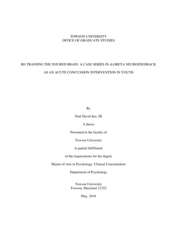
TOWSON UNIVERSITYOFFICE OF GRADUATE STUDIESRE-TRAINING THE INJURED BRAIN: A CASE SERIES IN sLORETA NEUROFEEDBACKAS AN ACUTE CONCUSSION INTERVENTION IN YOUTHByPaul David Ims, IIIA thesisPresented to the faculty ofTowson Universityin partial fulfillmentof the requirements for the degreeMaster of Arts in Psychology: Clinical ConcentrationDepartment of PsychologyTowson UniversityTowson, Maryland 21252May, 2018
Ims – Neurofeedback & ConcussionTOWSON UNIVERSITYOFFICE OF GRADUATE STUDIESTHESIS APPROVAL PAGEThis is to certify that the thesis prepared by Paul David Ims, III entitled “Re-training the InjuredBrain: A Case Series in sLORETA Neurofeedback as an Acute Concussion Intervention inYouth” has been approved by the thesis committee as satisfactorily completing the thesisrequirements for the degree Master of Arts in Psychology: Clinical Concentration.ii
Ims – Neurofeedback & ConcussionAcknowledgementsThis endeavor was supported by Chesapeake Neurology Associates and Harry Kerasidis,M.D., medical director of Chesapeake Neurology Associates, whose mentorship and support wasintegral to the completion of this project.iii
Ims – Neurofeedback & ConcussionAbstractRE-TRAINING THE INJURED BRAIN: A CASE SERIES IN sLORETA NEUROFEEDBACKAS AN ACUTE CONCUSSION INTERVENTION IN YOUTHPaul David Ims, IIIConcussion incidence rates are at epidemiological levels and rising (Giza & Hovda, 2001).Concussion symptoms caused by underlying cortical deregulation and functionaldisturbances (McCrory et al., 2017), which can be measured by quantitative EEG (Rapp etal., 2015), interrupt the daily functioning of the injured person. The principle investigatorhypothesized that neurofeedback would reduce symptoms and improve recovery timecompared to rest and recovery alone. Concussed youth completed a series of QEEG-guidedneurofeedback sessions. QEEG measurements, cognitive scores, and symptoms weretracked from injury to recovery and compared to individuals not receiving neurofeedback.This pilot study revealed a potential non-invasive treatment option for concussion whichwarrants further study.iv
Ims – Neurofeedback & ConcussionTable of ContentsList of Tables . viiList of Figures . viiiIntroduction .1Psychology and Brain Injury .1Concussion Defined .3Mechanism of Concussion Injury .5Concussion in the Developing Brain .6Current Approaches for Concussion Recovery and Rehabilitation .7Physical Rest and Graduated Return-To-Play Protocol .9Cognitive Rest and Return-To-School Protocol.10The Electroencephalogram .11De-Artifacting the EEG .15Quantitative EEG .18QEEG Database Parameters .22Standardized Low Resolution Electromagnetic Tomography .25Neurofeedback: EEG Biofeedback .31Neurofeedback Protocol Design .35Neurofeedback Session Structure .36QEEG, Neurofeedback, and Brain Injury .38Hypothesis.41Materials & Methods .42v
Ims – Neurofeedback & ConcussionSubject Population and Clinical Care . 42QEEG Data Collection and Analysis . 44Cognitive Testing . 46Neurofeedback Sessions . 50Results .51Case Study #1 – JR . 51Case Study #2 – RH . 62Case Study #3 – BN . 83Control Group . 108Discussion.114Elevated Delta and Theta After Injury . 114Recovery Indicated by Normaliation of Z-scores and Reduction in Affected Grey Matter Volume. 116Neurofeedback Intervention Contributed to Reduce Concussion Related Symptoms . 118Reduced Number of Abnormalities in Subjects Treated with Neurofeedback . 120Length of Recovery Time for Individuals Treated with Neurofeedback . 122Limitations .124Conclusions .126Appendix – IRB Approval .130References .131vi
Ims – Neurofeedback & ConcussionList of TablesTable 1 – Graduated Return-to-Play Protocol provided in McCrory et al. (2017) .9Table 2 – Return-to-School Protocol (McCrory et al., 2017) .11Table JR.1 – Results from JR’s WebNeuro Focus Test.56Table JR.2 – QEEG sLORETA images for Case JR after concussion and recovery .58Table RH.1 – QEEG sLORETA images for Case RH after concussion and recovery .80Table BN.1 – QEEG sLORETA images for Case BN after concussion and recovery .104Table 3 – Paired t-test results for PZV of control group.109Table 4 – Paired t-test results for control group percent changed grey matter volume .110Table 5 – Control group change scores.114Table 6 – JR change scores .121Table 7 – RH change scores.121Table 8 – BN change scores.122Table 9 – Summary of results for each case per hypothesis .123vii
Ims – Neurofeedback & ConcussionList of FiguresFigure 1 – Eye blink artifact during an eyes open EEG recording .17Figure 2 – Sample BrainDx QEEG Topographic Map .20Figure 3a – Sample BrainDx sLORETA imaging of delta activity .27Figure 3b – Sample BrainDx sLORETA imaging of delta activity .28Figure 3c – Sample QEEG Pro sLORETA imaging of delta activity.28Figure 3d – Sample QEEG Pro sLORETA imgaging of delta activity .29Figure 4 – sLORETA depiction of delta activity after concussion and after recovery.40Figure 5 – Sample XLNTbrain Cognitive Test Results .46Figure JR.1 – Post-Concussion sLORETA Imaging – Bamma Activity .53Figure JR.2 – Post-Recovery sLORETA Imaging – Bamma Activity .59Graph JR.1 – JR PZVmax after concussion and recovery .60Graph JR.2 – JR PZVmin after concussion and recovery .60Graph JR.3 – JR PIGMV after concussion and recovery .61Graph JR.4 – JR PRGMV after concussion and recovery .61Figure RH.1 – RH XLNTbrain cognitive test scores and symptoms after injury.63Figure RH.2 – Post-Concussion sLORETA Imaging – Delta Activity .64Figure RH.3 – Post-Concussion sLORETA Imaging – Alpha Activity .65Figure RH.4 – Post-Concussion sLORETA Imaging – Bamma Activity .66Figure RH.5 – Delta activity imaged at 2.5 Z-scores .68Figure RH.6 – Delta activity imaged at 2.0 Z-scores .68Figure RH.7 – Theta activity imaged at 2.5 Z-scores .69Figure RH.8 – XLNTbrain symptom tracker for Case RH .70Figure RH.9 – Post recovery cognitive test results and symptoms.71Figure RH.10 – Post-Recovery sLORETA Imaging – Delta Activity.72viii
Ims – Neurofeedback & ConcussionFigure RH.11 – Post-Recovery sLORETA Imaging – Alpha Activity .73Figure RH.12 – Post-Recovery sLORETA Imaging – Alpha Activity .74Figure RH.13 – Post-Recovery sLORETA Imaging – Beta Activity .75Figure RH.14 – Post-Recovery sLORETA Imaging – Bamma Activity .76Graph RH.1 – RH PZVmax after concussion and recovery .81Graph RH.2 – RH PZVmin after concussion and recovery .81Graph RH.3 – RH PIGMV after concussion and recovery .82Graph RH.4 – RH PRGMV after concussion and recovery .82Figure BN.1 – BN XLNTbrain cognitive test scores and symptoms after injury.84Figure RH.2 – BN Post-Concussion sLORETA Imaging – Delta Activity.85Figure RH.3 – BN Post-Concussion sLORETA Imaging – Theta Activity .86Figure RH.4 – BN Post-Concussion sLORETA Imaging – Alpha Activity.87Figure RH.5 – BN Post-Concussion sLORETA Imaging – Alpha Activity.88Figure RH.6 – BN Post-Concussion sLORETA Imaging – Beta Activity .89Figure RH.7 – BN Post-Concussion sLORETA Imaging – Bamma Activity .90Figure RH.8 – BN Post-Neurofeedback sLORETA Imaging – Delta Activity .94Figure RH.9 – BN Post-Neurofeedback sLORETA Imaging – Alpha Activity .95Figure RH.10 – BN Post-Neurofeedback sLORETA Imaging – Alpha Activity .96Figure RH.11 – BN Post-Neurofeedback sLORETA Imaging – Beta Activity .97Figure RH.12 – BN Post-Neurofeedback sLORETA Imaging – Bamma Activity .98Figure RH.13 – BN XLNTbrain cognitive testing collected after recovery .99Figure RH.13 – Symptom tracking during BN’s recovery .103Graph BN.1 – BN PZVmax after concussion and recovery .106Graph BN.2 – BN PZVmin after concussion and recovery .106Graph BN.3 – BN PIGMV after concussion and recovery .107Graph BN.4 – BN PRGMV after concussion and recovery .107Graph C.1 – Control group mean PZVmax after concussion and recovery .111Graph C.2 – Control group mean PZVmin after concussion and recovery .111Graph C.3 – Control group mean PIGMV after concussion and recovery .112Graph C.4 – Control group mean PRGMV after concussion and recovery.112ix
Ims – Neurofeedback & Concussion1INTRODUCTIONIn 2011, the Center for Disease Control (CDC) estimated 1.6 - 3.8 million sport- orrecreation- related concussions occur per year (Daneshvar et al., 2011). Concussion rates areincreasing in the adolescent population (Zhang et al., 2016). Current concussion recoveryguidelines emphasize the reduction of symptom-provoking behaviors promote recovery(McCrory et al., 2017). Electrophysiological assessment after concussion injury demonstratemeasurable differences in cortical functioning (Rapp et al., 2015). Dysfunctional patterns ofbrain activity related to concussion injury persist beyond concussion recovery defined by currentevidence-based guidelines (Kerasidis & Ims, 2017). Neurofeedback is the operant conditioningof brain waves as measured by the electroencephalogram (EEG) to promote neuroplasticity andself-regulation of brain activity (Collura, 2014), and stands to be investigated as an interventionfor acute sport- and recreation-related concussion in adolescents and children.The paper will begin by providing an operant definition of concussion injury, descriptionof the mechanism of concussion injury, and current evidence-based concussion managementpractices. Electroencephalogram (EEG) and quantitative EEG analysis techniques and metrics,including the relationship between brain activity and concussion, will be outlined, followed byan explanation of neurofeedback approaches and procedures. Subsequently, the hypothesis,study methods, and results will be presented for each case study. Finally, implications of theresults will be discussed, limitations outlined, and conclusions drawn.Psychology and Brain InjuryAs the brain is the physical medium to mental processing, impairment toneurophysiology exerts an effect on psychological functioning. Concussion presents acute and
Ims – Neurofeedback & Concussion2chronic mental health challenges (Broglio et al., 2016). In the days or weeks following injury,individuals with severe post-concussion symptoms are advised to rest and reduce cognitive andphysical activity that provokes symptoms (McCrory et al., 2017). Functional disturbances due toinjury in brain regions associated with emotional regulation may be directly responsible forchanges in mood or emotional lability. Unable to attend school or other social events,withdrawal from society for extensive time during recovery may contribute to depression(Broglio et al., 2016). For student athletes with intense academic workloads, anxiety mayincrease as time out of the classroom as past-due assignments build. Insomnia secondary toanxiety may impede sleep important to recovery, and concussion cases with sleep disturbancesymptoms further complicate efforts to rest. With make-up schoolwork mounting andinterruptions in activities of daily living, feelings of hopelessness may creep into the mind ofconcussed youth. In the PI’s clinical experiences, conversations with injured patients ofteninclude descriptions of “feeling crazy” because “life is out of control” and “wondering how toget back to normal.”The long-term effects of concussion may be seen in a host of psychological disorders.Personality, sleep, cognitive ability may be negatively influenced by brain injury, increasing therisk for suicide, PTSD, depression, and anxiety (Peskind et al., 2013). This comorbidity betweenbrain injury and mental illness is compounded by the fact that many concussions areunderreported, a symptom of the culture surrounding brain injury in sport or military service, orundiagnosed (Buck, 2011). As the PI has worked with individuals struggling with differentpsychological conditions, a suspect and undiagnosed concussion frequently lurks in the past,often forgotten by the patient or their parents.
Ims – Neurofeedback & Concussion3Throughout history, several historical figures suffered head injuries, which appearcorrelated with erratic personality and behavioral changes when evaluated in hindsight (Esty &Shifflet, 2014). When Phineas Gage famously survived a tamping rod blasting through his leftfrontal and temporal region in 1848, ensuing changes in Gage’s behavior after the destruction ofspecific brain regions offered unique insight to the impact of brain on psychology which areroutinely discussed in modern psychology classes. One high profile figure was King Henry VIIIof England, famous for a series of failed marriages, two of which ended in beheadings, andseparating England from the Catholic Church. Beloved as a young man, historical records notetwo major head injuries as an adult during two separate jousting accidents approximately twelveyears apart and ensuing problems of severe migraine headaches, impulsive spending, andemotional lability. Over the years, King Henry transformed from a handsome and benevolentgentleman to a greedy and temperamental ruler. Similarly, the lives of Mary Lincoln, ElvisPresley, and Howard Hughes spiraled out of control after each suffered documented head injuriesand related symptoms that contributed to dramatic changes in behavior and personality whichcontributed to their demise. Although the injuries were noted in time, past scientificunderstanding of the relationship between brain and behavior limited the connection between theinjuries and the individuals’ behavioral changes. Modern neuroscience and psychology haveinvestigated the correlation between brain structure and function and related deficits whencortical function is lost to varying degrees of brain injury.Concussion DefinedAccording to the Fifth International Conference on Concussion in Sport in Berlin,Germany in October, 2016, “concussion is a brain injury and is defined as a complex
Ims – Neurofeedback & Concussion4pathophysiological process affecting the brain, induced by biomechanical forces” (McCrory etal., 2017). When a secondary concussion injury occurs before the brain recovers from theprimary injury, the athlete is at risk for second impact syndrome which may lead to brainherniation in severe cases (Tator, 2013). Over time and many repeat concussions, post-traumaticdegeneration of the brain may develop into a condition known as chronic traumaticencephalopathy (CTE).Concussion involves a direct blow to the head or transmission of force from elsewhere onthe body (McCrory et al., 2017). The primary injury is known as the coup impact; contra-coupimpact occurs when momentum from the primary impact leads to impact on the opposite internalwall of the skull as the brain rocks back-and-forth inside the skull. Neurological impairmentmay develop immediately after injury or in the hours or days following injury. Clinicalsymptoms reflect functional disturbance rather than structural injury and typically resolve in asequential course. Concussion symptoms may include headache, cognitive impairment,emotional dysregulation, and sleep disturbances, and loss of consciousness; however, concussionmay occur without loss of consciousness.The term mild traumatic brain injury (mTBI) is often used interchangeably withconcussion (McCrory et al., 2017), but may not describe the same clinical syndrome that isconcussion injury as defined by the fifth consensus statement on concussion in sport. Rather,mTBI typically refers to a broader context of brain injury. Sport- and recreation- relatedconcussion is the focus of the present study, but the term mTBI may be used at times in reviewof relevant past research. The present study defines developmental time frames as the consensusstatement on concussion by McCrory et al.: adults as individuals over the age of 18, adolescentsas individuals between ages 13-18, and children as individuals under the age of 13.
Ims – Neurofeedback & Concussion5Mechanism of Concussion InjuryConcussion results in damage to the neuronal membrane, impairment of neuronalfunctioning, and ensuing disturbance of regional brain function (Giza & Hovda, 2001). Diffuseaxonal injury is a proposed mechanism of concussion injury. Rapid acceleration, deceleration, orrotation of the brain compromises the integrity of the neuronal membrane. The axon, theprojection from the neuronal cell body to the terminal, twists, stretches, or shears as a result ofbiomechanical injury. This physical insult leads to an ionic, metabolic, and physiologic chainreaction within the affected neuron or population of neurons.Trauma to the cerebral cortex induces unrelenting action potentials and the release ofexcitatory neurotransmitters, namely glutamate (Giza & Hovda, 2001). Due to the compromisedintegrity of the neuronal membrane; the sodium, potassium, and calcium electrochemicalgradients cannot be appropriately maintained. This results in a massive efflux of intracellularpotassium. Membrane ion pump activity, specifically the sodium potassium (Na-K) pump,increases to compensate for the potassium efflux and restore the ion gradients. The Na-K pumprequires energy in the form of adenosine triphosphate (ATP) to export three sodium ions out ofthe neuron and import two potassium ions. With membrane Na-K pump activity in overdrive,cellular ATP requirements increase dramatically and an energy crisis ensues as neuronalmitochondria are unable to keep up with the increased demand for ATP.Mitochondria produce ATP via cellular respiration and the citric acid cycle, otherwiseknown as the Krebs cycle (Stanfield & Germann, 2009). The citric acid cycle is the second stepof cellular respiration, the first step being the anaerobic process of glycolysis. Althoughglycolysis does not require oxygen, ATP is required to complete the process and produce the
Ims – Neurofeedback & Concussion6reactants for the citric acid cycle. Deregulation of the transmembrane ionic gradient results inincreased intracellular calcium, and increased intracellular calcium concentration impedesmitochondrial production of ATP (Giza & Hovda, 2001). Therefore, physical insult impairs theneuronal capacity for energy production and restoration of the ion gradient necessary to assumenormal neuronal functioning. As concussion impact occurs on the order of large brain regionsrather than a single neuron, this process affects massive populations of neurons.On a larger scale, regional cerebral blood flow (CBF) to the affected area decreases (Giza& Hovda, 2001), resulting in decreased glucose and oxygen delivery and waste removal.Positron emission tomography (PET) scans after concussion injury demonstrate decreasedglucose uptake that can last 2-4 weeks after injury (Giza & Hovda, 2001). Concussion injuryleaves the brain gasping for air, craving energy to restore homeostasis, and less able to receivethe oxygen and glucose required for recovery. The energy crisis caused by biomechanical injuryto the brain results in impaired neuronal functioning, and functional brain deficits related to thelocation of neurological impairment result in concussion related symptoms.Concussion in the Developing BrainHealthy brain development through childhood and adolescence has been studiedextensively with neuroimaging techniques. As the neuro-typical brain develops, highly usedfunctional connections are strengthened while less utilized neural pathways undergo synapticpruning, especially in the frontal lobe (Sowell et al., 2001). White matter volume increases withage as myelination improves speed of transmission and connectivity necessary for efficientneuronal communication (Geidd & Rapoport, 2010). Given the typical pattern of corticaldevelopment, external factors which alter development are of particular concern.
Ims – Neurofeedback & Concussion7Studies have demonstrated that biomechanical injury to the developing cortex andsubcortical structures may have persistent effects on brain function and structure (Toledo et al.,2012). Rat studies demonstrate that injury to the developing brain may lead to long termimpairment, although there may be minimal evidence of deficit at the time of the injury (Giza &Hovda, 2001). Human studies have demonstrated that concussion during adolescence may alterthe trajectory of maturation by impairing plasticity and cortical excitability (Meehan et al.,2017). In children, the effect of concussion on CBF may be more global compared to adults(Churchill, Hutchinson, Graham, & Schweizer, 2017). Although post-concussion metabolicchanges may be shorter in the immature and developing brain than adult brain, effects ofundermined plasticity and axonal integrity may be more dominant in the young brain (Giza &Hovda, 2014). Taken together, concussion injury to the young brain disrupts cortical plasticityand functioning which impair brain function, albeit subtle in some cases, as developmentcontinues.Current Approaches for Concussion Recovery and RehabilitationThe expected time frame of normal clinical recovery from concussion and relatedsymptoms is 10-14 days in adults and 4 weeks in children, and symptoms that extend past theserespective time points are labeled persistent symptoms (McCrory et al., 2017). Growingempirical evidence suggests that some youth, high school, and collegiate athletes require longerthan 10 days to recovery and return to sport (McCrory et al., 2017). Variability of concussionrecovery time and necessity for diverse approaches to rehabilitation is exemplified in the 2.5% ofconcussed individuals who remain symptomatic 45 days after injury although objective measures
Ims – Neurofeedback & Concussion8indicate recovery; meanwhile, 80-90% of individuals recover within 14 days withoutintervention (Broglio et al., 2016).According to a recent review of rehabilitation interventions for concussion, physical andcognitive rest are the most commonly used interventions (Broglio et al., 2016). The rationalebehind rest focuses on reducing concussion-related symptoms to reduce discomfort. Althoughrest is widely recommended, empirical support for rest versus an early return to light physicalactivity is wanting. Previous research suggests some athletes may benefit from rest while othersmay respond to light physical activity after injury. Due to the heterogeneity of concussion,Broglio et al. recommend a case by case approach regarding rest. The details of physical andcognitive rest plans suggested by McCrory et al. in the consensus statement will be describedbelow.Vestibular and oculomotor rehabilitation therapies have been increasingly investigated asinterventions for concussion and appear useful for concussed athletes whose symptoms involvethese systems (Broglio et al., 2016). If concussion disrupts normal vestibular sensoryprocessing, vestibular problems may ensue, such as benign paroxysmal positional vertigo,vestibulo-ocular reflex impairment, visual motion sensitivity, balance dysfunction, cervicogenicdizziness, and exercise-induced dizziness (Broglio et al., 2016). Impaired oculomotor functionmay play a role in concussion symptoms like blurred vision, difficulty reading, headache, andreading problems (Broglio et al., 2016). Interventions
TOWSON UNIVERSITY OFFICE OF GRADUATE STUDIES THESIS APPROVAL PAGE This is to certify that the thesis prepared by Paul David Ims, III entitled “Re-training the Injured Brain: A Case Series in sLORETA Neurofeedback as an Acute Concussion Intervention in Youth” has been approved by the
