Transcription
The Human SkeletonBone and Bone GrowthBone is living tissue, and, as such, can grow and remodel during aperson’s lifetime. The three types of bone cells are the osteoblasts, which areresponsible for bone growth; the osteoclasts, which are active in boneresorption; and the osteocytes, which serve a regulatory function, adjusting thecirculating levels of bone minerals. Because bone is malleable, it can bemodified through exercise, disease, injury, and diet.OsteologyOsteology is the study of the human skeleton, which includes all bonesof the body. It is important to know the correct descriptive terminology whenspeaking of the various bones and regions, i.e., the bone of the upper leg is thefemur, not the thigh-bone. Craniology is the study of the head and face. Thisportion of the body formerly received attention because it was thought toprovide more detailed information than the rest of the body with respect to theevolutionary trends in human physical morphology. However, in recent yearsthe postcranial body has been studied extensively for evidence of age at time ofdeath, estimate of stature and bodily proportions, and presence of injury anddisease.Although we will focus on the human skeleton, the same bones, withsome modification in shape, are found in non-human primates and othermammals, for example, the dog. Learning the 206 bones of the skeletonsounds like a formidable task, but many bones are paired, such as the right andleft femur (pl. femora), right and left parietals, and right and left ribs. If the nonpaired bones are identified first, the paired bones are much easier to recognize.The skeleton may be separated into two parts, the cranial (skull) portion(usually also includes the hyoid) and the postcranial portion, that is, all bonesbelow the skull. The axial region includes those bones of the trunk and thorax,
including the sacrum. The appendicular region includes the bones of theupper and lower limbs, shoulder and pelvic regions, hands, and feet.The next few pages will illustrate the bones of the cranial and thepostcranial skeleton as well as provide for you a list of general and directionaldefinitions. This list will introduce you to terminology that often is used inhuman skeletal studies.As you work with the diagrams and other materials, note the articulationsof the various bones. For example, observe that one nasal bone articulateswith (that is, meets or touches another bone by way of a suture, the junctureedge of each bone) the other nasal bone, one of two maxilla bones, and theunpaired frontal bone. Some bones, like the occipital bone, are easilyobserved, but others, like the vomer and ethnoid bones, are difficult to identifybecause they are part of the internal support structure of the nose and midfacialregion. These bones are illustrated in the diagram of a sagittal sectioning of theskull. Pterion refers to the region where the frontal, parietal, temporal, andsphenoid bones meet. Asterion refers to the region where the occipital,parietal, and temporal bones meet. Note that there is a right and left pterion andasterion region.Turning to the postcranial skeleton, note which bones are part of thepectoral (shoulder) girdle and the pelvic girdle. Use the articulated skeleton inthe laboratory as well as the diagrams provided to distinguish the bones of thehands (carpals, metacarpals, and phalanges) and feet (tarsals, metatarsals,and phalanges).Planes of OrientationIn studying the human body and skeleton, it is convenient to use certainproperly defined planes of orientation for descriptive purposes. It can readily beseen that an infinite number of possible planes can be thought to pass throughthe body in any direction. In osteometry the following planes are particularlyimportant.The Frankfurt plane is defined as the horizontal plane of the skulldetermined by the landmark called the porion (left or right) and the lowest point
on the inferior border on the left orbit (orbitale). It is often called the eye-earplane. This position roughly corresponds to that of the head of an individualstanding at attention and looking straight ahead. Proper orientation of the skullis important since some landmarks (i.e., opisthocranion), and themeasurements involved, will be in error without it.Except for the internal organs, the bodies of all vertebrates are bilaterallysymetrical along a median plane. This plane, called the median sagittal plane,passes from the sagittal suture of the skull downward, thus dividing the bodyand skull into symmetrical left and right halves.
Directional DefinitionsDorsalanatomy)the back or upper side (posterior in humanVentralthe under side, stomach side (anterior inhuman anatomy)Lateralto the side, right and leftAnterior, cephalic, or cranialnearer the front of the body (in bipeds thismeans ventral)Posterior, caudalthe tail end of the animal (inferior in anatomy)Medianmid-line of the body, also called sagittalCentralanimalthe part of a system nearest the middle of thePeripheralthe part nearest the surfaceProximalmass of the body, as the thighDistaltoesaway from the main mass of the body, as theSuperficialon or near the surfaceDeepsome distance below the surfaceSuperiorabove (anterior in animals)Inferiorbelow (posterior in animals)Medialtoward the mid-line of the bodyMid-Sagittal Planethe imaginary plane that transects the bodyalong the mid-point into the mirrored left andright sidesVick, Robbins, Smith; A Laboratory Manual of Methods and Techniques in PhysicalAnthropology
Vick, Robbins, Smith; A Laboratory Manual of Methods and Techniques in PhysicalAnthropology
General Descriptive DefinitionsThe skull consists of all of the bones that comprise the head. The cranium orcalvarium refers to the skull minus the mandible or lower jaw. The skull cap, orthe calvarium minus its base, is called a calva or calotte.Apertureopening on surface of space within a bone, e.g., nasal apertureBossa rounded eminence or bulging of bone, e.g., parietal bossesCanallong perforation in bone, e.g., occipital condyloid canalCapituluma small articular swelling, e.g., capitulum near head of ribCaputrounded articular eminence generally with a neck, e.g., head ofradiusCondylebony enlargement bearing an articulating surface, e.g., occipitalcondyleForamenshort perforation through bone, e.g., mental foramenCrestprominent border or margin, a distinct linear elevation or ridge, e.g.,supramastoid crestFissurenarrow slit through bone, e.g., superior orbital fissure on sphenoidboneFoveaa shallow pit on the bone, e.g., fovea on head of femurLinesroughened ridge on the bone, e.g., linea aspera on femurMeatusoutlet, opening in temporal bone, e.g., auditory meatusFossadeeper pit in single bone or formed by several bones, e.g.,mandibular fossa of temporal boneProcessa marked projection or prominence on a bone, e.g., mastoidprocessSinusclosed space within the bone, e.g., maxillary sinuses, frontalsinusesSpinea slender narrow or pointed bony projection, e.g., the spines ofthoracic vertebraeTuberclesmall bony tuber (projection), e.g., genial tubercle
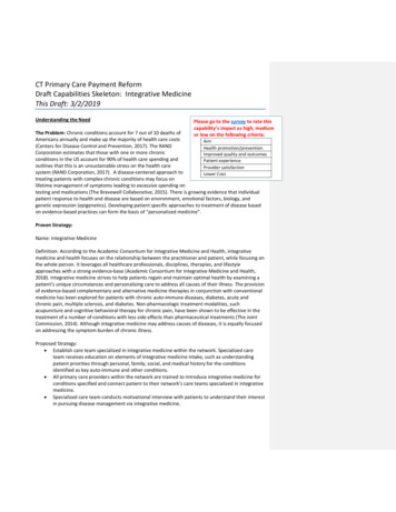
Tuberosity broad thick rough eminence, e.g., ischial tuberosityVick, Robbins, Smith; A Laboratory Manual of Methods and Techniques in PhysicalAnthropologyEvidence Used in Determination of Sex(Sexual Dimorphism)Humans vary in their degree of sexual dimorphism (difference in bodyform between male and female). Humans vary approximately 10-12% betweenmale and female. Sexual dimorphism varies in form mostly by body size andfeatures such as robustness and muscularity. Sexual dimorphism can be used todetermine the sex of unknown individuals. In general, males are larger and morerobust, with heavier muscle markings and larger teeth. Generally, in humans, theskull, long bones, and pelvis are all used to estimate sex. The best diagnosticevidence is the pelvis, however.PELVISMALEFEMALEGreater SciaticNotchNarrow anglerelatively deepWide anglerelatively shallowPreauricularSulcusInfrequent, sometimes absentCommon, mostly presentPubic Symphysis(less reliable)Deeper in malesObturatorForamen(less reliable)Relatively large and ovalRelatively small and triangularSubpubic AngleUnder 90 degrees, narrowGenerally 90 degrees or higher,widePairedSometimes absent, but if present ,single and in the midlineSKULL
Orbital RimSharpnessRounded and dull to the touchDistinct and sharp edgedTemporal RidgesMuscle attachmentslarger, more ruggedMuscle attachments slightNuchal RidgesMuscle attachments larger, more ruggedMuscles attachments slightSupraorbitalRidgesLarger, heavierSmaller or absentMastoid ProcessMedium to large (thumb size), usuallyprojecting below the below of the skullSmall, (little finger size), usuallydoes not project below the skullPosteriorZygomatic RootContinues into supramastoid crest aboveauditory meatus, heavier, greater lengthUnderdeveloped, lighter and morecompressedMandibleHeavier jaw, more “square” chin that hastwo points connected by more or less astraight line. The gonial angle is generally 125 degreesMore narrow and “pointed”(singleprominence), ramus is more gracile.The gonial angle is generally 125degreesSkull OverallRougher, larger, more ruggedSmoother, smaller, more rounded,adolescent-likeHeavier; heavy muscle markingcompared to female; larger head ofhumerus; larger head of femurLight muscle markings; smallerbones and joints in generalOTHERPostcranialGeneral
Vick, Robbins, Smith; A Laboratory Manual of Methods and Techniques in PhysicalAnthropology
Brothwell, Digging Up Bones
The narrow sciatic notch is a good indicator that the pelvis belongs to a male.This wide subpubic angle is a good indicator that this pelvis belongs to a female.
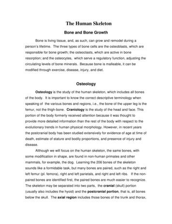
Evidence Used in Determination of AgeSkeletal EvidenceLines of evidence used in the determination of age involve examination ofepiphyseal union, closing of cranial sutures, and changes in the pubic symphysis.The epiphysis is a cartilaginous area of bone growth located near the ends oflong bones. As individuals mature, these epiphyses gradually ossify and join thediaphysis, in a timed sequence. In flat bones, such as the skull, growth occursfrom the center of the bone. Upon maturation, growth stops and the suturesgradually close. The pubic symphysial face also changes over time.Times of epiphyseal union of long bones vary somewhat due to nutritionand individual variability and can therefore serve only as an approximateindicator of age up to about 25-30 years. The sutures of the skull begin closing atapproximately 17 years of age; finally, in very old age, the sutures are completelyfused. Suture fusing begins on the inside of the skull and proceeds to theoutside. Average ages for suture closing have been determined. However,because of inter-individual variation, one should be very cautious in using sutureclosure data for age estimation. Stages in the aging of the pubic symphysis havealso been determined. This measure is more useful than other measures sincethe changes extend into later life.A.B.C.D.E.F.G.H.Fused by 40 years of ageFused by 65 years of ageFused by 72 years of ageFused by 80 years of ageFused by 40 years of ageFused by 50 years of ageFused by 80 years of ageFused by 65 years of ageA. Fuses between 18 and 25 years of ageWolfe, 1983
Age Estimation of Immature SkeletonsIn general, immature remains are those of individuals who are less than 20years old. Complete fusion of all major epiphyses has occurred and all teethhave erupted. Of course, the age of occurrence of these events varies dependingon sex, race, nutrition and other factors.I.Appearance of Ossification CentersA. Appearance of ossification centers occurs from birth to 15 years.B. The centers themselves rarely survive in archeological/forensiccontext because of their fragile nature.II.Epiphyseal UnionA. Most commonly used in teenage years (10-20 years)B. Standards available for humerus, clavicle, scapula, hip, elbow,hand, wrist, foot, ankle and knee.C. Epiphyseal union should be considered a process rather that aevent.D. A range of four years is seen between fusion onset in earlymaturing and completion of fusion in late-maturing individuals.E. Females are an average of two years in advance of males inepiphyseal union.F. Radiographs of the epiphysis provide earlier age estimates.III.Bone SizeA. In individuals from the prenatal period to about 6-7 years, the lengthof the long bone diaphyses can be used to estimate age.B. Growth rates vary greatly among groups and sexes.
C. For prenatal to birth skeletons, use long bone length to get anestimate of body length, then use body length to get estimate ofage.D. For postnatal skeletal remains, the diaphyseal length can be useddirectly to estimate age.IV.Dental calcification and eruptionA. Most accurate age indicator in subadults.B. Dental development largely controlled by genetic factors and is,therefore, relatively less susceptible to environmental factors.C. Most studies provide calcification and eruption data for specificgroups (e.g. white, black, Indian, male, female)D. Age estimates are available for calcification of deciduous (milk) andpermanent teeth.
Epiphyseal UnionMale and Female age ranges in years for complete fusion of epiphyses
Brothwell, Digging Up BonesDental EvidenceGeneral Dentition Information
Teeth provide much useful information to the physical anthropologist. Dueto their hardness, they very often fossilize. Teeth can give us insights about thelife ways of the individuals to whom they belonged. For example, eruption andwear patterns can provide information about age. Moreover, teeth can providedata on nutrition and diet.Occlusal surface top of crown that comes in contact with teeth of theopposing jaw when the mouth is closedCrownpart of the tooth above the alveolusNeckjoins crown and root(at the gum line)Rootanchors the tooth in the boneBuccalcheek side of toothLingualtongue side of toothMesialtoward the midline of chinDistalaway from the midline of chinGroovenatural valleys between cusps or other tooth partsAlveolussocket in the maxilla and mandible in which the root ofThe tooth is anchoredCuspa pronounced elevation on the crown surface of thetoothBicuspida tooth with two cusps (premolars)Incisora tooth with one cutting edgeMESIALCROWNBUCCALLINGUALNECKDISTALROOTVick, Robbins, Smith; A Laboratory Manual of Methods and Techniques in Physical Anthropology
Human DentitionDeciduous Maxillary DentitionIncisorIncisorCanineMolarMolarPermanent Maxillary MolarMolarVick, Robbins, Smith; A Laboratory Manual of Methods and Techniques in PhysicalAnthropology
Dentition Eruption SequenceBirth8 Years9 Months10 Years2 Years12 Years4 Years15 Years6 Years21 Years(after Schour and Massler)
Evidence Used in Determination of RaceRace is much harder to determine, as there is much interindividualvariation within the various ethnic groups. The following characteristics may beused to aid in racial identification.AsianAsian skulls have a flat, moon-like face. This is caused partly bythe fact that the cheek bone protrudes forward. Asians usually have whatis called an edge-to-edge bite; this occurs when teeth of the opposing jawtouch each other when the mouth is closed. The incisors are generallyshovel-shaped, and the malars (or zygomatics) are robust and flaring.There is usually no crowding of the teeth.
AfricanAfrican skulls generally have rounded foreheads, and a wide nasalopening that lacks the nasal sill seen in Caucasoids. Africans typically have whatis called an over-bite; this happens when the top teeth protrude farther than thebottom teeth. The incisors are generally blade-form, and the malars are smalland retreating. There is usually no crowding of the teethEuropeanThe European skull comes to a point along the midline and cheek bonesdo not extend forward. The nose in narrow and high bridged, with a nasal sillthat dams the nasal opening. Europeans have a “flat” face in the dental area,which is opposite of the African face. The incisors are generally blade-form, andthe malars are small and retreating, just as the African dentition. There isfrequently crowding of the teeth, particularly the impacted third (3rd) molars.Burns, Forensic Anthropology Training Manual
INDIVIDUAL BONES OF THE CRANIUM(FRONTAL VIEW)SAGITTAL SUTURECORONAL SUTUREPARIETALTEMPORALLINEFRONTALPTERION L BONES OF THE CRANIUM(BASAL VIEW)Vick, Robbins, Smith; A Laboratory Manual of Methods and Techniques in Physical Anthropology
INDIVIDUAL BONES OF THE CRANIUM(BASAL VIEW)PALATINEPARIETALMAXILLAMAXILLAEZYGOMATIC YLOIDPROCESSFORAMENMAGNUMPARIETALOCCIPITALVick, Robbins, Smith; A Laboratory Manual of Methods and Techniques in Physical Anthropology
INDIVIDUAL BONES OFTHE CRANIUM(LATERAL VIEW)CORONAL SUTUREPTERIONREGIONWORMIAN PITALETHMOIDLACRIMALTEMPORALNASALFRANKFORT (EYE – EAR) EAUDITORY MEATUSMAXILLA(ALVEOLARREGION)MASTOID PROCESSZYGOMATIC* INION – ALSO CALLED EXTERNALOCCIPITAL PROTUBERANCEVick, Robbins, Smith; A Laboratory Manual of Methods and Techniques in Physical Anthropology
SAGITTAL SECTION LATERAL TOTHE CENTER(ILLUSTRATING INTERNAL STRUCTURE OF THESKULL)NASALSUPERIOR NASALCONCHASPHENOPALATINEFORAMENETHMOIDMIDDLE NASALCONCHALACRIMALINFERIOR NASAL CONCHAOCCIPITALPTERYGOID PLATE OFSPHENOID BONEPALATINEMAXILLASAGITTAL SECTION OF THE SKULL(ILLUSTRATING INTERNAL STRUCTURE OF THE SKULL)NASAL PROCESSOF FRONTALCRISTA GALLI (ETHMOID)SELLA TURCICA (SPHENOID)ETHMOIDVOMERMAXILLAINTERNAL PTERYGOIDPLATE (SPHENOID)INFERIOR NASAL CONCHAPALATINEPALATOMAXILLARY PROCESSVick, Robbins, Smith; A Laboratory Manual of Methods and Techniques in Physical Anthropology
BONES OF THE SKELETONCRANIUMCERVICAL VERTEBRAECLAVICLESCAPULASTERNUMHUMERUSTHORACIC VERTEBRAELUMBAR VERTEBRAEULNAPELVIS (ILLIUM)RADIUSSACRUMPELVIS (PUBIS)COCCYGEAL BIAFIBULAMETATARSALSTARSALSPHALANGES
Vick, Robbins, Smith; A Laboratory Manual of Methods and Techniques in PhysicalAnthropologyBibliographyBass, W. M. 1989. Human Osteology: A Laboratory and Field Manual of theHumanSkeleton, 3rd ed. Columbia, MO: Missouri Archaeological Society.Brothwell, Dr. 1992. Digging up Bones, 3rd ed. Ithaca, New York: CornellUniversity Press.Burns, Karen Ramey. 1999. Forensic Anthropology Training Manual. UpperSaddle River, New Jersey: Prentice-Hall.Byers, Steven N. 2002. Introduction to Forensic Anthropology: A Textbook. APearson Education Company 75 Arlington Street Boston, MA: Boston,MA.Schour, l. and M. MasslerThe development of the human dentition. Journal of the American DentalAssociation 28:1153-1160.Vick, Laura, L. Robbins, and L. Smith. 2003. A Laboratory Manual of MethodsandTechniques in Physical Anthropology. Raleigh, NC: Department ofAnthropology, Peace College.
What and How Many?When one is dealing with bony remains, one must first determine to whatspecies (one or more than one) the remains belong. In other words, is only onespecies represented? Or are you dealing with more than one species? Anotherquestion involves the minimum number of individuals (MNI) represented in theremains. Clues such as shape, size, side, and number are used to answerthese question. In addition, color, degree of weathering, etc., can also offervaluable information.One of the first steps in analysis involves sorting. Place bones of thesame type together as, for example, all femurs versus all humeri. If homologousbones have very different shapes, they probably represent different species.Next look at size; two differently sized humeri will indicate at least two differentindividuals. With good understanding of the skeletal structure and relative sizedifferences of the various bones, size can also be used as a clue even when onehas non-homologous bones. Now check for side. An animal can only have oneleft femur or one left innominate bone (left half of the pelvis).Color and weathering are less valuable clues since part of a skeleton mayhave been exposed to sunlight while another part has been covered by dirt ordebris. However, these clues can sometimes help one in assigning remains toone or more than one individual.Demonstration / Exercise“Taphonomy Exercise” bags will be assigned to student groups. Each bagcontains some rhesus monkey bones (very similar to human bones) as well assome other bones. You will use clues such as size, size, shape, number,color, and weathering to determine the answers to the questions below.
After your group is ready to “present” your conclusions, check these withthe instructor or lab assistant.)Examine carefully the bones found in your bag. Bag ID NumberHow many species do you observe? at least
the postcranial body has been studied extensively for evidence of age at time of death, estimate of stature and bodily proportions, and presence of injury and disease. Although we will focus on the human skeleton, the same bones, with some modification in shape, are found in non-human