Transcription
Physica Medica 68 (2019) 132–145Contents lists available at ScienceDirectPhysica Medicajournal homepage: www.elsevier.com/locate/ejmpReview paperA guide to90a,⁎Y radioembolization and its dosimetrybcS. Peter Kim , Claire Cohalan , Neil Kopek , Shirin A. Engera,d,eTaMedical Physics Unit, McGill University, Montreal, Quebec H4A 3J1, CanadaDepartment of Physics and Biomedical Engineering, CHUM, Montreal, Quebec H2C0X1, CanadaDepartment of Radiation Oncology, McGill University Health Centre, Montreal, Quebec H4A 3J1, CanadadDepartment of Oncology, McGill University, Montreal, Quebec, H4A 3J1, CanadaeResearch Institute of the McGill University Health Centre, Montreal, Quebec H3H 2L9, CanadabcARTICLE Clinical BackgroundRadioembolization gains continuous traction as a primarily palliative radiation treatment for hepatic tumours. Aform of nuclear medicine therapy, Yttrium-90 containing microspheres are catheter guided and injected into theright, left, or a specifically selected hepatic artery. A multitude of comprehensive planning steps exist to ensure athorough and successful treatment. Clear clinical and physiological guidelines have been established and nuclearimaging is used to plan and verify dose distributions. Radioembolization’s treatment rationale is based on tumour and blood vessel dynamics that allow a targeted treatment approach. However, radioembolization’s dosimetry is grossly oversimplified. In fact, the currently utilized clinical dosimetric standards (e.g. partitionmethod) have persisted since the 1990s. Moreover, the multitude of radioembolization’s intertwining components lies disjointed within the literature. Particularly relevant to new readers, this review provides a methodicalguide that presents the treatment rationale behind every clinical step. The emerging dosimetry methods and itsfactors are further discussed to provide a comprehensive review on an essential research direction.1. IntroductionRadioembolization, selective internal radiation therapy (SIRT),intra-arterial radiation therapy, or trans-arterial radioembolization(TARE) are all various names for the same clinical procedure.Radioembolization is a specific type of nuclear medicine therapy usedto treat primary or metastasized hepatic tumours. It is administeredwhen other minimally invasive treatments have failed and is primarilypalliative. During treatment, a catheter is used to guide and injectYttrium-90 (90Y) containing microspheres into the right, left, or a specifically selected hepatic artery. Due to unique hepatic blood flow, themicrospheres are distributed preferentially to the tumour arteries,where they are permanently deposited. The tumour is then irradiatedby the particles emitted by 90Y.90Y is a pure beta-emitter with a half-life of 64.04 hours that disintegrates into stable zirconium-90 (90Zr) by emitting beta particleswith a maximum energy of 2.27 MeV and an average energy of0.937 MeV. Within soft tissue, the released beta energy has a maximumpenetration range of 11 mm with an average range of 2.5 mm. Since 90Ydeposits most of its energy within the first few millimeters, its therapeutic dose is concentrated at the microspheres location [1]. Currently, there are two commercially available 90Y microsphere products,⁎the glass (Therasphere; BTG plc, UK) and resin microspheres (SIRspheres; Sirtex Medical Limited, North Sydney, Australia). Both of thesespheres are biocompatible, but not biodegradable. Theraspheres haveaverage diameters of 20–30 µm with a standard activity that may rangefrom 3 GBq to 20 GBq per vial. Within a vial of 3 GBq, there are around22,000 to 72,000 microspheres per milligram, resulting to an averageactivity of 2500 Bq per microsphere [2,3]. In contrast, the diameters ofSIR-spheres range from 20 to 60 µm with an average activity of 50 Bqper microsphere. Normally, each resin vial has a standard activity of3 GBq and a range of 30–50 million microspheres [2,4]. Highlightsbetween the two differing microsphere products can be seen in theEuropean Association of Nuclear medicine (EANM) guidelines [5].Contained within pre-packaged vials, both types of 90Y microspheresare delivered, then their activity is measured right before treatment.Radioembolization involves multiple clinical steps spanning overnumerous multidisciplinary fields. There exists a plethora of articles onthis treatment modality, yet there is a lack of a study that comprehensively describes the rationale behind the entire radioembolizationprocess. Overall, this review has two objectives. The first aim is toprovide a full scope of radioembolization that ties together the rationaleand background behind every treatment step. Building on the discussedbackground, the second aim is to provide a comprehensive review byCorresponding author.E-mail address: shingyu.kim@mail.mcgill.ca (S.P. eived 5 February 2019; Received in revised form 15 September 2019; Accepted 17 September 2019Available online 28 November 20191120-1797/ 2019 Associazione Italiana di Fisica Medica. Published by Elsevier Ltd. All rights reserved.
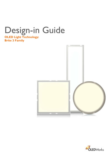
Physica Medica 68 (2019) 132–145S.P. Kim, et al.presenting the most relevant literature related to patient specificradioembolization treatments and its newly emerging dosimetry.steps that include imaging with a triple phase computed tomography(CT) and/or magnetic resonance imaging (MRI) to assess the liver forliver patency, extrahepatic disease, and tumoural and non-tumouralvolumes [6]. Since radioembolization is artery based, pre-treatmentworkups include an angiography as well as planar scintigraphy imaging(planar imaging) and single photon emission computed tomography(SPECT) co-registered with CT (SPECT/CT) for injected 99mTechnetiummagroaggregated albumin (Tc-MAA). Tc-MAA images are used to verifyvessel mapping and to visualize any additional arteries that may lead toextrahepatic microsphere distribution. Tc-MAA distribution is also usedto calculate what is called the lung shunting percentage or lungshunting fraction (LSF). The LSF estimates the total 90Y microspheredeposition within the lungs and quantifies the risk of developing radiation pneumonitis, a serious side effect. Consequently, radioembolization has lung safety thresholds; if the LSF is too high, patientsare deemed ineligible for treatment.During the treatment itself, an interventional radiologist places acatheter percutaneously via a patient’s femoral artery and guides it tothe correct hepatic artery under x-ray fluoroscopy. Connected to thecatheter, a vial containing 90Y microspheres is infused into the body.Differences exist in the administration of the two different microsphereproducts. Due to their higher embolic tendency (See 3.8 Treatment),90Y resin microspheres are successively infused with saline and 5%dextrose whereas glass microspheres are infused by saline alone [7–10].Recently, an alternate transradial approach has been deemed feasible.This approach involves a catheter placement via a patient’s radial arteryand demonstrates advantages such as patient preferability and lowercost, albeit with greater technical challenges [11]. Directly after treatment, the patient has follow-up imaging with SPECT/CT or PET/CTscans to check for the microsphere distribution and to verify the dose.The review of the different treatment steps will be discussed in detail inthe coming subsections.2. MethodsAs a multi-disciplinary and multi-step treatment, the topics of emphasis were purposefully constrained to patient-based studies andimage-based dosimetry; therefore, only 97 of around 130 originalsources were listed to provide a comprehensive and succinct discussionon radioembolization. To provide the understanding and rationale behind each treatment process, background articles on liver vasculature,related nuclear imaging modalities, current clinical dosimetry methods,novel clinical studies, and review articles were included. Search wordssuch as “patient based radioembolization”, “imaged based radioembolization”, “post-image dosimetry” “radioembolization”, “PET/CT”, “SPECT/CT”, “angiography”, and “primary radioembolizationtreatments” were inputted into specific databases. The most commondatabases used were Pubmed and WorldCat.3. Results and discussionA relatively complete picture of radioembolization can be obtainedby reading from the list of suggested articles within Appendix ATable 1. It is important to note that each of the suggested articles assumes a certain level of familiarity and only provides insight into selective segments of this treatment. In other words, none of the presented studies provide a comprehensive overview of every treatmentstep. Furthermore, dose response effect studies based on clinical dosimetric methods are not included in this review due to the rationale thatemerging image based dosimetry methods are more dosimetrically representative. However, a previous review has amassed such results [6].3.2. Patient eligibilityRadioembolization requires planning that involves patient evaluation and pre-treatment imaging, the treatment itself, and finally, shortlyafter the treatment is completed, post-treatment verification. Themultiple clinical steps are listed in Fig. 1. To start, patients are clinicallyexamined for relative and absolute contraindications such as insufficient liver function or extensive and untreated portal hypertension[5]. After a clinical evaluation, patients undergo further preliminaryPrior to radioembolization, patients must be deemed suitable fortreatment. Radioembolization is used as a primarily palliative treatment, i.e. to preserve the functional capacity of the hepatic tissue [12].Eligibility stems from the existing functionality and the potential radiation tolerance of a patient’s hepatic parenchyma. Such predictionsare divided into three sections indicating when a patient should havetreatment, when patient treatment is possible, or when a patient is ineligible for treatment. These indications are respectively labeled indications, relative contraindications, and absolute contraindications.Patient eligibility can be particular to different steps of the treatmentplan. In many cases a patient may be eligible under the initial clinicalevaluation, but later become ineligible during the pre-treatment imaging workup step of radioembolization [7].In general, most articles refer to the RadioembolizationBrachytherapy Oncology Consortium (REBOC) guidelines and manufacturing recommendations when discussing radioembolization patientselections [3,4,13]. The EANM has also complied patient selectionguidelines for the treatment of liver cancer and metastases [5]. Theimportant patient indications include an unequivocal and measurableliver tumour based on CT/MRI scans, a liver-dominant tumour burden,and a life expectancy of at least 3 months [7,13].Radiation dose to organs at risk is assessed through pre-treatmentimaging with an angiography and Tc-MAA to avoid severe side effectssuch as radiation pneumonitis, gastrointestinal ulceration, and/or gastrointestinal bleeding due to extrahepatic microsphere deposition [14].Patients are further ineligible for treatment if they have factors such asa compromised portal vein, are pregnant, or have liver failure [7,13].The treatment eligibility has relative contraindications as well, whichare based on a case-by-case basis and left to the discretion of the physician. Some factors include patients with a limited hepatic reserve,poor kidney function, or an Eastern Cooperation Oncology GroupBasic Radioembolization Steps3.1. Treatment overviewSTEPRATIONALE1. Clinical EvaluationDetermines PatientEligibility2. CT/MRI ScansAssess Tumourcharacteristics3. AngiographyMaps out the hepaticarteries4. Tc-MAA ImagingPredicts microspheredistribution and aids indosimetric planning5. 90Yttrium TreatmentAn angiograph-likeprocedure where 90Ymicrospheres are injected6. Post-Image VerificationVerification of distributionand absorbed doseFig. 1. Clinical steps involved in the90Y radioembolization process.133
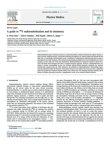
Physica Medica 68 (2019) 132–145S.P. Kim, et al.Table 1ECOG performance ranking description.GradeECOG Performance Status012345Fully active, able to carry on all pre-disease performance without restrictionRestricted in physically strenuous activity but ambulatory and able to carry out work of a light or sedentary nature, e.g., light house work, office workAmbulatory and capable of all self-care but unable to carry out any work activities; up and about more than 50% of waking hoursCapable of only limited self-care; confined to bed or chair more than 50% of waking hoursCompletely disabled; cannot carry on any self-care; totally confined to bed or chairDead(ECOG) performance status of 2–4 [7,13,15]. Grading levels of theECOG status are explained in Table 1. However, age and prior surgicalresection are not considered to be contraindications, making radioembolization appealing for palliative therapy [16].With restricting clinical factors well set for radioembolization, thereare ideal patients who are predicted to have a good tolerance to thetreatment. These patients normally show an ECOG status of less than 2,normal bilirubin levels, normal liver synthetic function (albumingreater than 3 mg/dL), a lack of ascites, and less than 50% tumourburden [7]. It has been shown that ECOG status rather than a tumour’sclinical stage is the more reliable factor in a patient’s treatment tolerance [15]. For metastatic tumours, lower bilirubin levels were alsofound to be reliable indicators for radioembolization tolerance [15,16].Overall criteria and specificities of eligibility are presented in Table 2.For 90Y treatment, it should be stated that the institutional and corresponding product eligibilities should always be consulted [3,4].the gastrointestinal tract while the hepatic arteries provide the oxygenated blood [17]. Branches of the portal vein further divide and passbetween hepatic lobules and eventually end up as sinusoids. Lobules arehexagonally functional units of the liver, otherwise known as hepaticparenchyma, and contain hepatocytes, bile of canaliculi, sinusoids, anda central vein. Hepatic tumours almost exclusively derive their bloodsupply from the hepatic arteries while the normal parenchyma is perfused through the portal veins. For a more thorough physiological investigation on hepatic perfusions, reviewing Van de Wiele (2012) issuggested [19]. With preferential hepatic perfusion being the basis forradioembolization, select hepatic artery catherization provides a wayfor the microspheres to target malignant hepatic tumours while sparingthe normal tissue [18]. Therefore, a background on the most commonhepatic vasculature is given.The standard hepatic vasculature scheme starts with the celiac axis[20]. The first major branch off the aorta, the celiac artery then trifurcates into the left gastric artery (LGA), splenic artery, and thecommon hepatic artery (CHA). From there, the CHA continues to bifurcate into the proper hepatic artery (PHA) and the gastroduodenalartery (GDA) and more distally the PHA divides into the right and lefthepatic arteries at the hilar plate. Moreover, both hepatic arteriescontinually branch and decrease in size to perfuse the hepatic parenchyma and eventually make up the microvasculature of the hepatocytes, which is later discussed in 3.11 Liver Microcirculation & Distribution. Fig. 2 illustrates an example of a patient’s hepaticvasculature.3.3. Standard liver vasculatureAs the name implies, radioembolization is built around the hepaticvasculature where the injected radioactive microspheres travel andpermanently localize within the hepatic arteries. Macroscopically, hepatic vascularization is comprised of a dual blood supply where theportal vein provides 75–80% of the hepatic blood supply and the hepatic arteries provide 20–25% of the blood [17,18]. The portal veinprovides partly deoxygenated but nutrient rich blood to the liver fromTable 2Patient Eligibility Specifications.Ideal Patient1. ECOG 22. No ascites3. Less than 50% tumour burden4. Albumin 3 mg/dl5. Bilirubin 2 mg/dlIndications1. CT/MRI established Tumour2. Liver Dominant Tumour3. Life Expectancy 12 weeksRelative Contraindications1. Excessive tumour burden without hepatic reserve2. Compromised Portal Vein when super-selective catherization cannot be performed3. Prior radiotherapy4. ECOG 2–45. Creatine 2.5 mg/dl6. Abnormal bone marrow function7. Bilirubin levels ( 2 mg/dl) with no reversible causeAbsolute Contraindications*Clinical Evaluation1. Ascites and/or other symptoms of liver failure2. Pregnant3. Capecitabine last 2 months or planned to be administered in the futureTc-MAA Imaging1. 30 Gy predicted to lungs or 20% LSF**2. Any extrahepatic deposition within the gastrointestinal tract3. Extensively compromised Portal Vein4. For repeated treatments, a 50 Gy cumulative lung dose should not be surpassed**Fig. 2. A digital subtraction angiography is shown for a patient being plannedfor right lobe radioembolization. The different parts of the hepatic vessels arelabeled: 1. Catheter in the celiac trunk, 2. Micro-catheter that is extended to 3.The common hepatic artery, 4. A gastroduodenal artery that is coil embolized,5. Proper hepatic artery, 6. Left hepatic artery, 7. Right hepatic artery.* Steps are based on Fig. 1.** See 3.9 Current Clinical Dosimetric Methods for Gy value explanations.134
Physica Medica 68 (2019) 132–145S.P. Kim, et al.3.4. Angiographyadministration. An adult activity consists of 185 MBq Tc-MAA suspended in normal saline. If the whole liver is treated, the right and lefthemilivers will be injected with 111 MBq and 74 MBq, respectively[23].After injection, a gamma detection system produces a whole-bodyimage. Planar imaging may be used to assess Tc-MAA depositions, butstudies suggest a tomographic imaging modality such as a SPECT scanor a combination of SPECT and CT to provide a more accurate check ofthe Tc-MAA bio-distribution [13]. It was found that planar imagingmight not provide adequate detection, especially when extrahepaticsites or a non-homogenous Tc-MAA distribution is present. However,planar imaging is still clinically adopted although SPECT/CT has beenproven to be more accurate for determining the distribution of Tc-MAAand the quantification of its LSF [25–27].The Tc-MAA image is then compared to the previously assessedangiograph where the injected particle distribution is compared to thepre-planned vasculature. Ideally, the Tc-MAA microspheres are allcontained within the imaged angiographic vasculature; however, theTc-MAA microsphere distribution and the angiographically predicteddistribution can differ [23]. There are multiple causes for this discrepancy. A catheter may have been misplaced distally or past thebranching point that excludes the part of the liver to be treated. Newparasite vessels may have sprouted between the time the angiographywas performed and the time Tc-MAA was injected. A hepatic artery mayhave been missed during the angiographic treatment. If any of theabove scenarios are illustrated, a second angiography and a subsequentTc-MAA injection is recommended to confirm the new vascularization[28]. Additionally, extrahepatic activity may be seen from Tc-MAA. Tocombat unwanted extrahepatic activity, solutions such as additionalcoiling, more distal placement of the catheter, and/or super selectivecatherization (radiation segmentectomy) during treatment are furthersuggested. Coiling would physically block the 90Y microspheres frompassing through a specific vessel. More distal catheter placement prevents less reflux of microspheres, if any, due to the catheter physicallybeing farther from any arterial branching. Similarly, super-selectivecatherization keeps microsphere localization within specifically perfused segments within the liver (for further detail see 3.8 Treatment).On top of overall vessel discrepancies, the distribution within thesame vasculature may have unusual Tc-MAA accumulations in bothextrahepatic and intrahepatic sites [23]. To plan a safe treatment,awareness of this heterogeneous distribution is needed to attribute aspecific distribution to a cause. A variety of factors such as presence ofnecrotic lesions, prior trans-arterial chemoembolization (TACE) treatments, invasion of tumour within hepatic arteries, aggregation of TcMAA particles, or variable flow dynamics within the liver may result inheterogeneous distribution. Proficient knowledge on the causes of discrepant Tc-MAA distributions and the necessary steps to combat them isrequired for an accurate radioembolization treatment plan and later, ifcalculated, for its dosimetry.In an ideal scenario, radioembolization microspheres will localizesolely within the hepatic tumour’s vasculature. In reality, radioembolization microspheres localize within both the healthy and tumourliver tissue. Many variations exist within the overall hepatic vasculaturethat may lead to additional complications. In fact, hepatic vessel variations are quite common and can be expected in 45% of patients [21].If such vessel variations do exist, the variable hepatic vasculature maycause extrahepatic deposition, which results in the irradiation of nonhepatic healthy tissue.Due to such variations in vasculature, an angiography is used tomap patient-specific arteries [20,22]. The goal of an angiographicevaluation is three fold: it is to guide the delivery catheter positioning,evaluate pre-treatment blood flow, and to determine the variant arteries that may lead to extrahepatic microsphere deposition [8,13]. Vanden Hoven et al. (2014) found that only 49% of patients had theiraberrant hepatic arteries correctly identified from a standard CT scanand only 86% during angiography [20]. Thus, several modificationswere recommended to the standard process including, but not limitedto the use of a multiphase liver CT before an angiography, the use of Carm cone beam CT, catheter-directed CT angiography (CTA), and anevaluation of SPECT/CT co-registered images from Tc-MAA [20,23].Once an angiography has been performed and possible variantvessels identified, coil embolization (also called coil occlusion or skeletonization) is recommended [8,13,15,23]. Coil embolization involvesa catheter and a metal cable that has attached collagen fibres. The metalcable is pre-formed as a spiral that is set straight. When released from acatheter, the pre-straightened coil spirals into its natural form thatoccludes most of the aberrant vessel. The attached collagen fibres theninduce thrombosis, which blocks the rest of the vessel and preventsunwanted microsphere deposition. Coil embolization in turn directsmicrosphere flow to the targeted region. Depending on the variant arteries, the size of the arteries, and possible angiographic preferences,coil embolization is a calculated choice. On one hand, prophylacticembolization is seen as the safer option as the dangers of grave clinicalcomplications such as gastrointestinal ulceration and bleeding outweigh the dangers of coil embolization [15]. On the other, coil embolization is avoided in the most experienced centers due to complicationsand limited benefits of an embolized treatment [24]. At our institution,coil embolization is administered when necessary such as when shuntsto the gastrointestinal track are visible. Fig. 2 further demonstrates anangiogram and the relevant vessels for a patient being treated forradioembolization.Coil embolization’s effectiveness depends on the patient’s coagulation speed and the effectiveness of the actual embolization. In somepatients, their de-coagulation speed may be faster than others when theinduced thrombosis is cleared up and blood flow is resumed (recanalization). In others, the initial coils may not have been packed tightenough to induce thrombosis in the first place. This problem is easilyremedied through additional packing of coils. Moreover, some patientsmay have new vessels (collaterals) develop leading to resumed bloodflow back to the targeted organ. Whatever the case, the best preparationmethod must be determined and evaluated for a patient undergoingradioembolization treatment.3.6. The lung shunt fraction (LSF)Theoretically, the 90Y microspheres should embolize within thetumour’s hepatic terminal arteries (See 3.11 Liver Microcirculation &Distribution). However, arteriovenous blood vessel shunts may provide90Y microspheres a direct vascular path to the lungs, which may thencause lung irradiation [13]. To predict this, Tc-MAA distribution is usedto calculate the LSF. Estimated by the ratio of counts in the lungs to thesummed counts in the lungs and liver, the LSF uses planar imaging orSPECT/CT count data for its calculations. In other words, the LSF is thefraction of Tc-MAA particles that made its way into the lungs due toarteriovenous shunts. It is worth noting that there are variations in size,density, and number between Tc-MAA particles and 90Y microspheres;however, Tc-MAA particles are still used to predict the undesirable 90Ymicrosphere deposition within the lungs. [23].Depending on the imaging modality used, there exist limitations3.5. Tc-MAA pre-treatment imagingAfter an angiography, Tc-MAA is used to predict the potential distribution of 90Y microspheres. Tc-MAA is normally provided in a vial of10 ml solution containing 4.8 x10⁶ aggregated albumin particles where90% or more are between 10 and 90 µm in diameter. The particles maymeasure outside such parameters, but still have a maximum range from0 to 150 µm [8]. With a short physical half-life of 6 hours and a continual bio-degradation of 99mTc from MAA, it is recommended that theTc-MAA imaging to be done at least within 60 minutes of135
Physica Medica 68 (2019) 132–145S.P. Kim, et al.that can affect the calculation of the LSF [29]. Planar imaging haslimitations that include uncorrected attenuation effects between theliver and lungs, lack of anatomical references for contouring, and asingle standard lung mass value of 1 kg. These limitations combine tonegatively affect the LSF calculations. In regards to SPECT/CT, a misregistration between the SPECT and CT data or truncated lungs due to alimited SPECT/CT field of view (FOV) will produce inaccurate LSFcalculations.control during the regenerative phase after treatment, providing a logical step before surgical resection [34].The last radioembolization option is whole liver treatment and isrequired when many diffuse tumours are localized within both of thelobes. There are two methods of administering the same dose to thewhole liver: sequentially through bi-lobar treatment or singularlythrough simultaneous treatment of both lobes. If treated sequentially,one lobe is treated first and after at least 30 days with sufficient liverregeneration the second lobe is treated [15]. When comparing the two,sequential radioembolization has resulted in fewer adverse side effectssuch as lower overall bilirubin levels and lower risks of developingradioembolization induced liver disease (REILD) [33]. Therefore, sequential liver treatment has become the more standard recommendation.If feasible, more segmented therapy might be performed. If a singular artery branch is identified to perfuse the targeted tumour(s) thena more selective treatment can be used to infuse the microspheres. Thisserves to avoid extrahepatic deposition as well as to make treatmentmore spatially localized, providing higher dosages to the tumour andless intrahepatic radiation to the normal tissue. However, radioembolization is currently used as a primarily palliative treatmentmaking more segmented treatments less likely and procedures such assequential liver treatment the norm. Regardless, every 90Y treatmentshould be delivered as selectively as possible to reduce irradiation ofthe normal liver parenchyma [5].Further considerations are necessary to ensure an optimal or accurate radioembolization treatment. Lau et al. (2012) have provided 90Yactivity recommendations depending on the number of tumours, patient specifications, and type of treatment [12]. The same authors havealso recommended threshold values to prevent REILD. Other considerations include similar catheter placement during 90Y microspheretreatment as during Tc-MAA injections, otherwise up to 30% of differing activity may be measured and the resulting microsphere distribution may differ [37]. For SIR-spheres, a higher number of microspheres is injected to reach the same prescribed activity as eachmicrosphere has less activity (50 Bq) compared to each Therasphere(2500 Bq) [2,5]. The greater number of SIR-spheres may cause an embolic effect called flow stasis, at which point microsphere administration should stop regardless of an incomplete administration of the totalprescribed activity [8]. Since the initial prescribed calculations arebased on the volume of the target, accurate volume measurements arenecessary. Hepatic volume measurements should only include the tumour volume perfused by the targeted arteries [5,15]. The treatmentitself should completely administer the dose throughout the whole tumour, which is achieved by injecting microspheres into all the arteriesthat perfuse the different parts of the tumours.3.7. Liver segmentation schemesThe Brisbane nomenclature is considered the standard liver segmentation scheme. Introduced and accepted by the InternationalHepato-Pancreato-Biliary Association (IHPBA), this standard was created to resolve the ambiguities presented by Couinard and Healey [30].According to the Brisbane nomenclature, there are three orders of division based on the liver’s internal anatomy: two hemilivers, sections orsectors, and segments [30]. The first-order division divides the left andright hemiliver by a plane called the midplane of the liver, commonlyknown as Cantlie’s line. The second-order divisions divide the liver intosmaller sections or sectors and have two distinct
Feb 05, 2019 · a cH4A3J1,Canada . palliative. During treatment, a catheter is used to guide and inject Yttrium-90 (90Y) containing microspheres into the right, left, or a spe- . vasculature where the injected