Transcription
Rectal biopsy for Hirschsprung's diseaseRectal biopsy for Hirschsprung's disease: a review oftechniques, pathology, and complicationsEleanor Dorothy Muise, Robert Anthony CowlesNew Haven, USAData sources: We searched databases includingPubMed, Google Scholar, and Scopus for the followingkey words: Hirschsprung's disease, rectal biopsy,pathology, ganglion cell, nerve trunk hypertrophy,pediatric constipation, and selected publications writtenin English that were relevant to the scope of this review.Results: Based on the data presented in the literature,we reviewed 1) biopsy techniques for the diagnosis ofHirschsprung's disease, addressed inadequate biopsies,and complications from rectal biopsy, and 2) pathologicand histologic interpretation of biopsy specimens for thediagnosis of Hirschsprung's disease.Conclusion: A well-executed rectal biopsy with expertpathologic evaluation of the specimen remains the goldstandard for the diagnosis of Hirschsprung's disease andis the subject of this review.World J Pediatr 2016;12(2):135-141Author Affiliations: Section of Pediatric Surgery, Department of Surgery,Yale University School of Medicine, New Haven, CT, USA (Muise ED,Cowles RA)Corresponding Author: Robert Anthony Cowles, MD, Section ofPediatric Surgery, Yale School of Medicine, 333 Cedar Street, FMB 131,PO Box 20862, New Haven, CT 06520, USA (Tel: 203-785-2701; Fax:203-785-3820; Email: robert.cowles@yale.edu)doi: 10.1007/s12519-015-0068-5Online First November 2015 Children's Hospital, Zhejiang University School of Medicine, China andSpringer-Verlag Berlin Heidelberg 2015. All rights reserved.World J Pediatr, Vol 12 No 2 . May 15, 2016 . www.wjpch.comKey words: ganglion cell;Hirschsprung's disease;nerve trunk hypertrophy;pediatric constipation;rectal biopsyIntroductionHirschsprung's disease (HD) most often presents asfunctional intestinal obstruction in the neonatalperiod. The most consistently reported clinicalsymptom is delayed passage of meconium, and this is seenin up to 90% of patients diagnosed with HD. Classically,neonates with HD are noted to have abdominal distentionand forceful passage of gas and meconium followingdigital rectal examination.[1-4] In 10%-25% of neonates,HD initially presents with serious complications such asHirschsprung-associated enterocolitis. Less frequently,older children who are eventually found to have HDinitially present with chronic, severe, and unexplainedconstipation. While a diagnosis of HD is unlikelyin older children presenting with chief complaint ofchronic constipation, if there is any suspicion for HD,a rectal examination and rectal biopsy should be doneto evaluate for this disorder with the ultimate goal ofnot missing the diagnosis.[5-7] The differential diagnosisfor a patient presenting with constipation is broad, andthe astute pediatrician must consider surgical as well asmedical etiologies (Table 1).When a detailed neonatal history is obtained fromparents of children diagnosed with HD, most recall an onsetof infrequent stools within the first month of life, regardlessof the eventual age at presentation.[8-10] A rectal biopsyshould be recommended when a careful history andfocused physical examination reveal the classic triad ofdelayed passage of meconium, abdominal distention,and vomiting, though the presence of all three is notnecessary for the evaluation of HD. It should be notedthat the diagnosis of HD is a particular challenge atthe extremes of the pediatric age group. For example,in premature infants with obstructive symptoms, only5% are eventually diagnosed with HD.[11] Similarly,older children present a diagnostic challenge as their135Review articleBackground: Hirschsprung's disease (HD) is oneof the most common congenital anomalies of colorectalfunction, affecting approximately 1 in 5000 live births,with a 4:1 male predominance. HD is characterizedby aganglionosis that is most often limited to therectosigmoid, but can extend proximally along the colonand, in rare instances, reach into the small intestine.A clinical history of delayed passage of meconiumbeyond 48 hours after birth, physical exam findings ofabdominal distention and vomiting, and a contrast enemademonstrating a transition zone are highly suggestive ofHD.
World Journal of PediatricsReview articlecaliber, created by distention of the normally innervatedbowel proximal to the narrowed and functionally obstructedaganglionic segment. Identification of a clear transitionzone in the colon strongly suggests the diagnosis of HD,and can help direct the surgical approach.[2] In cases wherethere is a clinical suspicion of HD even a normal-appearingcontrast enema cannot reliably eliminate the possibility ofHD, as a transition zone may not be noted, especially in thesetting of ongoing rectal irrigations.[12] HD can be describedas very short segment affecting only the distal rectum,classical or short segment affecting the rectosigmoid, or longsegment affecting the more proximal bowel. Approximately60%-85% of patients have short segment HD.[13]Anorectal manometry has also been discussed in theliterature as a possible noninvasive method to demonstratea normal anorectal inhibitory reflex in children withfunctional constipation. Though it may be more complexto set up than diagnostic imaging, in programs that utilizemanometry to demonstrate a normal anorectal inhibitoryreflex, a rectal biopsy may be avoided in a child with lowsuspicion for HD based upon manometric data.[14]parents may not recall a neonatal stooling history, andthe incidence of functional constipation in this age groupis much higher. Patients presenting to the pediatricsurgeon or pediatric gastroenterologist for evaluation ofconstipation who do not admit to a history of delayedpassage of meconium, abdominal distention, or vomitingmay not require rectal biopsy to rule out HD, as theincidence of HD would be predicted to be extremelylow given the absence of these cardinal symptoms.Any procedure carries risks, and by following theseguidelines, Lewis et al[4] estimated that up to 36% ofsuction rectal biopsies (SRB) could be avoided.During the evaluation of a neonate with delayedpassage of meconium, who is suspected of having HD,diagnostic radiology including a contrast enema is animportant adjunct to diagnosis. A plain abdominal X-raymay have been obtained by the newborn nursery orneonatal intensive care unit, and would likely reveal anonspecific bowel gas pattern or may show signs of distalintestinal obstruction with dilated loops of bowel. Contrastenema may demonstrate a transition zone, or change inTable 1. Differential diagnosis of constipation in childrenVariables MedicalNewborn PrematurityFunctional ileus of prematurity, AXR demonstrates gaseousdistention without air fluid levels, usually self limitedInfantile botulismRare but life threatening paralysis from neurotoxin in clostridiumbotulinum, linked to honeySurgicalHirschsprung's diseaseDelayed passage of meconium, abdominal distention, andvomiting herald diagnosis, congenital absence of ganglion cellson rectal biopsy, requires pull throughMeconium ileus (cystic fibrosis)Contrast enema may be diagnostic and therapeutic, clinical diagnosisof cystic fibrosis confirmed with blood tests for immmunoreactivetrypsinogen and DNA analysis for mutations in CFTRAnorectal malformationPrenatal diagnosis on ultrasound possible, positioning of anus iskey in physical exam, operative repair to eliminate fistula andpreserve sphincter functionToddlerFunctional constipationHirschsprung's diseaseand child Must exclude organic causes, diagnostic symptoms includeDelayed diagnosis may occur especially in patients with a veryincontinence, stool withholding, painful bowel movements,short aganglionic segment, consider anorectal manometry andfecaloma, large diameter stoolsrepeat biopsy to avoid missing diagnosisCow's milk intoleranceAnorectal malformationFood protein-induced enteropathy, allergic basis suggested forConstipation and functional disorders of elimination presentsymptomslong after intiial repair, and remain a challenge for surgeons andCeliac diseasegastroenterologistsBlood tests for IgA anti-tissue transglutaminase and IgG deaminated Intestinal pseudoobstructiongliadin peptides or classical biopsy shows duodenal mucosalSurgical involvement may include central venous access for TPN,atrophy, life long gluten free dietcecostomy tube placement, or colostomy/ileostomyLead poisoningNeurologic disordersLead colic, occurring with low levels of ingestion can presentSurgical involvement may include central venous access for TPN,with vomiting and abdominal pain as wellcecostomy tube placement, or colostomy/ileostomyHypothyroidismIntestinal neuronal dysplasiaCongenital or acquired hypothyroidism, diagnosed by bloodHistologic features of hyperganglionosis and abnormal AChElevels of TSH and free T4staining, controversial subset of variant Hirschsprung's disease,Intestinal pseudoobstructionsurgical involvement may include cecostomy tube placement orRare congenital or acquired condition caused by neuropathic orcolostomy/ileostomymyopathic disorders, colonic transit time prolongedInternal anal sphincter achalasiaExtremely low birth weightAbsent rectosphincteric reflex on anorectal manometry as inNeurodevelopmental delay and neurosensory impairmentHirschsprung's disease, but ganglion cells present on biopsyNeurologic disordersResulting in intestinal hypotoniaIntestinal neuronal dysplasiaIgA: immunoglobulin A; IgG: immunoglobulin G; AChE: acetylcholinesterase; AXR: abdominal X-ray; CFTR: cystic fibrosis transmembraneconductance regulator; TSH: thyroid stimulating hormone; TPN: total parenteral nutrition.136World J Pediatr, Vol 12 No 2 . May 15, 2016 . www.wjpch.com
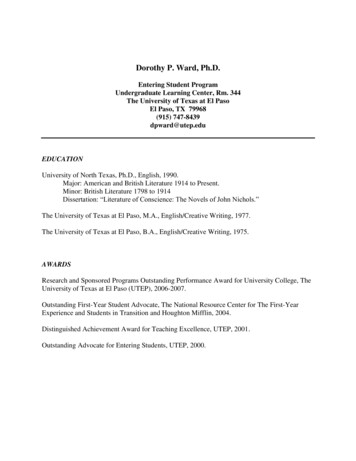
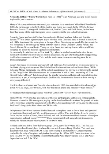
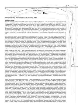
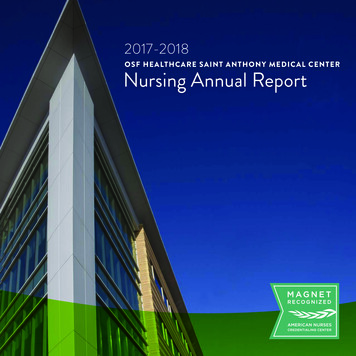
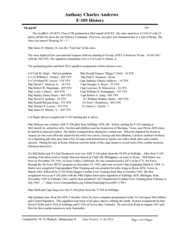
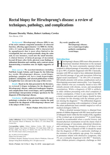
Rectal biopsy for Hirschsprung's diseaseThe gold standard test for the diagnosis of HDis rectal biopsy with careful evaluation of the biopsyspecimen by an experienced pathologist (Fig. 1). Tissueobtained from the posterior wall of the rectum 2 cmabove the dentate line (Fig. 2) in patients with HD willdemonstrate an absence of ganglion cells (Fig. 3) inthe submucosal plexus and hypertrophic nerve trunksin the submucosal plexus and myenteric plexus.[1,15]Histochemical staining for acetylcholinesterase (AChE)and immunohistochemical staining for calretinin mayaid in the diagnosis, though staining with hematoxylinand eosin (HE) may be sufficient for documenting thepresence or absence of ganglion cells (Figs. 4-6).[16]Rectal biopsy techniquesClinical practice varies, but due to the possibilityof obtaining insufficient tissue for diagnosis or thepossibility of taking a biopsy of the physiologicaganglionic region, most practitioners obtain 2-3 rectalbiopsy specimens per procedure.[16] An SRB can beperformed at the bedside without the use of generalxInternal sphincterExternal sphincterMucosaPerianal skinInternal rectal venousplexusRectal biopsy site 2 cm abovethe dentate lineTransition zoxe of physiologicaganglionosisDextate lineExternal rectal venous plexusAnal vergeFig. 2. Illustration of the rectum with site of rectal biopsy.Fig. 1. Suction rectal biopsy from a patient with Hirschsprung's disease(hematoxylin and eosin staining, original magnification 200). Athigher power and after examination of 50 sections no ganglion cellswere identified and nerve trunk hypertrophy was present (not seen at thismagnification).Fig. 4. Suction rectal biopsy of a patient without Hirschsprung'sdisease demonstrates normal acetylcholinesterase activity in themuscularis mucosa staining normal appearing nerve trunks (indicatedabove by red arrows) (original magnification 40).Fig. 3. A single normal ganglion cell (indicated above by red arrow) issufficient to rule out Hirschsprung's disease (original magnification 400).Fig. 5. Suction rectal biopsy of a patient with Hirschsprung's diseasedemonstrates abnormal acetylcholinesterase activity in the muscularismucosa highlighting hypertrophied nerve trunks in the muscularismucosa (indicated above by red arrows) (original magnification 40).World J Pediatr, Vol 12 No 2 . May 15, 2016 . www.wjpch.comFig. 6. Suction rectal biopsy of a patient without Hirschsprung'sdisease demonstrates normal calretinin activity in the lamina propriawith normal appearing nerve trunks in the muscularis mucosa(indicated above by red arrows) (original magnification 40) . Rectalbiopsy in a patient with Hirschsprung's disease would show an absenceof calretinin staining (not shown).137Review articleRectum
World Journal of Pediatricsanesthesia, though it often results in smaller tissuespecimens compared with full thickness biopsy (FTB).Though larger tissue specimens may be obtained, FTBrequires general anesthesia in the operating room andsuture closure of the biopsy site (Table 2).Review articleSuction rectal biopsyA myriad of techniques and devices to obtain a SRB havebeen described. A simplified procedure is as follows:position the patient in lithotomy position, insert thelubricated biopsy instrument along the posterior wall ofthe rectum, position the opening for the cutting blade 2cm above the dentate line and aim the blade posteriorly,apply suction, and trigger the cutting blade to obtaintissue specimen (Table 2). Different instrumentation setsrecommend different amounts of suction be applied toobtain a biopsy. Our particular instrument, the rbi2 (AusSystems, Australia), recommends calibration to 300 mmH2O prior to use, and then withdrawing the plunger to3-5 mL (using a 10 mL syringe), or ideally 150 cm H2Owhen using the manometer.[17] This is comparativelyless pressure than previously required by initial devicesthat classically requires 20 mL of pull back in a 60 mLsyringe, due to its completely air tight system that is ableto produce biopsy specimens at a lower, more uniformpressure.[17,18] Although practices vary by institution andphysician, typically 2-3 specimens are obtained at eachsession. Suction may be applied manually via syringe(preferred) or mechanically via wall suction, al
Hirschsprung's disease Delayed passage of meconium, abdominal distention, and vomiting herald diagnosis, congenital absence of ganglion cells on rectal biopsy, requires pull through Meconium ileus (cystic fi brosis) Contrast enema may be diagnostic and therapeutic, clinical diagnosis