Transcription
DocumentMaDiagnostic Imaging Guidelines:Getting to YesMagellan Health Services, Inc.April 2007
Diagnostic Imaging Guidelines:Getting to YES!Proprietary Notice 2007 Magellan Health Services, Inc. All rights reserved.The information contained and compiled herein (the “Information”) is the property of Magellan HealthServices, Inc. (“Magellan”) and is disseminated solely under license. It is never sold. The Information isprotected by copyright, trade-secret law and other laws. The copying, reproduction, republication or transferof the Information (in whole or part) by licensees or others without the express written permission ofMagellan is unlawful and will subject the violator to civil and criminal penalties.The Information may include imaging exam indicators (“Indicators”). Indicators are provided solely toqualified medical professionals and solely for informational purposes. Treatment decisions and othermedical decisions should be made only by qualified medical personnel in consultation with their patients andshould not be based, in whole or part, upon the Indicators. Distribution or dissemination of Indicators (bylicensees or others) other than to qualified medical personnel for any purpose whatsoever is strictlyprohibited.Magellan does not warrant the Indicators or other Information as to completeness or accuracy and disclaimsall warranties, express or implied, including any warranties of merchantability or fitness for any purpose. Magellan Health Services, Inc. 2007All Rights ReservedProprietary & ConfidentialPage 2
Diagnostic Imaging Guidelines:Getting to YES!ContentsProprietary Notice .2Dates of Revisions/Additions To Guidelines .6Diagnostic Imaging Guidelines, Getting to YES!.8Example of Decision Process .9Head and Neck Section . 11Brain, MRI – Codes 70551, 70552, 70553 (Includes Pituitary gland) . 11Brain, CT - Codes, 70450, 70460, 70470 (Includes Hearing Canal). 14Head MRA w/o Contrast–70544; MRA w/ Contrast–70545. 16Head MRA w/o & w/Contrast –70546 . 16Head, CT Angiogram – 70496. 18Neck MRAngiogram w/o Contrast 70547; . 19Neck MRA w/Contrast – 70548 . 19Neck MRAngiogram w/o & w/Contrast – 70549 . 19Neck CTA 70498 . 21Orbit, Face, and Neck MRI – 70540. 22Orbit, Sella, and Posterior Fossa MRI – 70480, 70481, 70482 . 22Orbit, Face, Neck MRIw/Contrast – 70542 . 22Orbit Face, Neck w/o & w/Contrast – 70543 . 22Temporal Bone, Mastoid, (Outer, Mid, or Inner) Ear CT –70480, 70481, 70482. 24Temporomandibular Joint (TMJ) MRI - 70336. 26Face, Neck and Orbit MRI – 70540. 27Face, Neck and Orbit CT w/Contrast– 70542. 27Face, Neck and Orbit CTw/o & w/Contrast- 70543 . 27Face and Sinus CT – 70486 70487 70488 . 27Paranasal Sinus CT – 70486 70487 70488 . 28Neck, Face, and Orbit MRI – 70540. 30Neck, Face and Orbit CT w/Contrast – 70542. 31Neck, Face, and Orbit CT w/o & w/Contrast - 70543 . 31Neck Soft Tissue CT – 70490, 70491, 70492. 31Brain Functional MRI (fMRI) 70554,70555 . 32Chest and Cardiac Section. 32Chest CT - 71250 71260 71270 . 33Chest MRI - 71550. 35Chest MRA – 71555 . 37Chest CTA - 71275 . 37Cardiac/Coronary CTA – 0148T . 38Breast MRI – uni lateral 77058, bi lateral 77059. 40Nuclear Cardiac Imaging / Myocardial Perfusion Imaging –78460, 78461 78464, 78465. 42MUGA / Gated Wall Motion Study - 78472 . 45Abdomen and Pelvis Section . 46Abdomen CT – 74150 74160 74170. 46Abdomen MRI – 74181 . 48Abdomen MRI w/Contrast – 74182. 48 Magellan Health Services, Inc. 2007All Rights ReservedProprietary & ConfidentialPage 3
Diagnostic Imaging Guidelines:Getting to YES!Abdomen MRI w/o & w/Contrast - 74183 . 48MRCP S8037 . 50Magnetic Resonance Cholangiopancreatography. 50CT Colonscopy (Virtual Colonoscopy) 0067 T. 52Abdominal MRA – 74185, CTA – 74175 Abdominal CT 74160 . 54Abdomen/Pelvis CT Combo: Abdo – 74150, 74160, 74170, Pelvic – 72192, 72193, 72194 . 56Pelvic CT – 72192 72193 72194 . 58Pelvic MRI - 72196. 60Spinal Imaging Section. 61Cervical Spine MRI – 72141 72142 72156 . 61Cervical Spine CT – 72125, 72126, 72127. 63Thoracic Spine MRI – 72146, 72147, 72157, CT – 72128, 72129, 72130. 64Lumbar Spine MRI - 72148 72149 72158. 65Lumbar Spine CT – 72131 72132 72133. 67Joint Imaging Section. 69Shoulder MRI – 73221. 69Joint MRI of Upper Extremity w/Contrast – 73722 . 69Joint MRI of Upper Extremity w/o & w/Contrast CT – 73200 73201 73202 . 69Elbow MRI Wrist MRI - 73221. 71Joint MRI of Upper Extremity w/Contrast- 73722 . 71Joint MRI of Upper Extremity w/o & w/Contrast - 73723. 71CT – 73200 73201 73202 . 71Knee MRI – 73721. 72Joint MRI of lower Extremity w/Contrast- 73722. 72Joint MRI of Lower Extremity w/o & w/Contrast - 73723 . 72CT – 73700 73701 73702 . 72Hip MRI - 73721 72196. 74Joint MRI of Lower Extremity w/Contrast – 73722. 74Joint MRI of Lower Extremity w/o & w/Contrast - 73723 . 74Pelvis (for bilateral hips). 74CT – 73700 73701 73702 . 74Ankle/Foot MRI - 73721 . 75Joint MRI of Lower Extremity w/Contrast – 73722. 75Joint MRI of Lower Extremity w/o & w/Contrast 73723. 75CT - 73700 73701 73702 . 75Non-Joint Extremity Imaging Section. 76Upper Extremity (non-joint) MRI – 73220. 76Upper Extremity (non-joint) w/o Contrast – 73218. 76Upper Extremity (non-joint) w/Contrast - 73219 . 76Lower Extremity (non-joint) MRI – 73720 . 76Lower Extremity (non-joint) w/o Contrast – 73718 . 76Lower Extremity (non-joint) w/Contrast - 73719. 76Upper Extremity CT – 73200 73201 73202 . 76Lower Extremity CT – 73700 73701 73702 . 76Extremity MRA Imaging Section . 78Lower Extremity MRA/MRV – 73725. 78 Magellan Health Services, Inc. 2007All Rights ReservedProprietary & ConfidentialPage 4
Diagnostic Imaging Guidelines:Getting to YES!“Runoff” CT Angiography -- 75635. 78Bone Marrow MRI Imaging. 79Bone Marrow MRI – 77084. 79PET (Positron Emission Tomography) Imaging Section. 80PET Cardiac Scan - 78459. 82PET Scan, Metabolic, Brain (Seizures and Tumors) 78608 . 83PET Scan, Metabolic, Brain (Alzheimer’s) 78608 . 84PET Scan, Tumor imaging - Head and Neck 78811, 78812, 78813, 78814, 78815, 78816. 86PET Scan, Tumor Imaging - Lymphoma -- 78811, 78812, 78813, 78814, 78815, 78816 . 87PET Scan, Tumor Imaging – Melanoma --78811, 78812, 78813, 78815, 78816. 88PET Scan, Tumor Imaging – ColoRectal -- 78811, 78812, 78813, 78814, 78815, 78816 . 89PET Scan, Tumor Imaging – Lung Cancer -- 78811, 78812, 78813. 90Non-Small Cell or Solitary Pulmonary Lesion --78814, 78815, 78816 . 90PET Scan, Tumor Imaging – Esophagus -- 78811, 78812, 78813, 78814, 78815, 78816 . 91PET Scan, Tumor Imaging – Thyroid --78811, 78812, 78813, 78814, 78815, 78816. 92PET Scan, Tumor Imaging – Cervical Cancer -- 78811, 78812, 78813, 78814, 78815, 78816. 93PET Scan, Tumor Imaging - Breast Imaging -- 78811, 78812, 78813. 78814, 78815, 78816. 94 Magellan Health Services, Inc. 2007All Rights ReservedProprietary & ConfidentialPage 5
Diagnostic Imaging Guidelines:Getting to YES!Dates of Revisions/Additions to GuidelinesGetting To YES! Guideline TitleCover PageProprietary NoticeTable of ContentsDates of Revisions/Additions to GuidelinesRead Me FirstHead and NeckBrain, Pituitary, Posterior Fossa, IAC MRIBrain, Pituitary, Posterior Fossa, IAC CTHead MRAHead CTNeck MRA/CTNeck CTAOrbit MRI/CTTemporal/Mastoid Bone MRI/CTTMJ/Mandible MRI/CTFace MRI/CTParanasal Sinus MRI/CTNeck, Face and Orbit MRINeck, Face and Orbit CTBrain Functional MRI (fMRI)Chest and CardiacChest CTChest MRIChest MRA/CTA.Cardiac/Coronary CTABreast MRINuclear CardiologyMUGAAbdomen and PelvisAbdominal CTAbdominal MRIMRCPCT Colonscopy (Virtual Colonoscopy)Abdominal MRA /CTAbdominal/Pelvic ComboPelvic CTPelvic MRISpinal ImagingCervical MRICervical CTThoracic MRI/CTLumbar MRILumbar CTJoint Imaging/Miscellaneous OrthoShoulder MRI/CTElbow /Wrist MRI/CTKnee MRI/CTHip MRI/CT Magellan Health Services, Inc. 2007All Rights EWXXXXXXNEWXXXXXXXXXXXXXProprietary & ConfidentialPage 6
Diagnostic Imaging Guidelines:Getting to YES!Getting To YES! Guideline TitleAnkle/Foot MRI/CTNon-Joint Extremity MRI/CT ImagingExtremity (non joint) MRI/CTExtremity MRA/CTA ImagingExtremity MRAPET ImagingPET (Positron Emission Tomography) Imaging Section “Read Me First”PET Cardiac ScanPET Scan – Metabolic Brain (Seizures and Tumors)PET Scan – Metabolic Brain (Alzheimer’s)PET Scan – Tumor Imaging - Head and NeckPET Scan – Tumor Imaging - LymphomaPET Scan – Tumor Imaging - MelanomaPET Scan - Tumor Imaging - ColoRectalPET Scan-Tumor Imaging - Lung Cancer,Non-Small Cell or Solitary Pulmonary LesionsPET Scan – Tumor Imaging - EsophagusPET Scan – Tumor Imaging - ThyroidPET Scan - Tumor Imaging - Cervical CancerPET Scan – Tumor Imaging - Breast Imaging Magellan Health Services, Inc. 2007All Rights ary & ConfidentialPage 7
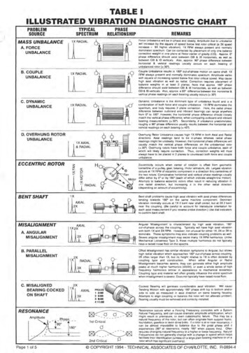
Diagnostic Imaging Guidelines:Getting to YES!Diagnostic Imaging Guidelines: Getting to YES!With due respect to Fisher and Ury, authors of the very popular book, Getting to Yes: Negotiating WithoutGiving In, this document is designed to establish a pathway to clinical consensus on the use of a single orcombination of Diagnostic Imaging examinations.2The reader/user is urged to keep in mind that the guidelines that follow are not only intended to aid in thearrival at clinical consensus with Magellan/NIA (National Imaging Associates) algorithms and/or Clinicalreviewers but also that they may be used as examples of “mainstream” medicine in clinical practice. Theyare intended to be “filters” that cover a specific study’s use in the vast majority of cases.Magellan/NIA understands that there will be unusual cases that will meet appropriate indications that arenot covered in the following document. Appropriate use is not limited to the following circumstances andmay be discussed and recommended at a peer-to-peer level.“Procedure-based guidelines” do not represent a complete episode of care; the decision process to obtain anImaging Study is assumed to be embedded in a continuum of care including pre-test assessment and theassumption that the test results will have a definite influence on post-test treatment. The reader will notethat the following document, as written, will reflect these imperatives.Note the following algorithm graphically demonstrates the process.2Fisher R, Ury W, Getting to Yes: Negotiating Without Giving In; 2nd Ed Penguin USA Dec. 1991. Magellan Health Services, Inc. 2007All Rights ReservedProprietary & ConfidentialPage 8
Diagnostic Imaging Guidelines:Getting to YES!Example of Decision ProcessCourtesy of Dexter Campinha-Bacote, MD. Magellan Health Services, Inc. 2007All Rights ReservedProprietary & ConfidentialPage 9
Diagnostic Imaging Guidelines:Getting to YES!Magellan/NIA criteria are the result of combined experience, current practice and extensive literaturereview. All revisions are the result of a scientific process of clinical consensus, approved by theMagellan/NIA Chief Medical Officer, Chief Executive Officer and Board of Directors. In each case ofapplication to an individual Health Plan they are also reviewed and approved by the plan Medical Directorconsistent with the plan’s internal quality and utilization management functions as well as all applicableaccrediting agencies including NCQA, URAC and the several States Departments of Insurance.Magellan/NIA reviewers use these guidelines in the day-to-day operation of the call center. Any MRI, CT,nuclear cardiac, PET or other study not found in this document will be referred for individual medical(peer-to-peer) review.We have included occasional literature references in this document. We have endeavored to providereferences to the latest or most classic information related to covered benefits as defined by typical healthplan Technical Assessment (TEC) Committees. Some will be recent while others will point to classic worksyet to be scientifically challenged.PLEASE NOTE Information provided during the review process is part of the patient’s medical record.Accuracy is paramount, as information provided may have a lasting impact on your patient’shealth ratings! General Medical Policy consists of medical guidelines and payment guidelines.Medical guidelines detail when certain medical services are medically necessary, and whether or not theyare investigational. (For more information concerning medical necessity and investigational criteria, pleasesee the health plan’s specific policies.) Our medical guidelines are written to cover a given condition for themajority of people. Each individual's unique, clinical circumstances may be considered in light of currentscientific literature. Medical guidelines are based on constantly changing medical science, and we reserve theright to review and update our policies periodically.Payment guidelines provide claims payment editing logic for CPT , HCPCS and ICD-9-CM coding.Payment guidelines are developed by clinical staff, and include yearly coding updates, periodic reviews ofspecialty areas based on input from specialty societies and physician committees and updated logic based oncurrent coding conventions.Benefits and eligibility are determined before medical guidelines and payment guidelines areapplied. Therefore, concurrence with these medical policies does not guarantee authorization,certification, explanation of benefits, or a contract. Benefits are determined by the group contractand the subscriber certificate that is in effect at the time services are rendered. Magellan Health Services, Inc. 2007All Rights ReservedProprietary & ConfidentialPage 10
Diagnostic Imaging Guidelines:Getting to YES!Head and Neck SectionBrain, MRI – Codes 70551, 70552, 70553 (Includes Pituitary gland)Pre-Test Clinical ConsiderationsMRI of the brain/head is a common procedure used for both diagnosis and follow-up of known orsuspected disease. It has largely replaced CT as the study of choice used to evaluate and follow a range of“diseases” from simple stroke to highly complex structural and metabolic conditions. It is a study that canbe appropriately utilized by skilled family practitioners and general internists as well as neuro-sciencespecialists. Over the past several years, elective imaging of the brain and head has moved toward MRI as theexamination of choice yet CT continues to occupy an important role in the diagnosis of acute injury,vascular, brain masses and bony diseases as well as the follow-up of known abnormalities. The attachedguidelines suggest the most appropriate use of a MRI brain study. Requests/indications not listed may resultin a physician-to-physician discussion.Magellan/NIA data indicates that most “discretionary” use of brain MRI is the result of inappropriatephysician/provider “demand” due to ill-defined headaches.Usually, the only two definite pre-test reasons for Neuro-Imaging in patients who present with generalized“headache” are: 3 Thunderclap HeadacheFocal neurologic signsOther Indications Headache; new and sudden, severe, persistent, unresponsive to medications (CT usually done foracute headache in the ER because of suspected bleeding)4 Visual disturbance Papilledema (swelling of the back of the eye) Neurological; weakness or paralysis, especially one sided; walking disturbance; cranial nerve or nerveimpairment, Bell’s Palsy, facial numbness Seizures (PET Scanning is gaining credence as the preferred examination for ill defined/persistentseizures)5 Nystagmus Tinnitus (constant “ringing” in one or both ears) Anosmia (loss of smell)6 7Frishberg BM; Rosenberg JH; et al. Evidence-Based Guidelines in the Primary Care Setting: Neuroimaging in Patients with NonacuteHeadache, 1999, The US Headache Consortium – American Academy of Neurology pp 8 and 16.34Lee S, Rao K, Zimmerman R (Ed). Cranial MRI and CT. (4th Edition). New York: McGraw Hill, 1999 p. 417-418.William H. Theodore, Kathey Kelley, Maria T. Toczek, William D. Gaillard Epilepsy Duration, Febrile Seizures, and Cerebral GlucoseMetabolism Epilepsia Vol. 45 Issue 3 Page 276 March 2004.56Amy Pruit, Primary Care Medicine, 1995 pp849.7Leopold, D.A. A perplexing olfactory loss. Arch. Otolaryngol Head Neck Surgery 126, 803. (2000). Magellan Health Services, Inc. 2007All Rights ReservedProprietary & ConfidentialPage 11
Diagnostic Imaging Guidelines:Getting to YES! Primary or metastatic tumor, new or follow-up (within reason, MRI will identify multiple orcomplex lesions)Trauma (MRI is commonly ordered but CT may be superior for depressed fracture)8Stroke or TIA9Multiple Sclerosis and other white matter disease10 11Arnold Chiari MalformationSuspected (r/o) bleeding, or vascular abnormalities12 13Syrinx, congenital or acquired (abnormal skull formation)14Suspected (r/o) congenital abnormality/developmental delayMeningitis or abscess15VasculitisEncephalopathy16Aneurysm or AV malformationHydrocephalus, primary or follow-up (usually CT)Craniosynostosis (recommend CT not MRI)AIDS17 18Endocrine abnormalityInvestigation of the internal auditory (hearing) canal is most often performed to evaluate a known orsuspected tumor such as an acoustic neuroma or cholesteatoma of the inner or middle ear. It isfrequently ordered in conjunction with a CT or MRI of the brain or head. In general, if “suspected” werecommend only the head examination be obtained but if “known” then we suggest a study of thespecific area. Documented sensorineural hearing loss Acoustic Neuroma OpticRothrock SG, Buchanan C, Green SM, et al. Cranial computed tomography in the emergency evaluation of adult patients without a recenthistory of head trauma: a prospective analysis. Acad Emerg Med 1997 Jul;4(7):654-61.89Raymond D. Adams & Maurice Victor, Principles of Neurology 1995.10 Rovaris M, Filippi M. The value of new magnetic resonance techniques in multiple sclerosis. Current Opinion in Neurology 2000Jun;13(3):294-54.PRISMS (Prevention of Relapses and Disability by Interferon -1a Subcutaneously in Multiple Sclerosis) Study Group : Randomised doubleblind placebo-controlled study of interferon -1a in relapsing/remitting multiple sclerosis. Lancet. 1998: 352: 1498- 504.1112 Rothrock SG, Buchanan C, Green SM, et al. Cranial computed tomography in the emergency evaluation of adult patients without a recenthistory of head trauma: a prospective analysis. Acad Emerg Med 1997 Jul;4(7):654-61.13Wardlaw JM, White PM. The detection and management of unruptured intracranial aneurysms. Brain 2000 Feb;123 (Pt 2):205-21.Pavlova NG, Konstantinove NN, Arutjunyan AV. Functional and biochemical criteria for investigation of brain development disorders. Int JDev Neurosci 1999 Dec;17(8):839-48.1415Moses S: Meningitis: Acute Bacterial Meningitis. 2001. Available at: http://www.fpnotebook.com/NEU112.htm. Accessed April 12, 2004.16Rosenberg S. Recent advances in the molecular biology of hepatitis C virus. J Mol Biol 2001; 313:451-64.17Rohit Bakshi, MD Medscape Neurology & Neurosurgery 2(1), 2000. 2000 Medscape Portals, Inc. Accessed April 2004.18Raymond D. Adams & Maurice Victor, Principles of Neurology 1995, pp561. Magellan Health Services, Inc. 2007All Rights ReservedProprietary & ConfidentialPage 12
Diagnostic Imaging Guidelines:Getting to YES!General MRI is better than CT, especially for the rear of the brain (chronic dizziness, hearing loss). In many cases MRI will be recommended as a substitute if a CT is requested unless there arecontraindications to MRI which may include motion, pacing devices and other metallic devices.19 CT is less expensive than a MRI, a much faster procedure and easier to schedule. It is a very good test forinitial study when the index of suspicion of complex disease is low. There is rarely a need both CT and MRI. For pituitary gland evaluation, MRI of the sella tursica or pituitary is the best study (microadenoma,prolactin tumor, others).20 Under certain circumstances MRS studies can be used to differentiate tumor/recurrence from radiationeffect and should be considered as an adjunctive study.21 MRS, however, is considered investigational by most plans. Orbits and/or sinuses are well demonstrated on brain MRI, and not as well on CT. Therefore if both are ofserious concern, consider a MRI to cover both. Under certain circumstances an ENT specialist mayspecifically want the bone detail of a CT. Not recommended for short term symptoms such as dizziness, tension headaches, hypertension. Temporal Bone, IAC or mastoid MRI: There is no separate CPT (billing) code for this procedure; theseareas are imaged in the exam for a Brain MRI.Combination studies - May be useful if they meet the following requirements: Brain/Cervical – Arnold Chiari, Multiple Sclerosis, demyelinating disease. Brain/Neck – confirmed carotid (blood vessel) occlusion of 60% (but only if candida
Aug 25, 2007 · Diagnostic Imaging Guidelines: Getting to YES! With due respect to Fisher and Ury, authors of the very popular book, Getting to Yes: Negotiating Without Giving In , this document is designed to establish a pathway to clinical consensus on the use of a single or combinat