Transcription
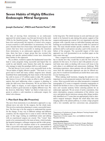
EditorialSeven Habits of Highly EffectiveEndoscopic Mitral SurgeonsInnovations00(0) 1–6 The Author(s) 2020Article reuse guidelines: sagepub. com/ journals- permissionsDOI: 10.1177/1556984519888456 journals. sagepub. com/ home/ invJoseph Zacharias1, FRCS and Patrick Perier2, MDThe idea of moving from sternotomy to an endoscopicapproach for mitral surgery was first put forward in the mid- 1990s by the Stanford group.1 The adoption of this idea hasbeen slow and presently the majority of mitral valve surgeryin most countries is still undertaken via sternotomy. Over thepast 2 decades there have been many individual surgeons andcenters that have been successful in making the transitionfrom sternotomy to an endoscopic approach. At the sametime, there has been a larger group that has resisted thischange and specific patient- related events have disinclinedthem further to this approach.We, as editors, wanted to capture the fundamental issues thatlead to some programs being successful and hoped that wecould try and identify some features that unify the surgeonswho manage to make the paradigm shift in a safe manner.The late Steven Covey wrote a very successful book on the“Seven habits of highly effective people.” This book is a verygood read in itself and many cardiac surgeons have alreadybenefitted from understanding the basic tenets of the book thathas sold in excess of 25 million copies to date. We unashamedly borrow Covey’s title to make an effort to distil 7 specificaspects that need addressing when a surgeon is planning tomake the shift away from sternotomy. We do not believe that“rules” apply in surgery, as each surgeon and their team arelikely to achieve good outcomes in slightly different ways. Wedo, however, think these “habits” are likely to help more surgeons to achieve a safe transition to endoscopic surgery formitral valve disease.1. Plan Each Step (Be Proactive)The move from sternotomy to an alternative approach is a significant move not only for the surgeon, but the whole team.This involves meticulous planning to create a successful transition. The first step is for the surgeon to understand the reasonsfor the change. It may vary in different settings and differentcountries, but the surgeon has to come to terms with the finalgoal. At this stage visiting an established center and talkingwith an experienced surgeon are crucial. This “vision” thenneeds to be communicated to the team at home, which includesthe hospital managers, colleague surgeons and the wider group.The idea of a win–win situation needs to be communicated.With a successful endoscopic surgery program, the number ofcases will increase in a department and all parties will be busierin the long term. The initial increase in costs and time per caseneeds to be factored in and, during this period, support of thesurgeon and program are crucial. It is very important to start theprogram with a specific team as individual learning and teamlearning are accelerated when the team is stable for a period oftime. The team should include specific anesthetic, scrub, andperfusion staff as each person can play a part in the success orfailure of the program. The successful impact of this teamapproach was well documented in an excellent paper on thetopic some time ago.2Once a surgeon has secured institutional support, he or shehas to decide how they would like to pick the first cohort ofpatients for this move. It is best to start with patients of a normal body mass index as dealing with excess soft tissue in theearly phase is an additional and unnecessary challenge. Withincreasing experience, obese patients who are likely to benefitmore from the endoscopic approach can also be offered surgery, but we would not recommend you tackle these cases earlyin your learning curve.When making small incisions, imaging the patient is veryimportant to avoid unexpected anatomy that leads to poor outcomes. Most successful units use either preoperative computedtomographic (CT) scans or magnetic resonance angiograms orHybrid lab facilities to have a clear understanding of both thoracic and vascular anatomy before selecting patients for anendoscopic approach. We are aware of aortic coarctation, iliacartery interruption, azygous continuation of the inferior venacava, and patent ductus arteriosus among other things incidentally identified on CT scans that can appropriately lead to alternative surgical strategies. Despite the small delay that extraimaging adds to the patient pathway, to create a large programwith a low mortality and low conversion rate, preoperative1Department of Cardiothoracic Surgery, Blackpool Teaching Hospitals NHSFoundation Trust, Blackpool, UK2Department of Cardiac surgery, Herz- und Gefäßklinik GmbH, BadNeustadt, GermanyCorresponding Author:Joseph Zacharias, Department of Cardiothoracic Surgery, BlackpoolTeaching Hospitals NHS Foundation Trust, 83 Whinney Heys Road,Blackpool FY3 8NR, UK.Email: drjzacharias@ gmail. com
2imaging is crucial to the success of most centers and should beincreasingly considered a standard of care.2. Decide on a Cannulation Technique(Begin With the End in Mind)A very important aspect of endoscopic surgery is identifyingalternative cannulation options both for arterial and venousaccess. Once preoperative imaging is complete, a large proportion of patients will be suitable for a femoral artery cannulation.Previous studies have raised concerns regarding peripheralcannulation and rightly so.3 Peripheral cannulation has to becombined with both preoperative imaging and intraoperativevisualization of guide wires with transesophageal echocardiography (TEE) during cannulation to make this a safe strategy.There will be a group of patients who are not suited to femoralartery cannulation, as identified on preoperative imaging. Thesepatients could be considered for either axillary cannulationorcarotid cannulation as described by appropriately experiencedaortic surgeons.4 In our experience, it is a very small group thatwill need central aortic cannulation but this also can be achievedif required.5 Some patients may have venous anatomical abnormalities that preclude femoral venous cannulation. Centralvenous cannulation is possible in selected cases but should berestricted to experienced centers. In our experience, mostpatients with a normal weight ( 80 kg) can have a single femoral venous cannula placed with the tip in the superior venacava to achieve adequate drainage of the patient. We would recommend an internal jugular cannula routinely in the early partof the learning curve and subsequently in larger patients andthose needing right heart procedures.We cannot stress enough, the importance of planning peripheral cannulation safely in each patient and this step can be theundoing of many surgeons in the early part of the learningcurve. Once peripheral cannulation is mastered, its benefitsextend to all aspects of cardiac surgery and make a lot of complex cardiac surgery safer. Good peripheral cannulation reducesthe stress at the end of the procedure and reduces the potentialfor bleeding from aortic suture sites.Innovations 00(0)surgeons who are used to placing clamps on the ascending aortathrough a sternotomy approach. This has to be combined witha cardioplegia cannula in the ascending aorta to deliver antegrade cardioplegia. The cardioplegia chosen should be the onebest suited to the department and the surgeon. There are excellent case series with the use of both crystalloid and blood plegiaand the principles of cardioplegia delivery should not be compromised. We have included a video of the application of anexternal clamp on the aorta performed by one of the authors(Supplemental Video 1).A less popular but equally effective option is one of an endovascular clamp. This does involve the insertion of a larger femoral cannula to reduce the flow pressure in the peripheralvessels due to space occupied by the catheter within the cannula. This technique received a bad reputation in the initialyears due to incidents of retrograde aortic dissection. This wasnoted to be strongly associated with high line pressures andintroducing a second inflow line in the contralateral femoralartery has virtually eliminated the risk of retrograde dissections.7 A guide for perfusionists is not to exceed 300 mmHgline pressure during cardiopulmonary bypass. The endoclampalso requires excellent TOE guidance and awareness of migration by monitoring both arterial pressures in both radial arteries, which are some of the factors that make this less popular.Another video shows an endoclamp being inflated with TOEmonitoring (Supplemental Video 2). As it is a single- use device,there is also a financial impact of using this approach.A further alternative that is predominantly used in the redosetting, but also by some groups in primary cases,8 is where theheart is allowed to fibrillate or beat on cardiopulmonary bypassand the procedure is done with coronary perfusion. This technique has previously been proven safeeven through a thoracotomy approach9 and is increasingly a popular alternative forendoscopic surgeons. An extra pump sucker is required to dealwith blood that trickles back into the left ventricle from thethebesian veins and additional time must be allowed for deairing the left ventricle before allowing it to eject back into thecirculation.4. Choose an Endoscope (Think Win–Win)3. Pick Your Myocardial Protection Strategy(Put First Things First)The key aspect for the explosion of cardiac surgery across theworld has been the introduction of cardiopulmonary bypass andof safe techniques in myocardial protection. The safety of myocardial protection techniques has improved to such an extent,that today it has proved extremely difficult to prove the advantage of off- pump surgery over on- pump arrested heart techniques.6 This should not be compromised in any way whensurgeons make the move to an endoscopic approach. To surgeons starting off, there are 3 clear alternatives to look at. Thefirst and most popular is the use of an external clamp on theascending aorta. This is intuitively the easier option for mostMany surgeons start to move away from a full sternotomy byinitially offering a hemisternotomy or a right minithoracotomy.This is the first step out of the comfort zone that we have beenused to and is a necessary step toward change. We encouragethese surgeons to take the next step and introduce an endoscopeinto the field not just to better illuminate the area of work, butto add magnification and better visual definition. With increasing experience, the surgeon will move from looking throughthe small incision to the large screen with superior clarity anddetail and this is when the real advantage of endoscopic cardiacsurgery comes into its own. With the endoscope, the utility incision does not need to match the surgeon’s interpupillary distance to allow binocular vision but can become smaller as theonly limitation to the size is the degrees of motion required for
3Zacharias and PerierTable 1. Pros and Cons of Endoscopy Choice.CostSize of portsDepth perceptionVisual comfortSurgeon tirednessManual dexterity aidLearning curveAssistant view2D HD endoscope3D endoscopeRobotic endoscopeHigh (x1)5 mmPoorGoodHighReasonableHard2DHigher (x2)10 mmExcellentBetterLowBetterEasy3DHighest (X10)12 mmExcellentBestLowestBestHardest2D2D, 2- dimensional; 3D, 3- dimensional; HD, high definition.the procedure. The teams that have moved to a totally endoscopic procedure see benefits in blood loss, length of stay, andrecovery times compared to sternotomy.10The type of endoscope used is of importance. Some centersproduce excellent results with a 2- dimensional (2D) endoscopebut this requires more mental energy and the learning curve canbe variable dependent on the individual involved.Many surgeons including the 2 authors have found the useof 3D endoscopy very helpful and there is evidence from otherspecialties that this is associated with a shorter learning curveand better depth perception.11 At present, all 3D endoscopesrequire a 10 mm port against a 5 mm port for a high- definition2D camera. We feel the extra 5 mm is a price worth paying butthis needs to be evaluated by each surgeon to compare the prosagainst the cons (Table 1). The surgeon needs to be comfortableworking off a large screen and moving from surgical loupes towearing different 3D glasses. (Figs. 1 and 2).Robotic surgery also provides the operating surgeon at theconsole with high- fidelity magnified and motion- stabilized 3Dvision but does leave the patient- side surgeon with 2D vision.The major advantage of robotic surgery is conferred by thearticulated arms, which aid complex intrathoracic motion.Some surgeons may find this helpful, and if so, they will againneed to carefully consider how it is implemented and how theFig. 1. Operative room setup.costs will be managed in their own institution. At present, thereare no randomized controlled trials or propensity- matched evidence showing an advantage of a robotic strategy over a minimally invasive one.12The endoscope has revived many surgical specialties.Urology and orthopedics have led the way and, lately, abdominal and thoracic surgery have moved to a predominantly endoscopic approach. This has had several advantages. First, it hasled to lower morbidity for patients and this, in turn, has permitted more patients to be considered suitable for a procedure.Secondly, it helps training junior surgeons as they see and learnmore from looking at a magnified image on a large screen thanthe occasional view over someone’s shoulder. This attracts abetter and more motivated trainee to the specialty. Finally, thishas to be seen as a win–win for a department as the more casesthat are referred the busier everybody becomes. In both ourinstitutions, our sternotomy surgeon colleagues are doing moreFig. 2. Surgeon and assistant using 3- dimensional eyewear duringsurgery.
4operations since we embarked on an endoscopic program asthere will always be a group of patients that need a sternotomyapproach. The departments benefit from the more motivatedjunior partners that this strategy attracts, and we have alsofound a better relationship with our industry partners to be anadded bonus.5. Master Mitral Valve Repair Techniques(Seek First to Understand, Then to BeUnderstood)Mitral valve repair is complicated and all surgeons who areinvolved in this subspecialty interest are on a learning curve, allof the time. The first and foremost principle should be to do thebest mitral repair irrespective of the incision. The easiest casesto deal with, through an endoscope, are mitral valves with pureannular dilatation. Leaflets that need a limited triangular resection are likely to be cases that are easy to deal with in the initialpart of the learning curve. With increasing experience, a surgeon can move to working on the subannular plane as judgingthe length of artificial chords can be tricky early in the learningcurve.There is a real need for technology to make this aspect easier. Currently the availability of premeasured chords helpssome surgeons deal with complex pathology and achieve excellent results with an endoscopic approach.13 Centers also achievesimilar results with partial bands and complete rings and bothoptions can be used through an endoscopic approach withexcellent long- term results.14 We would recommend surgeonsto use the techniques that they are most familiar with and notcompromise on the quality of mitral repair.Despite the best intentions of surgeons, mitral valve repairsdo fail sometimes. There is so far, no clear data to suggest thatmitral valves repaired through a sternotomy are less likely tofail than those done endoscopically. A randomized trial mayprovide some light on the subject in time. In the meantime, webelieve that patients who have had a sternotomy previously canhave an endoscopic approach and those who have had a failedendoscopic approach can have a low- risk sternotomy approachon the second sitting if required. Once again, a more collaborative attitude between these 2 approaches is a better way for thespecialty to progress.In due time we are likely to understand that we need to plana sternotomy approach for certain pathology and an endoscopicapproach for others. With increasing experience and bettertechnology this boundary will continue to shift in each institution over time.6. Be Aware of Postoperative Pitfalls(Synergize)Cardiac surgery is a pleasure to perform, although the joy issometimes tempered by the incidence of unpredictable complications. Over the years we have come to accept the manyInnovations 00(0)shortcomings of a sternotomy approach as a price to pay for ourway into the chest. Moving to an endoscopic approach brings anew set of complications that surgeons need to be aware of.With experience and careful attention to detail these compilations can be reduced to very low levels.One common problem after endoscopic surgery is the groinhematoma or seroma related to femoral vessel dissection.Moving the skin incision to above the groin crease and reducing the dissection to the front wall of the vessels can eliminatethis frustrating complication. Using newer technology thatallows percutaneous cannulation and dressings that have in- built suction may be an alternative to reduce these in the futurefor high- risk patients.Phrenic nerve palsy is rare, but can have devastatingsequelae, leaving asymptomatic patients with new breathlessness. Beware of the phrenic nerve when opening the pericardium, as traction too close to the phrenic nerve can lead to aneuropraxia that can take up to 6 months to recover from.Diathermy close to the nerve can lead to irreversible damageand should be avoided.There have been anecdotal reports of poor myocardial protection and this needs to be looked at in context of the experience of many centers where there have not been increasedreported use of intra- aortic balloon pump or assist devices.10,11If this problem is noted by a surgeon or at a center, a root causeanalysis may throw light on the specific aspect and hopefully aperiod of supervision from another center or surgeon will helpthem overcome this issue.A cerebrovascular event is another feared complication ofan endoscopic approach and previous reports have suggested ahigher incidence of this compared to sternotomy surgery.3 Wehave not found this to be a concern if patients are selected forthis approach with preoperative imaging.11,13 There have beenreports of unilateral pulmonary edema early in some case seriesthough the cause is still not yet fully understood.Cardiac tamponade is less likely after endoscopic surgery. Inthe very occasional case of bleeding from the atrial suture line,the blood overflows into the right chest due to the open pericardium on the right side. A regular chest X- ray is recommended,and any pleural effusions are best dealt with quickly if noticed.As a group of surgeons, we need to come together to understand each potential complication and work toward a strategyto avoid them going forward. Some of these complications mayneed help from industry partners but we need to work togetherwith the overall aim of making this approach both reproducibleand safe.7. Reflect and Learn (Sharpen the Saw)The most important advantage of an endoscopic approach hasto be the ability for surgeons to be able to record the procedurein real time. This can lead to reflective learning by reviewingthe video after the procedure. Most endoscopic surgeons spendsome time looking back at the operative video and often areable to comment on how they could do the procedure better
5Zacharias and Periernext time. This cycle leads to constant improvements and inturn leads to better outcomes. There is increasing evidence tosupport the long- suspected fact that surgical skill affects patientoutcomes.15 With increasing use of endoscopes we will be ableto not only learn from our own work but also from each other.Sharing videos between surgeons will become a powerful toolin transferring good practice and a pilot program is alreadyshowing benefits in pediatric cardiac surgery training in theUSA.16Constant improvement requires a great deal of insight.This is not always available and is often absent in those surgeons who probably would benefit the most. Recording anendoscopic procedure will be a first step in creating a transparent system where surgical skill can be commented uponand improved both during training and subsequently in lateryears.Recording devices have greatly helped improve theassessment and behavior of motorists and the police whentrialed.17 It is only a matter of time when it will be expectedof surgeons too. Future surgeons are more likely to be judgedby the quality of their surgical videos than the number ofpublications.A Mindset of AbundanceTaking a further leaf out of Stephen Covey’s book, we in cardiac surgery need to embrace endoscopic surgery by changingour mind frame from one of “scarcity” to “abundance.”Department leads need to see endoscopic cardiac surgery as acollaborative approach that needs developing within each team.Transcatheter technologies have threatened our specialty forsome time but it seems unlikely that they will ever replace theneed for cardiac surgery. Increasing number of surgeons andcenters are seeing their work reduced as more patients choose atranscatheter approach. We cannot expect patients who areolder and more frail than ever before to accept the morbidityassociated with a sternotomy in the coming years, especially aswe move to treat asymptomatic patients. This editorial capturesthe expertise of 2 busy endoscopic surgeons who have seentheir work grow in the past years. With the increase in globalpopulations and a more aging group of individuals, there issadly enough disease substrate to keep us all busy at work. Wecannot hope to compete against a catheter with a saw. We aremore likely to prevail with an endoscope.Declaration of Conflicting InterestsThe author(s) declared the following potential conflicts of interestwith respect to the research, authorship, and/or publication of thisarticle: Joseph Zacharias is a paid proctor for Edwards Lifesciences,Abbott, and Cryolife.FundingThe author(s) received no financial support for the research, authorship,and/or publication of this article.Supplemental MaterialSupplemental material for this article is available online.References1. Pompili MF, Stevens JH, Burdon TA, et al. Port- accessmitral valve replacement in dogs. J Thorac Cardiovasc Surg1996; 112: 1268–1274.2. Edmondson AC, Bohmer RM and Pisano GP. Disrupted routines:team learning and new technology implementation in hospitals.Adm Sci Q 2001; 46: 685–716.3. Falk V, Cheng DCH, Martin J, et al. Minimally invasive versusopen mitral valve surgery: a consensus statement of the International Society of Minimally Invasive Coronary Surgery (ISMICS)2010. Innovations 2011; 6: 66–76.4. Urbanski PP, Thamm T, Bougioukakis P, et al. Efficacy of unilateral cerebral perfusion for brain protection in aortic arch surgery.J Thorac Cardiovasc Surg. Epub ahead of print 21 February2019.5. Murzi M and Glauber M. Central versus femoral cannulationduring minimally invasive aortic valve replacement. Ann Cardiothorac Surg 2015; 4: 59–61.6. Benedetto U, Puskas J, Kappetein AP, et al. Off- pump versus on- pump bypass surgery for left main coronary artery disease. J AmColl Cardiol 2019; 74: 729–740.7. Casselman F, Aramendi J, Bentala M, et al. Endoaortic clamping does not increase the risk of stroke in minimal access mitralvalve surgery: a multicenter experience. Ann Thorac Surg2015; 100: 1334–1339.8. Ad N, Holmes SD, Shuman DJ, et al. Minimally invasive mitralvalve surgery without aortic cross- clamping and with femoral cannulation is not associated with increased risk of strokecompared with traditional mitral valve surgery: a propensityscore- matched analysis. Eur J of Cardio- Thoracic Surgery2015; 48: 868–872.9. Thompson MJ, Behranwala A, Campanella C, et al. Immediateand long- term results of mitral prosthetic replacement using aright thoracotomy beating heart technique. Eur J CardiothoracSurg 2003; 24: 47–51.10. Grant SW, Hickey GL, Modi P, et al. Propensity- matched analysis of minimally invasive approach versus sternotomy for mitralvalve surgery. Heart 2019; 105: 783–789.11. Anschuetz L, Niederhauser L, Wimmer W, et al. Comparison of3- vs 2- dimensional endoscopy using eye tracking and assessmentof cognitive load among surgeons performing endoscopic ear surgery. JAMA Otolaryngol Head Neck Surg 2019: 838. Epub aheadof print 25 Jul 2019.12. Hawkins RB, Mehaffey JH, Mullen MG, et al. A propensitymatched analysis of robotic, minimally invasive, and conventional mitral valve surgery. Heart 2018; 104: 1970–1975.13. Falk V, Seeburger J, Czesla M, et al. How does the use ofpolytetrafluoroethylene neochordae for posterior mitral valveprolapse (loop technique) compare with leaflet resection?A prospective randomized trial. J Thorac Cardiovasc Surg2008; 136: 1200–1206.
614. Seeburger J, Borger MA, Falk V, et al. Minimal invasive mitralvalve repair for mitral regurgitation: results of 1339 consecutive patients. Eur J Cardiothorac Surg 2008; 34: 760–765.15. Birkmeyer JD, Finks JF, O'Reilly A, et al. Surgical skilland complication rates after bariatric surgery. N Engl J Med2013; 369: 1434–1442.Innovations 00(0)16. Anderson BR, Kumar SR, Gottlieb- Sen D, et al. The CongenitalHeart Technical Skill Study: rationale and design. World J PediatrCongenit Heart Surg 2019; 10: 137–144.17. Turner BL, Caruso EM, Dilich MA, et al. Body camera footageleads to lower judgments of intent than dash camera footage. ProcNatl Acad Sci U S A 2019; 116: 1201–1206.
The late Steven Covey wrote a very successful book on the “Seven habits of highly effective people.” This book is a very good read in itself and many cardiac surgeons have already benefitted from understanding the basic tenets of the book that has