Transcription
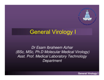
Chapter 5 - Lesson 4Virology TechniquesIntroductionVirology is a field within microbiology that encompasses the study of viruses and the diseases theycause. In the laboratory, viruses have served as usefultools to better understand cellular mechanisms. Thepurpose of this lesson is to provide a general overviewof laboratory techniques used in the identification andstudy of viruses.A Brief HistoryIn the late 19th century the independent work of DimitriIvanofsky and Martinus Beijerinck marked the beginning of the field of virology. They showed that the agentresponsible for causing a serious disease in tobaccoplants, tobacco mosaic virus, was able to pass throughfilters known to retain bacteria and the filtrate was ableto cause disease in new plants. In 1898, Friedrich Loeffler and Paul Frosch applied the filtration criteria to adisease in cattle known as foot and mouth disease. Thefiltration criteria remained the standard method usedto classify an agent as a virus for nearly 40 years untilchemical and physical studies revealed the structuralbasis of viruses. These attributes have become the basis of many techniques used in the field today.This electron micrograph depicts an influenza virusparticle or virion. CDC.BackgroundAll organisms are affected by viruses because virusesare capable of infecting and causing disease in all living species. Viruses affect plants, humans, and animals as well as bacteria. A virus that infects bacteriais known as a bacteriophage and is considered theChapter 5 - Human Health: Real Life Example (Influenza)Bacteriophage. CDC.1
most abundant biological entity on the planet. Manyanimal disease systems serve well as models for human disease. Many of the techniques used in the studyof viruses are the same whether they infect plants orwarm- or cold-blooded creatures because the techniques are based more on the virus studied rather thanthe species affected.Viruses are considered obligate intracellular parasitessince they require living host cells to replicate. Viruses take over or hijack the cellular synthesis machineryin order to reproduce. Viruses are submicroscopic andcontain either DNA or RNA as their genome and isenclosed in a protein shell called the capsid. Codedin the DNA or RNA genome of the virus is all theinformation needed for replication. Some viruses alsocontain lipids, carbohydrates, and special enzymesthat assist in their transmission and replication. Someviruses are enveloped; this envelope is acquired fromthe host cell either from the cytoplasmic or nuclearmembrane. This outer coat is immunogenic, (what theimmune system recognizes and what antibody producing cells respond to), and is necessary for the virusto invade a cell.Laboratory TechniquesSeveral different methods are used to study virusesand viral diseases, as the field is constantly changingwith the discovery of new methodologies and technologies. This section will provide a cursory overviewof the most commonly used techniques in diagnosticvirology and will conclude with a brief glimpse of virology in research.Diagnostic virology is concerned with identifying thevirus associated with clinical signs and symptoms.Procedures most commonly used include:1. Detection of a meaningful immune response tothe virus (antibody or cell-mediated) by immunologic assay(s)2. Identification of the agent by staining of specimens or sections of tissue (light and electron microscopy)3. Isolation and identification of the agent (cell culture or fertile eggs)4. Detection of viral nucleic acid (probes or amplification).2This illustration provides a 3D graphical representation of a generic influenza viron’s substructure. Aportion of the viron’s outer protein coat has beencut away. Dan Higgins, CDC.Detection of Immune ResponseOften, it is difficult to identify a virus in relation to thedisease observed, or when conducting a retrospectivestudy of a population to determine exposure to a virus,or when measuring the response of an individual to avaccine. In these cases, indirect methods of measureare needed, such as measuring antibody response tothe virus of interest. Several methods exist for thispurpose. A few of the most commonly used methodsinclude: Virus neutralization (VN)Hemagglutination inhibition (HI)Enzyme linked immunosorbent assay (ELISA)Indirect fluorescent antibody (IFA)Complement fixation (CF)Agar-gel immunodiffusion (AGID)Agar-gel precipitin (AGP)Latex agglutination (LA).The principles of these assays are fundamentally thesame, they depend upon antibody-antigen interactionsand consist of a known virus or viral protein, a patientsample (usually serum), and an indicator. If antibodies are present in the patient’s serum, they will bind tothe virus. If no antibodies are present, no binding willoccur. The indicator is observed to determine whetherthe sample is positive or negative for antibodies.Chapter 4 - Human Health: Real Life Example (Influenza)
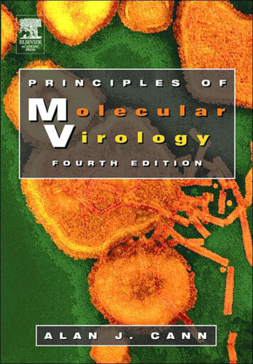
Hemagglutination (HA) and Hemagglutination Inhibition (HI) TestRed blood cellsstay in suspension Virus Red blood cells Virus Hemagglutination(clumping of red blood cells,cells remain suspended) Antibodies Red blood cellsform a pellet Red blood cells No hemagglutination(no clumping of red blood cells,the cells settle to the bottom of well)Virus NeutralizationIn the virus neutralization (VN) test, the sample of interest is incubated with the target virus and changesin cell culture are observed (called cytopathic effect,CPE). If the sample contains antibodies, it will preventthe virus from growing in the cell culture and no CPEwill be observed. If no antibodies are present in thesample, the virus will grow and CPE will be observed.Hemagglutination InhibitionCertain viruses have a protein on their surface that interacts with red blood cells and is able to attach tothem. This property is called hemagglutination andthe surface protein of the virus is hemagglutinin. Theinhibition or blocking of this activity is the basis ofthe hemagglutination inhibition (HI) test. The mostwell known virus with this property is the influenzavirus. Like the virus neutralization (VN) test, the patient’s serum sample is incubated with the virus ofinterest but instead of growing the virus in cells, redblood cells are added to the virus-serum mix. If antibodies are present, the hemagglutination activity willbe blocked; if no antibodies are present the virus willagglutinate (bind together). In this case the red bloodcells are the indicator.Chapter 5 - Human Health: Real Life Example (Influenza)Normal bovine kidney (BK) cellsBovine kidney (BK) cells showing cytopathic effects (CPE) of bovine herpesvirus.3
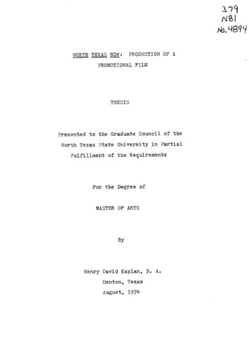
Enzyme-Linked Immunosorbent Assay (ELISA) / Enzyme Immunoassay Assay (EIA)EnzymeEnzymeEnzymeEnzymeCoat platewith virusor proteinAdd sampleIf antibody ispresent will bindto coated plateEnzymeEnzymeEnzymeEnzymeIf no antibodypresent will notbindIf no antibodypresent will notbind to coated plateNo antibody no colorIndicatoraddedIf antibodypresent willbindantibody colorEnzyme-Linked Immunosorbent AssayThe enzyme-linked immunosorbent assay ELISA is avery popular technique due to the ease of use and lowcost. The ELISA consists of plastic wells coated witheither the antigen (virus) of interest or a protein specific to the antigen (virus) of interest. The unknownsample (serum) is allowed to bind to the coated well,an antibody labeled with an enzyme is applied, an indicator is added, and then a color change is observed.The presence of color indicates the presence of antibodies and the absence of color indicates the absenceof antibodies.Agar-Gel Immunodiffusions (AGID)NegativeAbNegativeVirusAbAbPositiveLine ofIdenityThe agar-gel immunodiffusion (AGID), also referredto as an agar gel precipitin (AGP) test, involves thediffusion of virus and antibody through an agar (gelatin-like substance), which will form a line of identitywhere the antigen-antibody complexes form.Schematic of an agar gel immunodiffusion (AGID) or agargel precipitin (AGP) test. “Ab” represents a know antibodyto the known virus in the middle4Chapter 4 - Human Health: Real Life Example (Influenza)
The electron microscope is being used to examine a thin section of the variola virus, revealingsome of the structural features displayed by this pathogenic organism. CDC.Light and Electron MicroscopyLight MicroscopyViruses, unlike bacteria, are too small to be seen using a standard light microscope. Therefore, antibodieslabeled with an indicator, most frequently peroxidaseor fluorescence, designed to identify the virus of interest are used. This label then enables the visualizationof the virus cluster (because a single virus is too smallto see with a light microscope) with the light microscope, in the case of peroxidase, or an ultraviolet (UV)light microscope in the case of fluorescence.see the virus of interest. However techniques can beused to improve this, such as immune electron microscopy. With this technique the sample is incubatedwith an antibody against the virus of interest and theantibody-antigen reaction results in clumps of the virus, which allows for easier visualization. With theadvancements in molecular methodologies, electronmicroscopy is becoming less widely used.Electron MicroscopyAnother way to identify a virus is with the use of theelectron microscope. Since viruses are much smallerthan bacteria, a regular light microscope does not provide sufficient magnification to see them. The magnification of an electron microscope (50,000x magnified) provides the ability to see the viral particles. Theproblem with this method is the lack of sensitivity: aconcentration of approximately 106 (1,000,000) virusparticles per milliliter of fluid is required in order toChapter 5 - Human Health: Real Life Example (Influenza)Rotavirus particles as seen under an electron microscope.5
Microbiologist is inoculating 10-day of emryonated chicken eggs. CDC.Virus IsolationThe first step in identification of a viral infection ofteninvolves the ability to isolate the virus. The two mostcommonly used methods are cell culture and fertilechicken eggs. Several problems exist with this technology. One is that the success of isolation is dependent on a viable virus particle. Often, when trying toidentify a virus as the source of a disease, the virusis no longer in the process of reproducing itself andis not producing infectious offspring. This method isalso highly dependent on optimal collection and sample handling, as many viruses are not very stable outside of the host system. Another problem is the availability of suitable systems, such as the right cell lineto be able to grow the suspected virus. Not all virusesgrow in the same system and not all viruses grow incell culture, thus the need for a variety of options.The benefit of virus isolation as opposed to moleculartechniques is the possibility of identifying somethingnew in a species. Most molecular methods used todayrely on the development of an assay to a known target (virus) and are specific for the particular target ofinterest. Isolation methods are less specific and whatever can grow will.6Many viruses, when they grow in cell culture systems,will show characteristic changes in the cells from thecontrol (normal) cells. Virus isolation is often a veryslow and labor intensive process, thus alternativetests are constantly being developed. The most popular tests at this time are molecular methods, such aspolymerase chain reaction (PCR) and real-time polymerase chain reaction. With the increased availabilityof molecular tests, fewer diagnostic laboratories attempt virus isolations.Molecular MethodsThe area undergoing the most growth is in molecularmethods and techniques, particularly in relation to virology. Molecular methods do not rely on the presenceof a live virus like virus isolation procedures. Thesemethods detect a piece of the viral genome, makingthem more sensitive for the detection of viruses.Molecular techniques are similar across disciplinesthus techniques used in the study of viruses are similarto those used in other areas of microbiology such asbacteriology (bacteria) and mycology (fungus).Chapter 4 - Human Health: Real Life Example (Influenza)
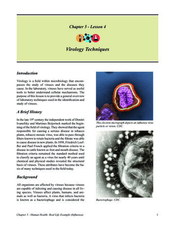
Reverse Transcriptase (RT) / Plymerase Chain Reaction (PCR)Reverse Transcription (RT)ReversetranscriptasePrimerPolymerase Chain Reaction (PCR)AmplificationRNA template5’3’3’5’Generation ofDNA copy (cDNA)of RNA template5’3’5’PrimercDNA5’3’Traditional molecular techniques are dot-blot, Southern blot, and in situ hybridization. These methods aredependent on the use of specific DNA or RNA probes.They are similar in sensitivity to some of the classicalmethods but are more tedious and expensive, and arenot routinely used in diagnostic laboratories.The technique that has probably had the greatest impact on the field of virology is the polymerase chainreaction (PCR, identification of DNA) and the reversetranscriptase polymerase chain reaction (RT-PCR,identification of RNA) along with the development ofbetter, more standardized methods of nucleic acid purification. The rapid advances and decrease in expenseassociated with nucleotide sequencing, commercialsynthesis of oligonucleotides (primers), and the availability of genetic sequences in public databases havecontributed to the increased appeal of these methods.The PCR involves the use of two primers designedto identify a target. In the presence of a DNA polymerase and other components required for DNA amplification, the target sequence of interest, if present,is amplified. The PCR requires double stranded DNA,Chapter 5 - Human Health: Real Life Example �3’3’5’thus it is useful for identifying DNA viruses. The discovery of reverse transcriptase, an enzyme capableof making a DNA copy of RNA, enabled the abilityto expand PCR to include a reverse transcription step(RT) which generates a double stranded target (RNADNA), which is then used in the PCR to identify RNAviruses.SummaryVirological techniques expand beyond diagnosticsinto the research laboratory. Many animal disease systems are used as models for human diseases. Usingseveral different types of molecular methods, such ascloning and inserting and deleting genetic information, viruses are being engineered in a variety of waysto improve human and animal health. Some of the results of these manipulations are improved vaccines,viruses engineered to carry genetic information foruse in gene therapy, as well as for use in cancer treatment. Virology is a constantly growing and dynamicfield with much left to discover.7
ReferencesQuestionsCastro, A. E., & Heuschele, W. P. (1992). Veterinarydiagnostic virology, A practioner’s guide. St. Louis, MO: Mosby-Year Book, Inc.1. Name three assays designed to identify the presence of antibodies in a patient’s serum.2. Name three assays designed to identify the presence of a virus.3. What is a limitation with virus isolation?4. What is an advantage for virus isolation?5. What is a benefit of molecular assays?MacLachlan, N., & Dubovi, E. J. (Eds.). (2011).Fenner’s veterinary virology (4th ed.). San Diego,CA: Academic Press, Elsevier Inc.White, D. E., & Fenner, F. J. (1994). Medical virology(4th ed.). San Diego, CA: Academic Press.8Chapter 4 - Human Health: Real Life Example (Influenza)
Diagnostic virology is concerned with identifying the virus associated with clinical signs and symptoms. Procedures most commonly used include: 1. Detection of a meaningful immune response to the virus (antibody or cell-mediated) by immuno-logic assa