Transcription
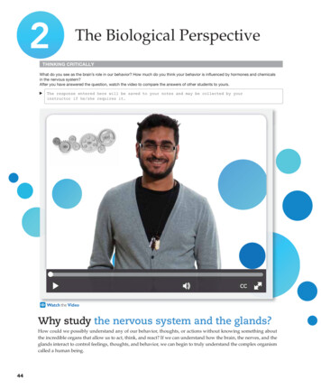
2The Biological PerspectiveTHINKING CRITICALLYWhat do you see as the brain’s role in our behavior? How much do you think your behavior is influenced by hormones and chemicalsin the nervous system?After you have answered the question, watch the video to compare the answers of other students to yours.The response entered here will be saved to your notes and may be collected by yourinstructor if he/she requires it.CCWatch the VideoWhy study the nervous system and the glands?How could we possibly understand any of our behavior, thoughts, or actions without knowing something aboutthe incredible organs that allow us to act, think, and react? If we can understand how the brain, the nerves, and theglands interact to control feelings, thoughts, and behavior, we can begin to truly understand the complex organismcalled a human being.44
Learning Objectives2.1Identify the parts of a neuron and thefunction of each.2.10Identify the different structures of thehindbrain and the function of each.2.2Explain the action potential.2.112.3Describe how neurons useneurotransmitters to communicatewith each other and with the body.Identify the structures of the brainthat are involved in emotion,learning, memory,and motivation.2.42.12Describe how the brain and spinalcord interact and respond to externalexperiences.Identify the parts of the cortex thatprocess the different senses and thosethat control movement of the body.2.52.13Differentiate the roles of the somaticand autonomic nervous systems.Name the parts of the cortex thatare responsible for higher forms ofthought, such as language.2.6Explain why the pituitary gland isknown as the “master gland.”2.142.7Recall the role of various endocrineglands.Explain how some brain functionsdiffer between the left and righthemispheres.2.15Identify some potential causes ibe how lesioning studies andbrain stimulation are used to studythe brain.2.9Compare and contrast neuroimagingtechniques for mapping the structureand function of the brain.45
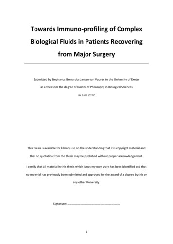
46CHAPTER 2Neurons and Nerves: Buildingthe NetworkThis chapter will explore a complex system of cells, chemicals, and organs that work togetherto produce behavior, thoughts, and actions. The first part of this complex arrangement is thenervous system, a network of cells that carries information to and from all parts of the body.The field of neuroscience is a branch of the life sciences that deals with the structure andfunctioning of the brain and the neurons, nerves, and nervous tissue that form the nervoussystem. Biological psychology, or behavioral neuroscience, is the branch of neurosciencethat focuses on the biological bases of psychological processes, behavior, and learning, and itis the primary area associated with the biological perspective in psychology.STRUCTURE OF THE NEURON: THE NERVOUS SYSTEM’S BUILDING BLOCK2.1 Identify the parts of a neuron and the function of each.nervous systeman extensive network of specializedcells that carries information to andfrom all parts of the body.In 1887, Santiago Ramón y Cajal, a doctor studying slides of brain tissue, first theorized that thenervous system was made up of individual cells (Ramón y Cajal, translation, 1995). Althoughthe entire body is composed of cells, each type of cell has a special purpose and function and,therefore, a special structure. For example, skin cells are flat, but muscle cells are long andstretchy. Most cells have three things in common: a nucleus, a cell body, and a cell membraneholding it all together. The neuron is the specialized cell in the nervous system that receivesand sends messages within that system. Neurons are one of the messengers of the body, andthat means that they have a very special structure, which we will explore in the Figure 2.1.DendriteAction potentialneuroscienceMyelin sheathNodea branch of the life sciences that dealswith the structure and function ofneurons, nerves, and nervous tissue.biological psychology orbehavioral neuroscienceSynapsebranch of neuroscience that focuses onthe biological bases of psychologicalprocesses, behavior, and learning.NucleusNeuronAxonneuronthe basic cell that makes up thenervous system and that receives andsends messages within that system.Axon terminalsdendritesSoma (Cell body)branchlike structures of a neuron thatreceive messages from other neurons.CCsomathe cell body of the neuron responsiblefor maintaining the life of the cell.axontubelike structure of neuron thatcarries the neural message from thecell body to the axon terminals, forcommunication with other cells.Figure 2.1The Structure of the NeuronThe parts of the neuron that receive messages from other cells are called thedendrites. The name dendrite means “tree-like,” or “branch,” and this structure doesindeed look like the branches of a tree. The dendrites are attached to the cell body, orsoma, which is the part of the cell that contains the nucleus and keeps the entire cell aliveand functioning. The word soma means “body.” The axon (from the Greek for “axis”) is afiber attached to the soma, and its job is to carry messages out to other cells. The end of
The Biological Perspectivethe axon branches out into several shorter fibers that have swellings or little knobs on theends called axon terminals (may also be called presynaptic terminals, terminal buttons, orsynaptic knobs), which are responsible for communicating with other nerve cells.Neurons make up a large part of the brain, but they are not the only cells that affectour thinking, learning, memory, perception, and all of the other facets of life that make uswho we are. The other primary cells are called glia, or glial cells, which serve a varietyof functions. While historically viewed as support cells for neurons, the expanded rolesof glia are still being discovered. And while they help maintain a state of homeostasis, orsense of balance in the nervous system, they are increasingly being better understoodas partner cells, not just support cells (Kettenmann & Ransom, 2013; Verkhratsky et al.,2014). Some glia serve as a sort of structure on which the neurons develop and work andthat hold the neurons in place. For example, during early brain development, radial glialcells (extending from inner to outer areas like the spokes of a wheel) help guide migrating neurons to form the outer layers of the brain. Other glia are involved in getting nutrients to the neurons, cleaning up the remains of neurons that have died, communicatingwith neurons and other glial cells, and insulating the axons of some neurons.Glial cells affect both the functioning and structure of neurons, and specific typesalso have properties similar to stem cells, which allow them to develop into new neurons,both during prenatal development and in adult mammals (Bullock et al., 2005; Gotz et al.,2015; Kriegstein & Alvarez-Buylla, 2009). Glial cells are also being investigated for theirpossible role in a variety of neurodevelopmental diseases like autism spectrum disorder,degenerative disorders such as Alzheimer’s disease, and psychiatric disorders including major depressive disorder and schizophrenia (Molofsky et al., 2012; Peng et al., 2015;Sahin & Sur, 2015; Verkhratsky et al., 2014; Yamamuro et al., 2015).to LearningObjectives 8.7, 14.9, and 14.14. Glial cells also play important roles in learning, behavior,and neuroplasticity by affecting synaptic connectivity and facilitating communicationbetween neurons in specific neural networks (Hahn et al., 2015; Martín et al., 2015).Two special types of glial cells, called oligodendrocytes and Schwann cells, generate a layer of fatty substances called myelin. Oligodendrocytes produce myelin for theneurons in the brain and spinal cord (the central nervous system); Schwann cells produce myelin for the neurons of the body (the peripheral nervous system). Myelin wrapsaround the shaft of the axons, forming an insulating and protective sheath. Bundles ofmyelin-coated axons travel together as “cables” in the central nervous system calledtracts, and in the peripheral nervous system bundles of axons are called nerves. Myelinfrom Schwann cells has a unique feature that can serve as a tunnel through which damaged nerve fibers can reconnect and repair themselves. That’s why a severed toe mightactually regain some function and feeling if sewn back on in time. Unfortunately, myelinfrom oligodendrocytes covering axons in the brain and spinal cord does not have thisfeature, and these axons are more likely to be permanently damaged.The myelin sheath is a very important part of the neuron. It not only insulates andprotects the neuron, it also speeds up the neural message traveling down the axon. Asshown in Figure 2.1, sections of myelin bump up next to each other on the axon, similarto the way sausages are linked together. The places where the myelin seems to bumpare actually small spaces on the axon called nodes, which are not covered in myelin.Myelinated and unmyelinated sections of axons have slightly different electrical properties. There are also far more ion channels at each node. Both of these features affectthe speed at which the electrical signal is conducted down the axon. When the electricalimpulse that is the neural message travels down an axon coated with myelin, the electrical impulse is regenerated at each node and appears to “jump” or skip rapidly from nodeto node down the axon (Koester & Siegelbaum, 2013; Schwartz et al., 2013). That makesthe message go much faster down the coated axon than it would down an uncoated axonof a neuron in the brain. In the disease called multiple sclerosis (MS), the myelin sheath isdestroyed (possibly by the individual’s own immune system), which leads to diminished47axon terminalsenlarged ends of axonal branches ofthe neuron, specialized for communication between cells.glial cellscells that provide support for the neurons to grow on and around, delivernutrients to neurons, produce myelinto coat axons, clean up waste productsand dead neurons, influence information processing, and, during prenataldevelopment, influence the generationof new neurons.myelinfatty substances produced by certainglial cells that coat the axons of neurons to insulate, protect, and speedup the neural impulse.nervesbundles of axons coated in myelin thattravel together through the body.
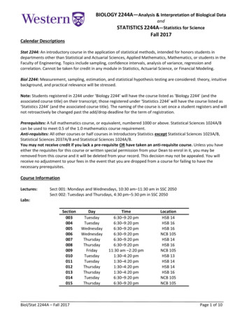
48CHAPTER 2or complete loss of neural functioning in those damaged cells. Early symptoms of MSmay include fatigue; changes in vision; balance problems; and numbness, tingling, ormuscle weakness in the arms or legs. Just as we are learning more about the expandedroles of glial cells, our knowledge about the structure and function of myelin is alsoexpanding far beyond myelin simply being an insulator of axons. Myelin thickness varies, and myelin distribution may vary along the length of an axon, likely affecting communication properties of those neurons and impacting larger neural networks (Fields,2014; Tomassy et al., 2014).GENERATING THE MESSAGE WITHIN THE NEURON: THE NEURAL IMPULSE2.2 Explain the action potential.Exactly how does this “electrical message” work inside the cell?diffusionprocess of molecules moving fromareas of high concentration to areasQH NQY EQPEGPVTCVKQP resting potentialthe state of the neuron when not firinga neural impulse.action potentialthe release of the neural impulse, consisting of a reversal of the electricalcharge within the axon.A neuron that’s at rest—not currently firing a neural impulse or message—is actuallyelectrically charged. Inside and outside of the cell is a semiliquid (jelly-like) solution inwhich there are charged particles, or ions. Although both positive and negative ions arelocated inside and outside of the cell, the relative charge of ions inside the cell is mostlynegative, and the relative charge of ions outside the cell is mostly positive due to bothdiffusion, the process of ions moving from areas of high concentration to areas of lowconcentration, and electrostatic pressure, the relative balance of electrical charges when theions are at rest. The cell membrane itself is semipermeable, meaning that some moleculesmay freely pass through the membrane while others cannot. Some molecules that areoutside the cell enter through tiny protein openings, or channels, in the membrane, whilemolecules inside the cell can pass through the same channels to the outside of the cell.Many of these channels are gated—they open or close based on the electrical potentialof the membrane—more about that in a minute. Inside the cell is a concentration of bothsmaller positively charged potassium ions and larger negatively charged protein ions.The negatively charged protein ions, however, are so big that they can’t get out, whichleaves the inside of the cell primarily negative when at rest. Outside the cell are lotsof positively charged sodium ions and negatively charged chloride ions, but they areunable to enter the cell membrane when the cell is at rest because the ion channels thatwould allow them in are closed. But because the outside sodium ions are positive andthe inside ions are negative, and because opposite electrical charges attract each other,the sodium ions will cluster around the membrane. This difference in charges creates anelectrical potential.Think of the ions inside the cell as a baseball game inside a stadium (the cell walls).The sodium ions outside the cell are all the fans in the area, and they want to get insideto see the game. When the cell is resting (the electrical potential is in a state called theresting potential, because the cell is at rest), the fans are stuck outside. The sodium ionscannot enter when the cell is at rest, because even though the cell membrane has all thesechannels, the particular channels for the big sodium ions aren’t open yet. But when the cellreceives a strong enough stimulation from another cell (at the dendrites or soma), the cellmembrane opens up those particular channels, one after the other, all down its surface,allowing the sodium ions (the “fans”) to rush into the cell. That causes the inside of thecell to become mostly positive and the outside of the cell to become mostly negative,because many of the positive sodium ions are now inside the cell—at the point where thefirst ion channel opened. This electrical charge reversal will start at the part of the axonclosest to the soma, the axon hillock, and then proceed down the axon in a kind of chainreaction. (Picture a long hallway with many doors in which the first door opens, then thesecond, and so on all the way down the hall.) This electrical charge reversal is known asthe action potential because the electrical potential is now in action rather than at rest.
The Biological Perspective49Neurotransmitter releaseNerve cell bodyntialpotectionDirection of aAction potentialNodes ofRanvierAxonterminalPositivesodium ionElectrical charge (millivolts)400-55-70ThresholdResting potentialResting potentialHyperpolarizationNegatively chargedinner cell membraneTimeeCCFigure 2.2The Neural Impulse Action PotentialVoltage is graphed at a given axonal node over 2 to 3 milliseconds (thousandths of a second). From an initial resting state, enough stimulation is received thatthe threshold of excitation is reached and an action potential is triggered. The resulting rapid depolarization, repolarization, brief hyperpolarization, and return toresting potential coincide with movement of sodium and potassium ions across the cell membrane.Each action potential sequence takes about one thousandth of a second, so the neuralmessage travels very fast—from 2 miles per hour in the slowest, shortest neurons to 270miles per hour in other neurons. (See Figure 2.2.)Now the action potential is traveling down the axon. When it gets to the end ofthe axon, something else happens: the message will get transmitted to another cell (thatstep will be discussed momentarily). Meanwhile, what is happening to the parts of thecell that the action potential has already left behind? How does the cell get the “fans”back outside? Remember, the action potential means that the cell is now positive insideand negative outside at the point where the channel opened. Several things happen toreturn the cell to its resting state. First, the sodium ion channels close immediately afterthe action potential has passed, allowing no more “fans” (sodium ions) to enter. The cellmembrane also literally pumps the positive sodium ions back outside the cell, kickingthe “fans” out until the next action potential opens the ion channels again. This pumpingprocess is a little slow, so another type of ion gets into the act. Small, positively chargedpotassium ions inside the neuron move rapidly out of the cell after the action potentialpasses, helping more quickly restore the inside of the cell to a negative charge. Now thecell becomes negative inside and positive outside, and the neuron is capable of “firingoff” another message. Once the sodium pumps finish pumping out the sodium ions, theneuron can be said to have returned to its full resting potential, poised and ready to do itall again.To sum all that up, when the cell is stimulated, the first ion channel opens andthe electrical charge at that ion channel is reversed. Then the next channel opens and thatcharge is reversed, but in the meantime the first ion channel has been closed and thecharge is returning to what it was when it was at rest. The action potential is the sequenceof ion channels opening all down the length of the cell’s axon.
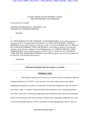
50CHAPTER 2So if the stimulus that originally causes the neuron to fire isvery strong, will the neuron fire more strongly than it would if thestimulus were weak?all-or-nonereferring to the fact that a neuroneither fires completely or does not fireat all.Neurons actually have a threshold for firing, and all it takes is a stimulus that isjust strong enough to get past that threshold to make the neuron fire. Here’s a simpleversion of how this works: Each neuron is receiving many signals from other neurons.Some of these signals are meant to cause the neuron to fire, whereas others are meant toprevent the neuron from firing. The neuron constantly adds together the effects of the“fire” messages and subtracts the “don’t fire” messages, and if the fire messages are greatenough, the threshold is crossed and the neuron fires. When a neuron does fire, it firesin an all-or-none fashion. That is, neurons are either firing at full strength or not firingat all—there’s no such thing as “partial” firing of a neuron. It would be like turning ona light switch—it’s either on or it’s off. Once the switch is turned to the on position, thelight will come on. When it’s turned to the off position, the light is off.So, what’s the difference between strong stimulation and weak stimulation? Astrong message will cause the neuron to fire repeatedly (as if someone flicked the lightswitch on and off as quickly as possible), and it will also cause more neurons to fire (as ifthere were a lot of lights going on and off instead of just one).NEUROTRANSMISSIONsynaptic vesiclessaclike structures found inside thesynaptic knob containing chemicals.2.3 Describe how neurons use neurotransmitters to communicate with eachother and with the body.neurotransmitterschemical found in the synaptic vesicles that, when released, has an effecton the next cell.SynapticvesiclesNow that we know how the message travels within the axon ofthe cell, what is that “something else” that happens when the actionpotential reaches the end of the axon?Once a neural signal reaches the axon terminals of a neuron, several events takeplace to allow neurons to communicatewith each other. These events are dependent upon key structures within a neuronand on the surface of adjacent neurons.NerveimpulseS E N D I N G T H E M E S S AG E TO OT H E RCELLS: THE SYNAPSESurface ofpostsynapticneuronAxon aptic(receiving) neuronSodium ionCCFigure 2.3The SynapseThe nerve impulse reaches the axon terminal, triggering the release of neurotransmitters from the synaptic vesicles. The molecules of neurotransmitter cross the synaptic gap to fit into the receptor sites that fitthe shape of the molecule, opening the ion channel and allowing sodium ions to rush in.Look once again atthe axon terminals in Figure 2.1. Figure 2.3shows an axon terminal enlarged to giantscale. Notice that the presynaptic terminalis not empty. It has a number of little saclike structures in it called synaptic vesicles. The word vesicle is Latin and means a“little blister” or “fluid-filled sac.”Inside the synaptic vesicles arechemicals suspended in fluid, whichare molecules of substances called neurotransmitters. The name is simpleenough—they are inside a neuron andthey are going to transmit a message.(Neurons have traditionally been viewedas containing a single type of neurotransmitter, but it is now accepted that neurons
The Biological Perspectivemay release more than one neurotransmitter. For simplicity and unless otherwise specified, our discussion throughout the text will assume a single, predominant neurotransmitter is being released.) Next to the axon terminal is the dendrite of another neuron(see Figure 2.3). Between them is a fluid-filled space called the synapse or the synapticgap. Instead of an electrical charge, the vesicles at the end of the axon (also called thepresynaptic membrane) contain the molecules of neurotransmitters, and the surface ofthe dendrite next to the axon (the postsynaptic membrane) contains ion channels thathave receptor sites, proteins that allow only particular molecules of a certain shape tofit into it, just as only a particular key will fit into a keyhole. Synapses can also occur onthe soma of the postsynaptic cell, as the surface membrane of the soma also has receptorsites.How do the neurotransmitters get across the gap? Recall the action potential making its way down the axon after the neuron has been stimulated. When that action potential, or electrical charge, reaches the synaptic vesicles, the synaptic vesicles release theirneurotransmitters into the synaptic gap. The molecules then float across the synapse,and many of them fit themselves into the receptor sites, opening the ion channels andallowing sodium to rush in, activating the next cell. It is this very activation that stimulates, or releases, the action potential in that cell. It is important to understand that the“next cell” may be a neuron, but it may also be a cell on a muscle or a gland. Muscles andglands have special cells with receptor sites on them, just like on the dendrite or soma ofa neuron.So far, we’ve been talking about the synapse as if neurotransmitters always causethe next cell to fire its action potential (or, in the case of a muscle or gland, to contract orstart secreting its chemicals). But the neurons must have a way to be turned off as well ason. Otherwise, when a person burns a finger, the pain signals from those neurons wouldnot stop until the burn was completely healed. Muscles are told to contract or relax, andglands are told to secrete or stop secreting their chemicals. The neurotransmitters foundat various synapses around the nervous system can either turn cells on (called an excitatory effect) or turn cells off (called an inhibitory effect), depending on exactly what synapse is being affected. Although some people refer to neurotransmitters that turn cells onas excitatory neurotransmitters and the ones that turn cells off as inhibitory neurotransmitters, it’s really more correct to refer to excitatory synapses and inhibitory synapses. Inother words, it’s not the neurotransmitter itself that is excitatory or inhibitory, but ratherit is the effect of that neurotransmitter that is either excitatory or inhibitory at the receptor sites of a particular synapse.NEUROTRANSMITTERS: MESSENGERS OF THE NETWORKThe first neurotransmitterto be identified was named acetylcholine (ACh). It is found at the synapses betweenneurons and muscle cells. Acetylcholine stimulates the skeletal muscles to contractbut actually slows contractions in the heart muscle. If acetylcholine receptor sites onthe muscle cells are blocked in some way, then the acetylcholine can’t get to the siteand the muscle will be incapable of contracting—paralyzed, in other words. This isexactly what happens when curare, a drug used by South American Indians on theirblow darts, gets into the nervous system. Curare’s molecules are just similar enoughto fit into the receptor site without actually stimulating the cell, making curare anantagonist (a chemical substance that blocks or reduces the effects of a neurotransmitter) for ACh.What would happen if the neurons released too much ACh? The bite of a blackwidow spider does just that. Its venom stimulates the release of excessive amounts ofACh and causes convulsions and possible death. Black widow spider venom is an agonist(a chemical substance that mimics or enhances the effects of a neurotransmitter) for ACh.ACh also plays a key role in memory, arousal, and attention. For example, AChis found in the hippocampus, an area of the brain that is responsible for forming new51synapse (synaptic gap)microscopic fluid-filled space betweenthe axon terminal of one cell and thedendrites or soma of the next cell.receptor sitesthree-dimensional proteins on thesurface of the dendrites or certaincells of the muscles and glands,which are shaped to fit only certainneurotransmitters.excitatory synapsesynapse at which a neurotransmittercauses the receiving cell to fire.inhibitory synapsesynapse at which a neurotransmittercauses the receiving cell to stop firing.antagonistschemical substances that blockor reduce a cell’s response tothe action of other chemicalsQT PGWTQVTCPUOKVVGTUagonistschemical substances that mimic orenhance the effects of a neurotransmitter on the receptor sites of thenext cell, increasing or decreasing theactivity of that cell.
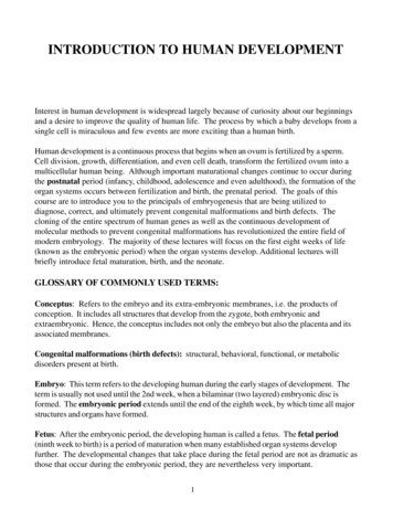
52CHAPTER 2The venom of the black widow spidercauses a flood of acetylcholine to bereleased into the body’s muscle system,causing convulsions.memories, and low levels of ACh have been associated with Alzheimer’s disease, themost common type of dementia.to Learning Objective 6.13. We will focus moreon agonists and antagonists later in the chapter.Dopamine (DA) is a neurotransmitter found in the brain, and like some of the otherneurotransmitters, it can have different effects depending on the exact location of itsactivity. For example, if too little DA is released in a certain area of the brain, the resultis Parkinson’s disease—the disease that is currently being battled by actor Michael J.Fox, and that affected the late former boxing champ Muhammad Ali (Almasay, 2016;Ahlskog, 2003). If too much DA is released in other areas, the result is a cluster of symptoms that may be part of schizophrenia (Akil et al., 2003).to Learning Objective 14.13.Serotonin (5-HT) is a neurotransmitter originating in the lower part of the brainthat can have either an excitatory or inhibitory effect, depending on the particular synapses being affected. It is associated with sleep, mood, anxiety, and appetite. For example, low levels of 5-HT activity have been linked to depression.to LearningObjective 14.9.Although ACh was the first neurotransmitter found to have an excitatory effectat the synapse, the nervous system’s major excitatory neurotransmitter is glutamate.Like ACh, glutamate plays an important role in learning and memory and may alsobe involved in the development of the nervous system and in synaptic plasticity (theability of the brain to change connections among its neurons). However, an excessof glutamate results in overactivation and neuronal damage and may be associatedwith the cell death that occurs after stroke or head injury or in degenerative diseaseslike Alzheimer’s disease and Huntington disease (Julien et al., 2011; Siegelbaum et al.,2013).Another neurotransmitter is gamma-aminobutyric acid or GABA. Whereas glutamateis the major neurotransmitter with an excitatory effect, GABA is the most common neurotransmitter producing inhibition in the brain. GABA can help calm anxiety, for example, by binding to the same receptor sites that are affected by tranquilizing drugs andalcohol. In fact, the effect of alcohol is to enhance the effect of GABA, which causes thegeneral inhibition of the nervous system associated with getting drunk. This makes alcohol an agonist for GABA.to Learning Objective 4.13. Table 2.1 below lists someneurotransmitters and their functions.A group of substances known as neuropeptides can serve as neurotransmitters orhormones or influence the action of other neurotransmitters (Schwartz & Javitch, 2013).Table 2.1 Some Neurotransmitters and Their FunctionsNeurotransmittersFunctionsAcetylcholine (ACh)Excitatory or inhibitory; involved in arousal, attention, memory,and controls muscle contractionsNorepinephrine (NE)Mainly excitatory; involved in arousal and moodDopamine (DA)Excitatory or inhibitory; involved in control of movement andsensations of pleasureSerotonin (5-HT)Excitatory or inhibitory; involved in sleep, mood, anxiety,and appetiteGamma-aminobutyric acid(GABA)Major inhibitory neurotransmitter; involved in sleep and inhibitsmovementGlutamateMajor excitatory neurotransmitter; involved in learning, memoryformation, nervous system development, and synaptic plasticityEndorphinsInhibitory neural regulators; involved in pain relief
The Biological Perspective53You may have heard of the set of neuropeptides called endorphins—pain-controllingchemicals in the body. When a person is hurt, a neurotransmitter that signals pain isreleased. When the brain gets this message, it triggers the release of endorphins. Theendorphins bind to receptors that open the ion channels on the axon. This causes the cellto be unable to fire its pain signal, and the pain sensations eventually lessen. For example, you might bump your elbow and experience a lot of pa
Biological psychology, or behavioral neuroscience, is the branch of neuroscience that focuses on the biological bases of psychological processes, behavior, and learning, and it is the primary area associated with the biological perspective in psychology. STR