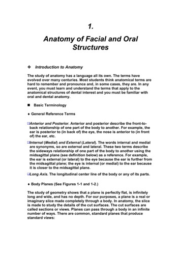
Transcription
1What Yoga Therapists Should Know About the Anatomy of Breathing Leslie Kaminoff and The Breathing Project, Inc.IntroductionThere are staggeringly vast numbers of people in this country suffering frombreathing related disorders; particularly when factoring in related maladies such ashypertension, back pain and depression.Not surprisingly, similarly large numbers of people are seeking out alternativeapproaches to healing. A 2002 CDC study1 on Complementary and Alternative therapiesfound that the most popular method of natural healing (other than prayer and nutritionalsupplements) is deep breathing exercises, with 12 percent of the population practicing.Another 8 percent of Americans say they practice meditation and 5 percent practice yogafor natural healing. We can only expect that these numbers have increased significantlyin the past four years.These trends indicate that for many millions of people, the way they breathe playsa significant factor in disorders that affect their health, as well as providing the means torestoring that health.In terms of sheer numbers, no profession has a greater influence over the waypeople are trained to breathe than yoga teachers, and the demand for our services hasincreased exponentially over the past decade.If yoga teachers have such an effect on how the public is trained to breathe, whatfactors influence the way yoga teachers are trained to teach breathing? The key factor, itseems, is tradition. Oft-repeated, outdated imagery along with inaccurate or unclearanatomical and physiological information have persisted in teacher training programs forhalf a century in spite of enormous advances in breath science.OverviewIn this article, I will summarize the four most common confusions about breathing that1U.S. Dept. of Health and Human Services: Complementary and Alternative MedicineUse Among Adults: United States, 2002. Advance Data No. 343. 20 pp. (PHS) 20041250 icine.htm
2have been compiled from a review of relevant literature.The emerging field of Yoga Therapy offers its practitioners a singular opportunityto provide the public with accurate and useful information about breathing. In order toaccomplish this, I will present basic definitions, anatomical information and analogiesthat can help to dispel each of the common confusions that have surrounded breathingmethodology.To conclude the article, I will summarize my anatomical information whilehighlighting correlated principles from the Yoga Therapy tradition of T.Krishnamacharyaand T.K.V. Desikachar.Four Common Confusions about BreathingLast year, as part of my preparation for producing “The Future of Breathing”symposium at Kripalu Center for Yoga and Health, I wanted to review and evaluatetraditional breathing information objectively. With the support of Kripalu, and the skilledresearch of Danna Faulds, we conducted a review of the breathing-related source materialfor the major Yoga teaching traditions. This survey revealed a number of flawedassumptions and outright errors related to breathing and breath anatomy that haveremained both consistent and largely unchallenged through most of the history of Yogateaching in America. Most of this confusion can be classified into the following fourbroad categories:Confusion #1: Context droppingThis common error most often appears as either an explicit or implicit suggestionthat there is a “right” or “proper” way to breathe without stating the context that givesrise to that breathing method. Context refers to the conditions unique to each individual’shistory, condition and goals. Context also refers to activity and body position – all ofwhich significantly affect breathing patterns.Since individual intentions, body type, shape and orientation all create differentconditions for breathing, it’s clear that no one pattern could suffice to deal with all ofthem. In other words, there is no one right way to breathe that will work under allconditions, and implying that there is only encourages people to create breathing habits
3that make their systems less adaptable to change.My simple, comprehensive definition of breathing as shape-change will help todispel this confusion, and clarify the context in which breathing patterns arise.Confusion #2: False dichotomy between diaphragmatic, non-diaphragmaticbreathingThis error arises from the commonly stated bromide that “belly” breathing equalscorrect diaphragmatic breathing, and “chest” breathing equals incorrect nondiaphragmatic breathing. The idea that “correct” breathing involves the proper use of thediaphragm is true enough, but to equate diaphragmatic breathing exclusively withabdominal movement, and ribcage expansion with non-diaphragmatic (accessory)breathing is incorrect, because the diaphragm is capable of creating chest as well as bellymovement.This error arises from the lack of recognition that the diaphragm can mobilize theribcage without the aid of the accessory muscles, and it leads to teachers making theseemingly helpful observation: “You’re not using your diaphragm.” Saying this to a nonparalyzed person is essentially the same as telling them they are dead – for it is therhythmic contraction of the diaphragm that is the tangible manifestation of Pranaexpressing itself through a human form.A corollary result of this confusion is that many students’ breathing patterns areevaluated only by the location of shape change in the body, i.e.: belly breathing is good,chest breathing is bad. In reality, it is possible for breathing to manifest as tense,disordered belly movement, or relaxed, integrated chest movement. An excessive focuson the region of shape change as an indicator of “correct breathing” can blind us to manyother, more relevant qualities of the breath.My analysis of the 3-dimensional action of the diaphragm’s muscle fibers, and mysubsequent metaphor comparing the diaphragm to the engine of a car will help to clarifythis confusion.Confusion #3: Confusion between respiratory shape changes and regionalventilation
4Here is a passage from a book on pranayama by one the world’s most respectedteachers, but it could have come from any yoga book:Respiration may be classified into four types:High or clavicular breathing, where the relevant muscles in the neck mainlyactivate the top parts of the lungs.Intercostal or midbreathing, where only the central parts of the lungs areactivated.Low or diaphragmatic breathing, where the lower portions of the lungs areactivated chiefly, while the top and central portions remain less active.In total or pranayamic breathing, the entire lungs are used to their fullestcapacity.2Here, the author speaks of “lung activation,” which could be interpreted correctly(which is rare) or incorrectly (which is far more common).The correct interpretation refers to the way lung tissue follows the ribcage anddiaphragmatic breath movements (see “The Diaphragm’s Relations: OrganicConnections” later in this article).The incorrect interpretation of “lung activation” is to equate it with local airmovements in the upper, middle and lower portions of the lungs (regional ventilation).Simply stated, this error results from confusion between the concept of “breath” and theconcept of “air.”Air moves into and out of the lungs via the pathway of the bronchial tree. Thispathway is not affected by the sequence of shape change in the cavities of the chest andabdomen. These differing breathing patterns refer to some of the ways in which wemanipulate the accessory breathing muscles in order to produce specific respiratory shapechanges, but that is not the same thing as isolating the ventilation in the correspondingregions of the lungs.In other words, contrary to what most teaching language implies, “bellybreathing” does not fill the base of the lungs, “intercostal breathing” does not fill themiddle of the lungs, and “clavicular breathing” does not fill the tops of the lungs.Understanding that the accessory muscles “steer” the direction of the breath helps2Light on Pranayama, by B.K.S. Iyengar, (New York: Crossroad, 1981) p. 21
5to clarify this confusion.Confusion #4: Deep Breathing and More Oxygen is always a good thingTo read many yoga and breathing books, one could get the impression that deepbreathing and oxygenation are the holy grails of health, well-being and enlightenment.The assumption is that the more carbon dioxide you get rid of and the deeper you breathe,the more oxygen you get in, and the healthier you’ll be. The fact is, not enough carbondioxide is dangerous, deep breathing is only occasionally appropriate, and too muchoxygen is toxic.Breathing patterns should always be linked to your body’s metabolic needs.3 Ifyour level of activity requires a larger than usual supply of oxygen, deeper or more rapidbreathing is perfectly appropriate. Those same patterns of breath, however, if applied to aresting state of metabolic activity would produce blood alkalosis (hyperventilation).Your body has homeostatic mechanisms that prevent a toxic excess of oxygenfrom building up in the tissues.4 The idea that one can improve health by increasing O2concentrations in the blood is physiologically incorrect, and shouldn’t be confused withthe immense relief that accompanies a deep, freeing breath pattern. In fact, freeing thebreath allows respiratory activity to more closely match body metabolism by releasingexcessive, oxygen-hungry tension from the breathing musculature.Your body is many times more sensitive to changes in blood levels of carbondioxide than it is to oxygen. Carbon dioxide plays a critical role in helping hemoglobintransport oxygen from your blood to your body’s tissues. If you don’t have enough CO2in your blood, the O2 gets held too tightly by the hemoglobin and not enough oxygen willbe released into your tissues. The idea that one can improve health by ridding oneself ofexcess CO2 is physiologically incorrect, and shouldn’t be confused with the simple act ofexhaling more effectively (which is a prerequisite for a deep inhale).Understanding that healthy breathing is linked to metabolic activity and normalCO2 levels will help to clarify some of these issues.3The Psychology and Physiology of Breathing by Robert Fried Ph.D. (New York:Plenum Press 1993) p. 344ibid: Fried p. 29
6Dispelling Confusion: Simple Definitions, Anatomy and ImageryBreathing DefinitionThe Oxford American Dictionary defines breathing as: “the process of taking airinto and expelling it from the lungs.” This is a good place to start, but let’s define the“process” being referred to.Movement in the two cavitiesA simplified image of the human body divides the torso into twocavities, the thoracic and abdominal. These cavities share someproperties, and have important distinctions as well. Both contain vitalorgans: the thoracic contains the heart and lungs; the abdominal containsthe stomach, liver, gall bladder, spleen, pancreas, small and largeintestines, kidneys, bladder, among others.Both cavities are bounded posteriorly by the spine. Both open atone end to the external environment - the thoracic at the top, and theabdominal at the bottom. Both share an important structure, thediaphragm - the roof of the abdominal cavity and the floor of the thoracic.Another important shared property is that they are mobile – they change shape. Itis this shape-changing ability that is most relevant to breathing, because withoutmovement, the body cannot breathe at all.Change in the Abdominal Cavity: Shape, Not VolumeAlthough both the abdominal and thoracic cavities change shape, there is animportant structural difference in how they do so.The abdominal cavity changes shape like a flexible fluid-filled structure such as awater balloon. Think of what it’s like to hold a water balloon and imagine what happenswhen you squeeze one end of it - the other end bulges. This is because water is noncompressible. Your hand’s action only moves the fixed volume of water from one end ofthe flexible container to the other. The same principle applies when the abdominal cavityis compressed by the movements of breathing; a squeeze in one region produces a bulge
7in another. This is because in the context of breathing, the abdominal cavity changesshape, but not volume.In context of life processes other than breathing, the abdominal cavity doeschange volume. If you drink a gallon of liquid or eat a big meal, the overall volume ofthe abdominal cavity will increase due to expanded abdominal organs (stomach,intestines, bladder). It’s useful to note that any increase of volume in the abdominalcavity will tend to produce a corresponding decrease in the volume of the thoracic cavity.This is why it’s harder to breathe after a big meal, before a big bowel movement, or whenpregnant.Change in the Thoracic Cavity: Shape And VolumeIn contrast to the abdominal cavity, the thoracic changes both shape and volume;it behaves like a flexible gas-filled container, similar to an accordion bellows. When yousqueeze an accordion, you createa reduction in the volume of thebellows and air is forced out, andwhen you pull the bellows open,its volume increases and the air ispulled in. This is because theaccordion is compressible andexpandable. The same is true ofthe thoracic cavity, which unlike the abdominal cavity andits contents - can change itsshape and volume.To sum up the distinctionbetween the two cavities asregards breathing: the abdominal cavity changes shape but not volume, and the thoraciccavity changes shape and volume.Volume and Pressure
8As in the example of an accordion bellows, volume changes in the thoracic cavityresult in movement of air. Volume and pressure are inversely related -- when volumeincreases, pressure decreases, and when volume decreases, pressure increases. Since airalways flows towards areas of lower pressure, increasing the volume inside an accordion- or the thoracic cavity - will decrease pressure and cause air to flow into it. This is aninhale.Pressure/Volume Shift and Shape ChangeLet’s now imagine the thoracic and abdominal cavities as an accordion stacked ontop of a water balloon; movement in one will necessarily result in movement in the other.Recall that during an inhale, the thoracic cavity expands its volume. This pushesdownward on the abdominal cavity, which changes shape as a result of the pressure fromabove.During relaxed, quiet breathing (such as while sleeping) an exhale is a passivereversal of this process. The thoracic cavity and lung tissue - which have been stretchedopen during the inhale - spring back to their initial volume, pushing the air out andreturning the abdominal cavity to its previous shape. This is referred to as a “passiverecoil.” It’s important to note that any reduction in the elasticity of these tissues willresult in a reduction of the body’s ability to exhale passively –leading to an increase ofmuscular breath effort and a host of respiratory problems.In breathing that involves active exhaling (such as blowing out candles, speaking,singing, as well as various Yoga exercises), the musculature surrounding the two cavitiescontracts in such a way that the abdominal cavity is pushed upward into the thoracic, orthe thoracic is pushed downward into the abdominal, or any combination of the two.An Expanded Definition of BreathingHere’s our expanded definition of breathing:“Breathing is the intaking and expelling of air in the lungs, caused bychanging the shape of the thoracic and abdominal cavities.”Defining breathing this way not only tells us what it is, but how we do it. This hasprofound implications for Yoga practice, as it can lead us to examine the supporting,
9shape changing structure that occupies the back of the body’s two primary cavities - thespine. This is why breathing and spinal movement are so intimately connected: flexionof the spine IS the shape change that reduces thoracic volume (exhale) and spinalextension IS the shape change that increases thoracic volume (inhale).Additionally, as we shall soon see, the musculature of the breathing mechanism ISthe musculature of postural support.Answer to Confusion #1: Breathing Occurs in a ContextGravity, posture, activity, habit, intention are just some of the factors that affectthe shape-changing activities of the body cavities (breathing). To imply that there is onecorrect pattern of shape-changing (such as belly bulging) is to divorce breathing from thereality in which it occurs: individual human bodies engaging in an infinite number ofactivities on a planet with a gravitational field.
10The goal of breath training is to free up the system from habitual, dysfunctionalrestrictions -- and the first thing we need to free the breath from is the idea that there’s asingle right way to do it. Integrated breathing means that the breathing mechanism isable to freely respond to the demands that we place on it in the wide variety of positionsand activities that comprise our daily lives.Breathing Shape Change is Three-DimensionalThe lungs occupy a 3-dimensional space in the thoracic cavity, and when thisspace changes shape to cause air movement, it changes shape 3-dimensionally.Specifically, an inhale involves the chest cavity increasing its volume from top-to-bottom, from side-to-side and from front-to-back, and an exhale involves a reduction ofvolume in those same three dimensions.Because thoracic shape change is inextricably linked to abdominal shape change,we can also say that the abdominal cavity changes shape (not volume) in threedimensions – it can be pushed or pulled from top-to-bottom, from side-to-side or fromfront-to-back. In a living, breathing body, there can be no thoracic shape change withoutabdominal shape change. This is why the condition of the abdominal region has such aninfluence on the quality of our breathing, and why the quality of our breathing has apowerful effect on the health of our abdominal organs.
11In order to understand how a single muscle – the diaphragm - is capable ofproducing all this movement (its actions), it is necessary to understand its definition,location, shape attachments and relations.The Diaphragm – a definitionJust about every anatomy book describes the diaphragm as the principal muscle ofbreathing. Let’s use our expanded definition of breathing, along with our “3-D”observation, to get a better understanding of this remarkable muscle:“The diaphragm is the principal muscle that causes three dimensional shapechange in the thoracic and abdominal cavities.”The Diaphragm – locationThe diaphragm divides the torso into the thoracic and abdominal cavities. It is thefloor of the thoracic cavity and the roof of the abdominal cavity. Its structure extendsthrough a wide section of the body – the uppermost part reaches the space between thethird and fourth ribs, and its lowest fibers attach to the front of the third lumbar vertebra;“nipple to navel” is one way I describe it.The Diaphragm – ShapeThe deeply domed shape ofthe diaphragm has evoked manyimages: jellyfish, parachute, helmetor mushroom. It’s important to notethat the shape of the diaphragm iscreated by the organs it encloses andsupports. Deprived of its relationshipwith those organs, its dome wouldcollapse, much like a stocking capwithout a head in it.It is also evident that thediaphragm has an asymmetricaldouble-domed shape, with the right
12dome rising higher than the left. This is because the liver pushes up from below the rightdome, and the heart pushes down from above the left dome.The Diaphragm’s Attachments – Origin and InsertionOrigin: The lower edges of the diaphragm’s circumference originate from threedistinct regions: the bottom of the sternum, the base of the ribcage, and the front of thelower spine. These three regions form a continuous rim of attachment for the diaphragm,and the only bony components of this rim are the back of the xiphoid process and thefront surfaces of the first three lumbar vertebrae. The majority of the diaphragm (over90%) originates on flexible tissue: the costal cartilage of ribs 6 thru 10 and the arcuateligaments which bridge the span from the 10th rib’s cartilage to the floating 11th and 12thribs, and from there to the spine.Insertion: All the muscular fibers of the diaphragm rise upward in the body fromtheir origins. They eventually arrive at the flattened, horizontal top of the muscle, thecentral tendon, into which they insert. In essence, the diaphragm inserts onto itself – itsown central tendon, which is fibrous non-contractile tissue.The Diaphragm’s Relations: Organic ConnectionsThe central tendon of the diaphragm is a point of anchorage for the connectivetissue that surrounds the thoracic and abdominal organs. The names of these importantstructures are easily remembered as the “Three P’s.” Pleura – which surround the lungs Pericardium – which surrounds the heart Peritoneum – which surrounds the abdominal organsEvery organ has a membrane that tightly enwraps it, called the visceralmembrane. Outside of the visceral is another layer that anchors the organ to the body.This outer membrane is the parietal membrane.It is the parietal membranes that attach the organs to the diaphragm and the innersurfaces of the thoracic and abdominal cavities. Thus, it should be clear that the shapechanging activity of these cavities has a profound effect on the movements of the organsthey contain. The diaphragm is the primary source of these movements, and the
13relationship of its healthy functioning to the wellbeing of the organs is abundantlyevident.The Diaphragm’s Action: BasicsIt is important to rememberthat the muscular fibers of thediaphragm are oriented primarilyalong the vertical (up-down) axis ofthe body, and this is the direction ofits muscular action. Recall that thehorizontal central tendon is noncontractile, and can move only inresponse to the contraction of themuscular fibers, which insert onto it.Like any other muscle, thecontracting fibers of the diaphragmpull its insertion and origin (the central tendon and the base of the ribcage) towards eachother. It is this action that is the fundamental cause of the three dimensional thoracoabdominal shape changes of breathing.As with any muscle contraction of the body, the movement it produces is aquestion of whether origin moves towards insertion, or insertion towards origin. Statedsimply, this will depend upon which end of the muscle is stable, and which is mobile.The Diaphragm’s Action: Origin/Insertion - Stable/MobileThe muscular action of the diaphragm is usually associated with a bulging5movement in the upper abdomen, which is commonly referred to as a “Belly Breath,” butthis is only the case if the diaphragm’s origin (the base of the ribcage) is stable, and itsinsertion (the central tendon) is mobile.5Even though most teachers refer to this diaphragmatic action as an “expansion” of the abdomen, this isincorrect. In the context of breathing, the abdominal cavity does not change volume – only shape; thereforeit is more accurate to refer to this movement as a “bulging” of the upper abdomen – for the same reason wewould say a water balloon is bulging when we squeeze one end of it.
14If the central tendon is stabilized, and the ribs are free to move, a diaphragmaticcontraction will cause an expansion of the ribcage6. This is a “chest breath, ” whichmany people believe must be caused by the action of muscles other than the diaphragm.This mistaken idea can create a false dichotomy between diaphragmatic and “nondiaphragmatic breathing.” The unfortunate result of this error is that many peoplereceiving breath training who exhibit chest movement (rather than belly movement) aretold that they are not using their diaphragm, which is false. Except in cases of paralysis,the diaphragm is always used for breathing. The issue is whether it is being usedefficiently or not.If it were possible to release all of the diaphragm's stabilizing muscles, and allowits origin and insertion to freely move towards each other, both the chest and abdomenwould move simultaneously. This rarely occurs, as the need to stabilize the body's massin gravity will cause many of the respiratory stabilizing muscles (which are also posturalmuscles) to remain active through all phases of breathing.7The recognition that the diaphragm can mobilize the ribcage without the aid of theaccessory muscles is a key element to understanding the integrated nature of breathingpractices in yoga – especially the bandhas. It is the singular action of the diaphragm thatis the prime mover of the thoracic and abdominal cavities. The specific patterns that arise67This is what happens during inhale while correctly applying Mula Bandha.This also explains why babies’ breath goes everywhere: they aren’t standing up yet!
15in Yoga asana, bandha or breathing practices result from the action of muscles other thanthe diaphragm that can change the shape of the cavities: the accessory muscles. In orderto better understand this principle, the analogy of a car and its engine is very useful.Answer to Confusion #2: The Diaphragm is the “Engine” of 3-D Shape ChangeThe engine is the prime mover of the car. All the movements that contribute to acar’s functioning are generated by the engine. In the same manner, the three dimensional,abdomino-thoracic shape change of breathing is primarily generated by the diaphragm.To say that diaphragmatic action is limited to the abdominal bulging commonlyreferred to as “belly breathing” is as inaccurate as asserting that a car’s engine is onlycapable of making it go forward – and that there must be some other source of power thatgoverns reverse movement. Just as this automotive error is linked to not understandingthe relationship of the car’s engine to its transmission, the breathing error is linked to notunderstanding the relationship of the diaphragm to the accessory muscles.Moreover, equating belly movement with proper breathing and chest movementwith improper breathing is just as silly as stating that a car is best served by only drivingforward at all times. Without the ability to reverse its movements, a car would eventuallyend up someplace it couldn’t get out of.Answer to Confusion #3: The Accessory Muscles “Steer” the Breath – not the AirSince the diaphragm, in anunobstructed state, will create 3-D shapechange in the thorax and abdomen,intentionally isolating the breath in onedimension requires us to block the otherdimensions of movement. I like to refer tothis as “steering” the breath.We don’t steer our car with itsengine. In order to control the power of theengine, and guide it in a particulardirection, we need the mechanisms of the
16transmission, brakes, steering and suspension.In the very same way, we don’t “steer” our breathing with the diaphragm. Aswith a car’s engine, all we directly controlabout the diaphragm is the speed/timing ofits function. In order to control the powerof the breath, and guide it into specificpatterns, we need the assistance of theaccessory muscles – the muscles other thanthe diaphragm that change the shape of ourthoracic and abdominal cavities.The accessory breathing musclesinclude the abdominal group, intercostalgroup, sternocleidomastoids, scalenes,pectoralis minor, serratus anterior, and ahost of other muscles that stabilize them.It is important to note that what weare “steering” with the accessory muscles isBREATH (shape-change), not AIR. Justbecause a particular region of the chest ismoving more than another does not meanthat there is more air going into the lung just beneath thatmovement. 8. A look at the structure of the bronchial treewill reveal the pathway of lung tissue ventilation. This is notaltered by the pattern of abdomino/thoracic shape-change.It’s understandable that we make this error, becausewe don’t have direct sensory awareness of lung tissue, butdo we have direct feedback from the breathing musculature– thus, it’s easy to confuse one with the other.8This is referring to what occurs in a single breath, which is a distinct issue from the chronic constrictionsin lung tissue due to injury, disease, or habit – all of which can affect the elasticity of the lung, and thus itsability to expand fully.
17Answer to Confusion #4: Healthy Breathing is Linked to Activity and CO2 LevelsThe shape, depth, rhythm and volume of our breath is a reflection of our habits,training, intentions, body position and state of mind – to name just few of the myriadfactors that influence our breathing.Faulty concepts about the breath can also be a significant source of breathingdifficulties, and I frequently encounter this in my Yoga Therapy practice. One of themost common patterns I observe is the trained yogi’s tendency to do deep, slow Ujayyibreathing even when lying supine on a treatment table. Since this pattern is associatedwith vertical postural support, I usually ask why they are doing Ujayyi in a context wherehorizontal release is more appropriate. The usual reply that they dodn’t even know theyare doing it, and they find it difficult to release the pattern even after several attempts.This pattern (among many others) is linked to a pervasive assumption in the Yogaworld that breathing should ALWAYS be deep and full. This single absurdity is perhapsresponsible for more dysfunction than any other I’ve encountered. As you read thesewords, quickly check your breathing. Is it deep or shallow? Unless you are reading thisjournal while taking a walk, or exercising on a treadmill, the answer should be quiet andrelaxed9. This is because your body is at rest, and doesn’t require a huge supply ofoxygen to fuel the minimal metabolic activity of sitting and reading.Yes, in Yoga we train ourselves to breathe deeply, and in a variety of unusualpatterns, but this is only for the purpose of exploring the full potential of our breathingmechanisms in order to uncover and dismantle habitual patterns that obstruct normalfunction.In other words, the end goal of practicin
What Yoga Therapists Should Know About the Anatomy of Breathing Leslie Kaminoff and The Breathing Project, Inc. Introduction There are staggeringly vast numbers of people in this country suffering from breathing related disorders; particularly when factoring in related
