Transcription
MEDICALBIOCHEMISTRY
THIS PAGE ISBLANK
Copyright 2006, 2002 New Age International (P) Ltd., PublishersPublished by New Age International (P) Ltd., PublishersAll rights reserved.No part of this ebook may be reproduced in any form, by photostat, microfilm,xerography, or any other means, or incorporated into any information retrievalsystem, electronic or mechanical, without the written permission of the publisher.All inquiries should be emailed to rights@newagepublishers.comISBN : 978-81-224-2300-6PUBLISHING FOR ONE WORLDNEW AGE INTERNATIONAL (P) LIMITED, PUBLISHERS4835/24, Ansari Road, Daryaganj, New Delhi - 110002Visit us at www.newagepublishers.com
To mmyyElder daughterLate NallurNallurii Kiranmayi Chowdary
THIS PAGE ISBLANK
PREFACE TO THE SECOND EDITIONI attempted to provide essential information on molecular basis of health and diseasethat is mainly related to life of surviving cell(s) in the first edition of the book. However lifecycle of cell(s) includes cell(s) birth and cell(s) death apart from survival. For the last coupleof years these frontier areas are advancing rapidly which is viewed by many as good signfor development of :(a) new therapy or therapeutics for cancer(b) immortalized cells.The latter fuels growth of biotechnology and pharmaceutical industries also. Hence, inthe second edition two chapters—1. Biochemistry of cell cycle (cell birth) 2. Biochemistry ofapoptosis (cell death) are added.As living organisms evolved from simple unicellular to highly complex multicellularmammals, several new systems and organs were developed. For example, blood which actsas vehicle or communication between various locations of body, immune system whichprotects body from intruders or foreign organisms.Parkinsonism, psychosis, depression, Schizophrenia, loss of taste and olfaction are dueto disturbances in nervous, taste and olfactory systems. Various organs present in bodyperform several organ specific functions which are essential for life. If functions of theseorgans are disturbed, diseases in which may culminate in death. So, in this edition biochemistryof blood including immune response in Chapter-32; molecular and cellular mechanism oflearning, memory, behaviour, taste and olfactory in biochemical communications Chapter;tests, procedures that are done in hospital biochemistry laboratory to assess functions ofliver, kidney in Chapter-33 and thyroid in Chapter-29 are detailed.Depending on disease a particular constituent of blood is either elevated or lowered.Diagnosis and prognosis of disease usually involves detection and measurement of variousblood constituents in hospital biochemistry laboratory. Therefore advanced techniques likehigh performance liquid chromatography (HPLC), affinity chromatography, and generaltechniques like centrifugation and dialysis, instruments from spectrophotometer to autoanalyzer and methods used for detection and quantitation of blood constituents likecarbohydrates, proteins, lipids, nucleic acids, enzymes, electrolytes etc., in health and diseaseare detailed in Chapter-34.Humans and other mammals are able to remove waste products, toxins, foreigncompounds from blood and organs in the form of urine. In disease, composition of urinevaries from that of healthy state. So, detection, quantitation of various constituents of urineis carried out in hospital biochemistry laboratory to confirm diagnosis of diseases. In Chapter(vii)
(viii)34, methods for detection and estimation of urine constituent under normal and diseasedconditions are also detailed.Most striking feature in this edition of the book is inclusion of biochemical aspects ofdiseases or disease causing organisms common to tropical countries like malaria, tuberculosis,peptic ulcer or gastritis, pneumonia, leishmaniasis, giardiasis, trypanosomiasis etc. Sincemost of the organisms are developing resistance to the existing drugs, there is need fordevelopment of new drugs which requires thorough biochemical knowledge of these diseasesas well as disease causing organisms. Apart from adding new chapters, all existing chaptershave been updated by adding new subject matter. References of each chapter updated byincluding reviews, books, research articles etc. Further, number of unsolved problems havebeen increased in most of the chapters.I hope this edition will be well received by teachers and students of various medical,dental, pharmacy, biotechnology, physiotherapy, medical laboratory technology, biomedicalengineering, life sciences under graduate and post graduate courses, Suggestions or commentsfrom teachers and students are welcome. I am grateful to Sri. R.K. Gupta, Chairman; SriSaumya Gupta, Managing Director of New Age International, New Delhi, for publishingsecond edition.N. MALLIKARJUNA RAO
PREFACE TO THE FIRST EDITIONThis book explains the fundamentals of biochemical (molecular) bases of health anddisease. Hence it meets medical and allied health sciences student’s needs. As a teacher ofmedical, dental, pharmacy, biomedical engineering and science students for the last twodecades, I know the problems faced by students in mastering (conceptualizing) the subjectwithin a limited time. Most of these students need a book for their routine day-to-day studywhich contains only the necessary information in a simple and concise way. Therefore, thisbook is written in simple language in such a way that a student with very little chemistryor biology background can easily follow the various aspects of biochemistry that are presented.This book is also useful for those who are specializing in biochemistry (M.Sc. or M.D.)because advances in frontier areas of biochemistry are presented in a systemic way. Ofcourse advances in other areas that are relevant to medical students are also included toa limited extent. An interesting feature of this book is that the medical and biologicalimportance of each chapter is highlighted in simple numbered statements. Further, in somechapters, diseases, drugs (treatments) or toxins of particular subject matter are describedunder medical importance heads. Further, each chapter’s text is designed to facilitate easyflow of information in an interesting, thought provoking and logical manner. Exercises(cases) given at the end of each chapter help in mastering of the subject by student andutilization of biochemical principles by the student in solving health problems. To enthusiasticstudents, references given at the end of each chapter provide additional information.There are 29 chapters in the book. First six chapters deal with the composition, structure,function and life cycle of cells and the goal of biochemistry; occurrence, chemistry, structureand functions of biomolecules like amino acids, peptides, proteins, enzymes, carbohydratesand lipids. This is then followed by chapters 7 and 8 that deal with membrane structure,various transporters that move biomolecules across membrane and disintegration of complexmolecules of food and absorption of resulting products, respectively.Chapters 9-12 deal with the production and utilization of energy in various pathways ofcarbohydrate, lipid and aminoacid metabolisms. Regulatory mechanisms of some of theimportant pathways are also outlined. Further synthesis of biologically (medically) importantcompounds including non-essential amino acids is detailed. In chapter 11 the ultimate wayof producing energy from all energy yielding compounds in the respiratory chain is described.Changes in the flow of metabolites into various pathways of carbohydrate, lipid and proteinmetabolism that occur among tissues in well fed state, diabetes and starvation are describedin chapter 13.Fundamentals of molecular biology i.e., occurrence, chemistry, structure, functions,metabolism of nucleotides, nucleic acids and control of gene expression as well as appliedmolecular biology i.e., recombinant DNA technology are detailed in chapters 14-20. Biomedical(ix)
(x)(chemical) aspects of two major health problems of the 20th century—cancer and AIDS arebriefed in chapter 21. In chapter 22 occurrence, chemistry, structure, functions and metabolismof porphyrins and hemoglobin are described.Clinically related topics like vitamins, minerals, macro nutrients, energy, nutraceuticalsof food, electrolytes, acid-base balance and detoxification are described in chapters 23-27.Chemistry, production, detection and uses of isotopes in biochemistry and medicine aredetailed in chapter 28. Chapter 29 deals with mechanisms of communication between cells.I hope both teachers and students of Biochemistry at undergraduate and postgraduatelevels use this book extensively and their suggestions to improve the book further are mostwelcome. I express my sincere thanks to New Age International, Publishers for publishingthe book.N. MALLIKARJUNA RAO
CONTENTSI'"fff ffto 1M S«t.md &Ii/ionI'"ff{oct to flu Fi,., Edition.,.ColiAminoAcids and Peptides3. Proteins ElUymes5. Carbohydrates6. Lipids,Membrane and Transport"."."."."""".".,,." 9"""''""Protein and Amino Acid MetabolilmIntegration of Metabolism'". 9Nucleotides363Nucleotide Metaboli.m'"""".Carbohydral ! MetabolilmLipid MetabolismU . Biological Qx;dation and Re8piralOt'y (''hainNudeicAcidsBiOlynthesis of Nucleic Acid,Protei n BiosynthesisReguLation of G.!ne E prenion20. Recombinant DNA Techr alogyCancer and Aids22. Porphyrin and HaemOjlobi n Metabolism23. Vitamirul".(u).'"8. Digestion and Absorption of Food « ii)Minerala 5, ,.'".90""'",n
(:ai)"""Energy, Nutrients. Medie;nft and 'Ibxins of Food26. Wate r. Electrolytes and Acid Bue Balanoo28.629Dernxification ofXenobiotiCI, 'l llOtopeB'"Biochemical C lmmuniutionl30. Bioehemistry or ApoptOlli,31. Biochemistry orCeu Cycle32. Biochemistry of Blood33. Organ Function "" 3 . Biochemical Tec:hnolocYG1."AnsWl'l11l of Exen:iselIndex.666""'"'",.'".no
1CHAPTERCELLCell is the universal functional unit of all forms of life. On the basis of differences in cellstructure, all life forms are divided into two major classes. They are prokaryotes andeukaryotes. Prokaryotes are simple cells and in most cases, individual cell itself is theorganism. They contain cell wall and cytosol is not divided into compartments. Examples forprokaryotes are bacteria, primitive green algae and archae bacteria. All other organisms arecalled eukaryotes. They are multicellular organisms. They are plants, animals, fungi, protozoa, uni-cellular yeast and true algae.MEDICAL AND BIOLOGICAL IMPORTANCE1. All higher living organisms including humans are made up of cells.2. Human body contains wide variety of cells that differ in structure and function.3. Human cell contains subcellular structures like nucleus, mitochondria, lysosomes andperoxisomes etc.4. Each subcellular structure the has unique shape and function.5. Some diseases are due to a lack of subcellular structures.6. Zellwegers syndrome is due to lack of peroxisomes.7. Lysosomal enzymes are involved in spreading of cancer.8. Lack of lysosomes or its enzymes results in lysosomal diseases.9. Growth of cells requires cell divisions. Cell cycle encompasses all the events of celldivision.10. Cells are not immortal. They have finite life span. Because of this humans are notimmortal.11. Cell death is crucial for shaping of organs during development and for recovery frominjuries.12. Biochemistry explores molecular mechanisms of normal cellular processes as well asdiseases.13. Mitochondria is involved in apoptosis.1
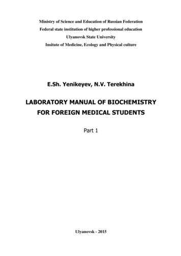
2Medical Biochemistry14. Endoplasic reticulum, lysosomes and golgi complex are involved in the integration ofpro-apoptic signals.MOLECULAR COMPOSITION OF CELLWaterWater accounts for about 70-75% of the weight of the cell. Other cellular constituents areeither dissolved or suspended in water.Organic Compounds1. Organic compounds accounts for 25-30% of the cell weight.2. They are nucleic acids, proteins, polysaccharides (carbohydrates) and lipids. Proteinsaccounts 10-20% of the weight of the cell. Nucleic acids account 7-10% of the cell weight.Polysaccharides usually account for 2-5% of the cell weight. About 3% of cell weight isdue to lipids. Lipids content may be higher in adipocytes or fat cells. Proteins mayaccount more of cell weight in cells like erythrocytes.3. Other low molecular weight organic compounds may account for 4% of cell weight. Theyare monosaccharides, aminoacids, fatty acids, purine and pyrimidine nucleotides, peptides,hormones, vitamins and coenzymes.Inorganic Compounds1. Inorganic compounds account for the rest of the cell weight.2. They are cations like sodium, potassium, calcium, magnesium, copper, iron and anionslike chloride, phosphate, bicarbonate, sulfate, iodide and fluoride.EUKARYOTIC CELL STRUCTURE AND FUNCTIONIn eukaryotes, cells aggregate to form tissues or organs and these are further organized toform whole organism. In humans, eukaryotic cells exist in large number of sizes and shapesto perform varieties of functions. For example, nerve cells differ from liver cell which differfrom muscle cell and they differ in function also. Though the eukaryotic cells differ in sizesand shapes they have certain common structural features. Further, eukaryotes containsubcellular structures and well defined nucleus. Cells are surrounded by membranes. Itseparates the cells from surrounding and it is called as plasma or cell membrane. The othersubcellular organelles are also composed in parts by membranes.A typical eukaryotic cell is shown in Figure 1.1.SUBCELLULAR STRUCTURES AND THEIR FUNCTIONSCell MembraneStructure1. The outermost structure of the cell that decides its contour is the cell membrane.2. It is a lipid bi-layer. It also consist of proteins and small amounts of carbohydrates(Figure 1.1 a).
Cell3Fig. 1.1 (a) Cell membraneFunctions1. It is fluid and dynamic.2. It is semi permeable, only selected compounds are allowed to pass through from outside. The selective permeability is responsible for the maintenance of internal environment of the cell and for creating potential difference across the membrane.3. The modification of the cell membrane results in formation of specialized structures likeaxon of nerves, microvilli of intestinal epithelium and tail of spermatids.NucleusStructure1. Centre of the cell is nucleus.2. It is surrounded by double-layer membrane of about 250-400 Å thick.3. The two layers of nuclear membrane are an outer and inner membrane (layer). The twomembranes fuse periodically to produce nuclear pores. Exchange of material betweennucleus and rest of the cell occurs through nuclear pores.4. The outer nuclear membrane continuous with other cytomembranes. In some eukaryoticcells, like erythrocyte nucleus is absent. In spermatozoa, nucleus accounts for 90% ofcell whereas in other cells nucleus accounts for less than 10% of the cell. In prokaryotes,nucleus is not well defined.Functions1. Nucleus is the information centre of eukaryotic cell. More than 90% of the cellular DNAis present in the nucleus. It is mainly concentrated in the form of chromosomes.
4Medical Biochemistry2. Human cell contains 46 chromosomes. These chromosomes are composed of nucleoproteinchromatin, which consist of DNA and proteins histones. Some RNA may also presentin the nucleus.3. In prokaryotes, the DNA is present as thread in the cytosol.NucleolusStructure and FunctionThese are small dense bodies present in the nucleus. Their number varies from cell to cell.There is no membrane surrounding them. They are continuous with nucleoplasm. Proteinaccounts for 80% of nucleolus remainder is DNA and RNA.NucleoplasmIt is also called as nuclear matrix. It contains enzymes involved in the synthesis of DNA andRNA.Cytosol, Cytoplasm or Cell SapStructure1. The extra nuclear cell content that possess both orgenelles and other material constitutes cytoplasm. Material other than subcellular components in the cytoplasm makes upthe cytosol or cell sap.2. Sometimes soluble portion of the cell is referred as cytosol. Cytoplasm accounts for70-75% weight of the cytosol.Functions1. Numerous enzymes, proteins and many other solutes are found in cytosol.2. Cytosol is the main site for glycolysis, HMP shunt, activation of aminoacids and fattyacid synthesis.MitochondriaStructure1. Are the second largest structures in the cell.2. Generally mitochondria are ellipsoidal in shape and can assume variety of shapes.3. The length of a mitochondrion is about 7 microns and has a diameter of 1 micron.4. Mitochondria consist of outer and inner membranes. The outer membrane is composedof equal amount of protein and lipids.5. The lipids are mainly phospholipids and cholesterol. The outer membrane functions asa limiting membrane and permeable to many compounds.6. The inner membrane consist of 75% protein and remainder is lipid.7. Cardiolipin is the important phospholipid of inner mitochondrial membrane.8. The inner membrane is convoluted to form number of invaginations known as cristaeextending to matrix (Figure 1.1b).9. These cristae are covered with knob like structures, which are composed of head piece,stalk and a base piece.
Cell5Functions1. The number of mitochondria ranges from 1-100 per cell depending on type of cell andits function. Several factors influence the size and number of mitochondria in cells. Inyeast, mitochondria is present in aerobic state and absent in anaerobic state. Exposureto cold increases mitochondria by 20-30% in liver cells.2. In highly metabolically active cells mitochondria are more and large.3. Location of mitochondria in cell also depends on types and functions of cell. In liver cellmitochondria are scattered. In muscles they are parellely arranged. Mitochondria inliver cell may range up to 2000 whereas in kidney they may range up to 300.4. Mitochondria is the power house of the cell. It is responsible for the production ofenergy in the form of ATP. The knob like structures function in electron transport andoxidative phosphorylation.5. Mitochondria also contain other energy producing pathways like citric-acid cycle, fattyacid oxidation and ketone-body oxidation.6. Some reactions of gluconeogenesis and urea cycle also occurs in mitochondria.Mitochondria is capable of synthesizing some of its proteins.7. Mitochondria contains some DNA known as mitochondrial DNA and ribosomes.8. Mitochondria which are essential for life because of their involvement in ATP production, also pay key role in programmed cell death of several types of cells. Duringapoptosis, mitochondrial membrane potential drops. This leads to permeabilization ofmitochondrial membrane. Cytochrome-C or mitochondrial proteins are released intocytosol which activates death enzymes. Further alterations in mitochondrial morphology also occur during apoptosis.9. In humans, mitochondria is derived from mother only. Hence, origin of mother ofhumans have been traced.10. Outer and inner mitochondrial membranes contain translocase enzymes. They are involved in sorting of nuclear encoded proteins into mitochondrial sub-compartments aswell as for their import into mitochondria. The inter mitochondrial membrane space ishome for several lethal proteins like pro-death enzymes.LysosomesStructure1. They are small vesicles present in cytoplasm.2. They are surrounded by a membrane. Lysosomes are called as ‘Suicidel bags’ of the cell.Functions1. Lysosomes are rich in hydrolytic enzymes, which are active at acidic pH. The lysosomalenzymes digest the molecules brought into the cell by phagocytosis.2. Macrophages are rich in lysosomes.Medical Importance1. Lysosomal enzymes are involved in bone remodelling and intracellular digestion.
6Medical Biochemistry2. Disease, shock or cell death causes rupture of lysosomes and release of enzymes. Insome organisms, lysosomal enzymes are responsible for cell death of larval tissues.3. Lack of one or more of lysosomal enzymes cause accumulation of materials in the cellresulting in lysosomal diseases.4. In some disease like arthritis and muscular dystrophy, lysosomal enzymes are releasedto cause uncontrolled destruction of surrounding tissues. Lysosomal proteases cathepsinsare involved in spreading of cancer (metastasis).5. As the age advances in digestible material an age pigment ‘lipofuscin’ occurs in somecells.6. Lysosomol cystine transporter cystinosin is defective in cystinosis, which is a lysosomoldisease. Hence, cystine transport into cytosol from lysosome is blocked.7. Lysosomes are involved in integration of pro-apoptic signals.PeroxisomesStructure1. Are also small vesicles surrounded by a membrane. They are also called as microbodies.Functions1. They contain enzymes of H2O2 metabolism. The concentration of protein in peroxisomesis very high and they may occur in crystallines form. The enzymes of H2O2 catabolismpresent in peroxisomes are peroxidase and catalase.2. Peroxisomes also contain other enzymes like D, L-amino acid oxidase, uric acid oxidaseand L-hydroxy fatty acid oxidation that generates H2O2. Glycerophospholipids are alsosynthesized in peroxisomes.Medical Importance1. Lack of peroxisomes result in Zellwegers syndrome.CytomembranesThere is an extensive network of membranes in the cytoplasm. These membranes are calledas cytomembranes. They are divided into endoplasmic reticulum and golgi complex or apparatus. The endoplasmic reticulum is further subdivided into rough endoplasmic reticulum(RFR) and smooth endoplasmic reticulum (SER).Rough Endoplasmic ReticulumStructure1. It is continuous with outer nuclear membrane.2. The cytoplasmic surface of rough endoplasmic reticulum is coated with ribosomes.Membrane enclosed channels of endoplasmic reticulam are called cisternae. The ribosomesare complexes of RNA and protein.Functions1. Ribosomes and rough endoplasmic reticulum are involved in protein synthesis.2. Protein synthesized, enters cisternae and later extruded.
Cell7Smooth Endoplasmic ReticulumStructure1. It is continuous with rough endoplasmic reticulum. It differs from RER by the absenceof ribosomes. When isolated SER is called as microsomes.Functions1. SER of intestinal cells is involved in formation of triglycerides.2. In the adrenal cortex, SER is the site of steroid formation.3. Cytochrome P450 dependent monooxygenases are present in liver cell SER.Golgi ApparatusStructure1. It consist of cluster of paired cytomembranes. The margins of these cytomembranes areflattened.2. It also contains several small vesicles, which are pinched off from the flattened marginsof membranes.Functions1. The golgi bodies are well developed in cells, which are involved in secretion. Materialproduced in the cell for export is processed by golgi body and is packaged as vesicle andis pinched off. The vesicles fuse with plasma membrane and their content is releasedto exterior by the process known as exocytosis. The digestive enzymes of pancreas andinsulin are produced and released in this way.2. Golgi apparatus helps in the formation of other subcellular organelles like lysosomesand peroxisomes.3. Golgi apparatus is involved in protein targeting. It directs proteins to be incorporatedinto membranes of other subcellular structures. It is also involved in glycosylation andsulfation of proteins.4. Golgi apparatus is involved in integration of proapoptic signal. It generates preapopticmediator ganglioside GD3.Medical ImportanceSome cases of diabetes are due to defective processing of insulin in golgi complex.Intracellular Ion ChannelsMembrane of endoplasmic reticulum, golgi complex and nucleus has ion channels. They areinvolved in transport of ions between cytosol and these intracellular components. Calciumand chloride ion channels which are involved in their transport from these components intocytosol are known.Vacuoles. Some animal cells contain vacuoles. They are membrane enclosed vesiclescontaining fluid. Mostly they contain nutrients.Cell Coat. Some mammalian cells contain thin coat known as cell coat on the outersurface of the cell membrane. The cell coat is flexible and sticky. It is composed ofmucopolysaccharides, glycolipids and glycoproteins. The adhesive properties of cell andorganization of tissue is controlled by cell coat.
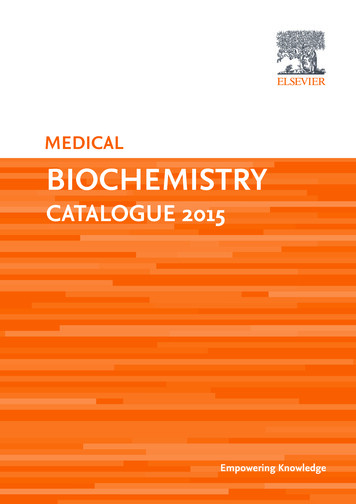
8Medical BiochemistryCytoskeletonsThese are filament like structures made up of proteins present in cytoplasm. Non-musclecells perform mechanical work with these intracellular network of proteins.(a) Microfilaments. They are actin like filaments. They form loose web beneath cellmembrane.(b) Myosin Fibres. Same as that of myosin of skeletal muscle.(c) Microtubules. Tubulin is the building block of microtubules. Dendrites, axons of nervecells and sperm cells contain microtubules. The sperm cell moves with the help offlagellum, a microtubule. These cyto skeletons are involved in the maintenance of cellshape, cell division, cell motility, phagocytosis, endocytosis and exocytosis.(d) Intermediate Filaments. They are not involved in movement of cell. They are stablecomponents of cytoskeleton. Neurofilament of neurons, glial filaments of glial cells andkeratin of epithelial cells are some examples of intermediary filaments.CELL CYCLEMEDICAL IMPORTANCE1. In all forms of life growth requires cell division.2. However, some cells divide even after growth like erythrocytes and epithelial cells ofintestine.Sequence of events associated with cell division occur in cyclic manner. Hence, cellcycle consist of sequence of events, which occur in cyclic manner during cell division. Thereare four stages (phases) in cell cycle. They are1. S (Synthesis)-Phase2. G1 (Gap 1)-Phase3. G2 (Gap 2)-Phase4. M (Mitosis)-PhaseSometimes, cell cycle is considered in two main events. They are mitosis and interphase which consist of G1, G2 and S-phases.1. S (Synthesis)-Phase: Division of a cell into two daughter cells requires duplication ofDNA. During S-phase concentration of DNA precursors increases nearly 10-20 folds. InS-phase DNA synthesis occurs. Period of DNA synthesis is almost constant in all adultcells. (1 Hour)2. G1 and G2-Phases: G1 and G2-phases are gaps or breaks in cell cycle. No special eventsoccur during these phases except the size of the cell may increase. However, there maybe many biochemical reactions taking place preparing the cell for division and checkingthat all appropriate steps are completed. The period of S1, G2 and M-Phases may rangefrom 12-18 hours. But the period of G1-phase varies, it can be few hours to months oreven years.3. Mitosis (M)-Phase: Many events take place in this phase of cell cycle. At the end mitosiscell divides into two daughter cells. The daughter cells are in G1-phase.
Cell9Check Points in Cell Cycle1. It is essential that during cell cycle, the synthesis of DNA, chromosomal segregationand cytoplasm division takes place in proper order. So, controls or check points withinthe cell cycle exist for all organisms.2. During cell cycle, oscillation of cell from mitosis to interphase is controlled by manycellular proteins. Further check points exist at the G1/S and G2/M boundaries of cellcycle. Cell cycle with check points is illustrated in Figure 1.2.Fig. 1.2 Four phases of cell cycle with check pointsCELL DEATHMEDICAL AND BIOLOGICAL IMPORTANCE1. Cells are not immortal i.e., they have finite life span. In the body, cells are formed anddestroyed. So, cells are in dynamic state.2. Cell division and cell death are two opposite processes required to maintain constanttissue volume (tissue homeostasis).3. Further cell death plays an important role in shaping tissues and organs during development or during recovery from injuries.4. Cell death may occur due to several external factors also.There are three types of cell death.1. Necrosis: It is also termed as cell murder. Cells undergo necrotic death if cell membrane is damaged or due to decreased oxygen supply and if energy (ATP) production isblocked.2. Apoptosis: This type of cell death occurs in tissue turnover. Individual cells or groupsof cells undergo this type of death. Aged cells in the body are removed by apoptosis.It is a genetically programmed cell death. In the initial stages of apoptosis, cellshrinks, followed by fragmentation and finally these fragments are eliminated byphagocytosis.
10Medical Biochemistry3. Atrophy: This type of cell death occurs in the absence of essential survival factors.Survival factors required by the cell are produced by other cells. Absence of nervegrowth factor leads to atrophy of nerves. It is also genetically programmed cell death.BIOCHEMISTRY, CELL AND DISEASEBiochemistry explains all cellular or biological events in chemical terms. The chemicalreactions that occur in biological systems are called biochemical reactions. Biochemistry alsoexplains how different sequences of biochemical reactions interact with each other for survival of cell (organism) under various conditions.When all the biochemical events occur in proper order, the cell or body remains normal. Blocks in biochemical events manifest as disease. So, every known (to be known)disease must (may) be due to blocks in biochemical events. The goal of biochemistry is toexplain all diseases in molecular terms. Therefore, biochemistry knowledge is requiredwhen one wishes to treat (cure) a disease. In addition, biochemistry suggests ways tomanipulate life forms for the benefit of mankind.REFERENCES1. Krstie, R.V. Ultra structure of mammalian cells, Springer-Verlag, Heidelberg, Germany,1979.2. Ernster, L. and Schatz, G. Mitochondria: a historical review. J. Cell Biol. 91, 227 (S) 235 (S), 1981.3. Rothman, J.E. The compartmental organization of golgi body. Sci. Am. 253(3), 84-95,1985.4. Duive. Microbodies in livings cells. Sci. Am. 248(5), 52-62, 1983.5. Bainton, D.L. The discovery of lysosomes. J. Cell Biol. 91, 665-675, 1981.6. Zimmerman, R.A. Ins and outs of ribosome. Nature 376, 391-392, 1995.7. Birchmeicr, W. Cytoskeleton structure and function. Trends Biochem. Sci. 9, 192-195,1984.8. Murray, A.W. and Kirschner, M.C. What controls cell cycle. Sci. Am. 264 (3)
This book is also useful for those who are specializing in biochemistry (M.Sc. or M.D.) because advances in frontier areas of biochemistry are presented in a systemic way. Of course advances in other areas that are relevant to medical students are also included to a limited extent. An interesting feature of this book is that the medical and .