Transcription
A Comprehensive Newborn Examination:Part II. Skin, Trunk, Extremities, NeurologicMARY L. LEWIS, MD, Dwight D. Eisenhower Army Medical Center, Fort Gordon, GeorgiaSkin findings are common during the newborn examination. Although these findings are often benign, it is importantto visualize the entire skin surface to distinguish these findings and appropriately reassure parents. The chest shouldbe observed for symmetric movement, pectus excavatum, pectus carinatum, prominent xiphoid, or breast tissue. Theinfant should be as relaxed as possible so that the physician can more easily detect any abdominal masses, which areoften renal in origin. A single umbilical artery may be associated with another congenital abnormality, especiallyrenal anomalies, and intrauterine growth restriction and prematurity. Signs of ambiguous genitalia include clitoromegaly and fused labia in girls, and bilateral undescended testes, a micropenis, or a bifid scrotum in boys. Sacraldimples do not warrant further evaluation if they are less than 0.5 cm in diameter, are located within 2.5 cm of theanal verge, and are not associated with cutaneous markers; dimples that do not fit these criteria require ultrasonography to evaluate for spinal dysraphism. Brachial plexus injuries are most common in newborns who are large forgestational age, and physical therapy may be required to achieve normal function. Patients with abnormal findings onOrtolani and Barlow maneuvers should be evaluated further for hip dysplasia. It is also important to assess newbornsfor tone and confirm the presence of normal primitive reflexes. (Am Fam Physician. 2014;90(5):297-302. Copyright 2014 American Academy of Family Physicians.)This is part II of a two-partarticle on the newbornexamination. Part I,“General, Head andNeck, Cardiopulmonary,”appears in this issue ofAFP on page 289.CME This clinical contentconforms to AAFP criteriafor continuing medicaleducation (CME). SeeCME Quiz Questions onpage 280.Author disclosure: No relevant financial affiliations.Part I of this two-part article discusses the assessment of generalhealth, head and neck, heart, andlungs.1 Part II focuses on assessingthe newborn’s skin, trunk and extremities,and neurologic function.SkinA variety of normal and abnormal lesionsmay be present on newborn skin (Table 1).2-6Although these findings are often benign, itis important to visualize the entire skin surface to distinguish these findings and appropriately reassure parents.2-6 It is common tosee bruising and petechiae, which typicallyresolve over time.Jaundice, which may cause yellowing ofthe skin and eyes, is common and typicallybenign. However, very elevated bilirubinlevels can lead to serious consequences, suchas kernicterus and acute bilirubin encephalopathy. Thus, bilirubin levels should beevaluated carefully according to AmericanAcademy of Pediatrics guidelines.7ChestChest movement should be symmetric.Asymmetry suggests an abnormality such aspneumothorax, cystic malformation of thelung, or diaphragmatic hernia. The physicianshould also observe for pectus excavatum,pectus carinatum, prominent xiphoid, andbreast tissue (may be present in girls or boysas a result of maternal hormone exposure), allof which are generally inconsequential findings but may pose cosmetic concerns. Supernumerary nipples can occur in the verticalline from the axilla to the pubic region (themilk line) and require no treatment.8 Widelyspaced nipples may be a sign of Turner syndrome.9 A prominent precordium may indicate a congenital heart defect.AbdomenWhen examining the abdomen, it maybe beneficial to use one hand to hold thelegs with the hips and knees flexed to helprelax the newborn, and use the other handto palpate the abdomen. General observation of the abdomen may reveal a scaphoidabdomen, which can indicate a congenital diaphragmatic hernia, or a distendedabdomen, which may indicate an intestinalobstruction. The presence of a gastroschisis(intestines protrude through the abdominalwall without a sac, usually to the right of theSeptember2014VolumeFamily90, Numberwww.aafp.org/afp AmericanFamily297Downloaded1,fromthe AmericanPhysician5website at www.aafp.org/afp.Copyright 2014American Academy of FamilyPhysicians.For thePhysicianprivate, noncommercial use of one individual user of the website. All other rights reserved. Contact copyrights@aafp.org for copyright questions and/or permission requests.
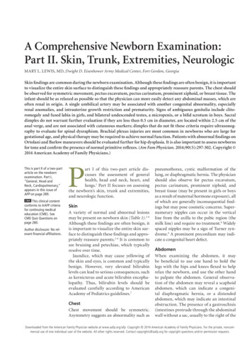
Newborn Examination: Part IISORT: KEY RECOMMENDATIONS FOR PRACTICEClinical recommendationRoutine renal ultrasonography to look for renal anomalies in newborns with isolated cases of a singleumbilical artery is not beneficial.Bilateral undescended testes, a micropenis, or a bifid scrotum should prompt investigation forambiguous genitalia.Newborns with a hypospadias should not be circumcised because the foreskin may be needed for repair.A sacral dimple is simple if it is less than 0.5 cm in diameter, located within 2.5 cm of the anal verge, andnot associated with cutaneous markers (discoloration or hypertrichosis). In the absence of these criteria,ultrasonography should be performed before three months of age to evaluate for spinal dysraphism.Girls born in the breech position should receive imaging to evaluate for hip dysplasia. Imaging shouldbe considered in newborns with a family history of developmental hip dysplasia and in newbornboys born in the breech position.EvidenceratingReferencesC11, 12C15CC1922C33, 34A consistent, good-quality patient-oriented evidence; B inconsistent or limited-quality patient-oriented evidence; C consensus, disease-orientedevidence, usual practice, expert opinion, or case series. For information about the SORT evidence rating system, go to http://www.aafp.org/afpsort.umbilicus) or omphalocele (abdominal contents protrude within a sac at the midline) isusually known before delivery and warrantsemergent surgical consultation.10 Diastasisrecti abdominis is caused by a relative weakness of the fascia between the two rectusabdominis muscles and usually resolves onits own over time. Linea nigra is a dark pigmented vertical line appearing from belowthe umbilicus to the pubic symphysis that iscaused by maternal hormone exposure anddisappears over time.The umbilical cord should be inspectedfor signs of infection and bleeding. It shouldcontain two arteries and one vein. A singleumbilical artery may be associated withanother congenital abnormality, especiallyrenal anomalies, and intrauterine growthrestriction and prematurity. In the past,renal ultrasonography was routinely performed in newborns with a single umbilical artery; however, routine renal imagingin isolated cases has not been proven beneficial.10-12 Umbilical hernias, which arecommon in newborns, rarely become incarcerated or strangulated, and most resolve bythe time the child reaches three years of age.The abdomen should also be auscultatedfor bowel sounds in all four quadrants, andthen palpated to feel for masses and organomegaly. It may be helpful to use the flats of thefingers rather than the tips. The liver is usually approximately 1 to 2 cm below the lowercostal margin. About one-half of abdominal masses are renal in origin, such as multicystic dysplastic kidneys, Wilms tumor,hydronephrosis, renal vein thrombosis,298 American Family Physicianwww.aafp.org/afpand ureteropelvic junction obstruction.Other masses include teratoma, neuroblastoma, ovarian torsion, ovarian cyst, and gastric duplication cyst.13GenitourinaryTerm female newborns have prominent labiamajora, whereas preterm female newbornshave prominent labia minora and clitoris.A white discharge or small amount of bloodmay be present and is a normal responseto maternal estrogen withdrawal. Separating the labia minora allows evaluation forpatency of the hymen. Signs of ambiguousgenitalia include clitoromegaly and fusedlabia. A hymenal tag may be present andusually requires no intervention.In male newborns, both testes should bedescended into the scrotal sac. Retractiletestes can be milked into the scrotum anddo not require intervention. Bilateral undescended testes, a micropenis (penile lengthfor a term newborn is 2.5 to 3.5 cm),14 or abifid scrotum should prompt investigationfor ambiguous genitalia. Findings suggestiveof ambiguous genitalia should be addressedurgently, and consultation with endocrinology, genetics, and urology is indicated.Gender should not be assigned until allinformation is obtained.15Evaluation of the scrotum may reveal aninguinal hernia or hydrocele (Figure 1). Aninguinal hernia occurs when the processusvaginalis fails to close, which leaves an opening for bowel to pass through and leads to abulge in the inguinal canal that may extendto the scrotum. The hernia can often beVolume 90, Number 5 September 1, 2014
Newborn Examination: Part IITable 1. Common Skin Findings in NewbornsFindingCharacteristicsLocationSalmon patch(nevus simplex)Port-wine stain(nevus flammeus)Macular pink-red capillary dilations, often bilateral, fades overtimeDark purple or red, capillary malformation, generally does notfade, ophthalmology referral needed if near the eyeHemangiomaBenign vascular tumor, caused by increased growth ofendothelial cells on blood vesselsOften benign and self-involutes, management depends onsize and locationFlesh-colored papules with erythematous base, containseosinophils, resolves in first weekPustules without erythema, contain neutrophils, rupture andleave hyperpigmented macules that may persist for monthsWhite papules, consist of epidermal cysts with keratinousmaterial, spontaneous rupture within first few weeks of lifeBlue-gray macules, up to 10-cm diameter, more common inblack, Native American, and Hispanic populations; usuallyfade by four years of age, but can be confused with abuseand should be documentedLight-brown macule, likely benign but may be an early sign ofneurofibromatosis or McCune-Albright syndromeReticular mottling of the skin caused by vascular response tocold, typically resolves with warmthForehead, upper eyelid, nape of the neck(stork bite)Nonspecific locationSturge-Weber syndrome: over thetrigeminal nerve (associated withseizures, glaucoma)Klippel-Trénaunay-Weber syndrome:extremityNonspecificErythema toxicumPustular melanosisMiliaMongolianspots (dermalmelanocytosis)Café au lait spotsCutis marmorataDiffuse (face and trunk)Diffuse (forehead, chin, neck, back)NoseCommon in sacral regionNonspecificUsually localized to the lower limbsInformation from references 2 through 6.reduced with pressure, and bowel sounds aresometimes auscultated.16,17 Inguinal herniasmay incarcerate and strangulate, and thusshould be surgically repaired. A hydroceleoccurs when the failed closure allows onlyfluid to pass through and usually transilluminates with light. Hydroceles often resolvewithin the first one to two years of life.16,17An American Academy of Pediatrics policy statement asserts that the health benefitsof male circumcision (e.g., prevention ofurinary tract infections, penile cancer, andtransmission of some sexually transmitted infections) outweigh the risks; however,the procedure should be performed only ifthe parents choose it.18 Hypospadias is theabnormal ventral placement of the urethralopening, which may be within the glans,the shaft of the penis, or scrotum (Figure 2).Newborns with hypospadias should not becircumcised because the foreskin may beused for repair.19 Chordee is a ventral curvature of the penis. It is a contraindicationto circumcision and warrants referral toSeptember 1, 2014 Volume 90, Number 5urology for correction. A buried penis (apenis that retracts into a fat pad) should notbe circumcised if the head is not exposedduring rest because it could become trappedin the fat pad with postoperative swelling.20,21Anus/RectumThe rectum should be examined for normalplacement and patency. A sacral dimple is considered simple if it is less than 0.5 cm in diameter, is located within 2.5 cm of the anal verge,and is not associated with cutaneous stigmata(e.g., hairy patches, hemangiomas). Classically, sacral dimples that do not fit these criteria have prompted ultrasonography beforethree months of age to evaluate for spinal dysraphism.22,23 Recent information has shownthat this may not be the best criteria for ultrasonography because evaluation for complexdimples without associated nonneurologiccongenital abnormalities is unlikely to revealan abnormality requiring surgical intervention. However, ultrasonography is a simple,noninvasive procedure and a good detectorwww.aafp.org/afp American Family Physician 299
AInguinalcanalCBIntestineSpermatic cordInguinalherniaHydroceleFigure 1. (A) Normal testis in the newborn examination. (B) An inguinal hernia occurs when the processus vaginalis failsto close, which leaves an opening for bowel to pass through and leads to a bulge in the inguinal canal that may extendto the scrotum. (C) A hydrocele occurs when the failed closure allows only fluid to pass through.Copyright Jordan Mastrodonato.of a tethered cord, if present.24 An imperforateanus may be a sign of a syndrome or may bean isolated finding. A VACTERL associationincludes vertebral/vascular, anorectal, cardiac, tracheoesophageal, radial/renal, andlimb anomalies. It is also commonly seenwith trisomies 18 and 21.ExtremitiesThe hands and feet should be inspected forsyndactyly and polydactyly. SupernumeraryGlanularAnterior 50%SubcoronalDistal penileMidshaftMiddle 30%Proximal penilePenoscrotalScrotalPosterior 20%PerinealFigure 2. Possible location of the urethral meatus in newborns withhypospadias.Copyright Jordan Mastrodonato.300 American Family Physicianwww.aafp.org/afpdigits arise from the lateral surface of a digit,most commonly on the ulnar aspect of the little finger, and vary from a complete digit witha nail bed and cartilage to a small amount ofpedunculated skin. These digits are usuallybenign but are removed for cosmetic reasons.Supernumerary digits without bone involvement usually can be tied off at the base,whereas digits with bone often need surgery,which should be performed by a skilled plastic surgeon. Polydactyly may be an isolatedabnormality but warrants a careful physicalexamination to investigate for other geneticabnormalities.25 A single palmar crease maybe a sign of trisomy 21 syndrome, but alsooccurs in 3% to 10% of persons without thesyndrome.26,27 Clinodactyly (curvature of thefifth digit) may be hereditary or a sign of avariety of syndromes, including Turner syndrome and trisomy 21 syndrome.28,29Various palsies may occur in associationwith brachial plexus injury. These injuriesare more common in newborns who arelarge for gestational age, especially thosewith shoulder dystocia or a prolonged delivery. An Erb palsy results from a tractioninjury to the C5 to C7 spinal nerve rootsand may cause the newborn to hold his orher arm medially rotated to the side with thehand pronated (“waiter’s tip”). A Klumpkepalsy involving lower segments of C8 andT1 results in paralysis of the entire arm withabsent reflexes. A variant of Klumpke palsy,known as Klumpke paralysis, produces aclaw hand, in which the forearm is supinatedand the wrist and fingers are flexed. Palsiesmay require physical therapy.30Volume 90, Number 5 September 1, 2014
ABFigure 3. Techniques to evaluate for hip dysplasia in newborns. (A) The Barlow maneuverinvolves adducting the hip while pushing the thigh posteriorly to see if can be dislocated. (B)The Ortolani maneuver involves abducting the hips while pushing the thigh anteriorly andrelocates reducible hips dislocated by the Barlow maneuver creating a clunk.Copyright Jordan Mastrodonato.Talipes equinovarus, or clubfoot, is a common orthopedic abnormality impacting oneper 1,000 births. This abnormality results inthe inversion and adduction of the forefoot,inversion of the heel and hindfoot, limitedextension of the ankle and subtalar joint,and internal rotation of the leg. The examiner should first determine if this is merely apositional deformity that is easily correctedwith manipulation and requiring no furthermanagement, or if it is truly talipes equinovarus necessitating an orthopedic consultation and usually casting.31 Pedal edema in thenewborn may indicate Turner syndrome.32When evaluating the hips for dysplasia,each hip should be examined separatelywith the other hand stabilizing the pelvis(Figure 3). The Barlow maneuver involvesadducting the hip while pushing the thighposteriorly to see if it can be dislocated. TheOrtolani procedure tests for posterior dislocation of the hips and is considered positiveif a clunk is heard when the femoral headrelocates anteriorly into the acetabulum. Ifthe results of the newborn hip examinationare equivocally positive (i.e., a soft click andmild asymmetry without an Ortolani or aBarlow sign), a follow-up hip examinationtwo weeks later by the child’s physician isrecommended. A previous article in American Family Physician includes a full description of the assessment for hip dysplasia.33Girls born in a breech position (risk ofdysplasia in this population is 120 per 1,000)should receive imaging (ultrasonography atsix weeks of age or plain radiography at fourSeptember 1, 2014 Volume 90, Number 5months of age). Imaging should be considered in newborns with a family history ofdevelopmental hip dysplasia (risk of 44 per1,000 in girls and 9.4 per 1,000 in boys) andin boys born in a breech position (risk of 26per 1,000).34NeurologicThe neurologic examination should assessthe primitive reflexes, which include the suck,grasp, root, primitive stepping, and Mororeflexes. The Babinski reflex should causedorsiflexion of the great toe on stimulation ofthe sole of the foot. In general, these reflexesbecome increasingly difficult to elicit beyondsix months of life, as the central nervous system matures and begins to allow voluntarymotor activity. The newborn’s muscular toneshould be evaluated by placing him or herin vertical suspension. When examining anewborn with hypotonia, it will feel as if thenewborn is slipping through the examiner’shands due to weakness in the shoulder girdleand legs. The newborn will also feel rigidwhen moving the extremities and will stayextended for a prolonged period while in vertical suspension.35Data Sources: A PubMed search was completed usingthe terms infant, newborn, developmental delay, developmental disturbance, and physical examination. Thesearch included meta-analyses, randomized controlledtrials, clinical trials, and reviews. We also searchedPOEMs (patient-oriented evidence that matters), ClinicalEvidence, the Cochrane database, and Essential EvidencePlus. Search dates: January 1, 2012, and May 2, 2014.The opinions and assertions contained herein are theprivate views of the author and are not to be construed aswww.aafp.org/afp American Family Physician 301
Newborn Examination: Part IIofficial or as reflecting the views of the U.S. Army MedicalDepartment or the U.S. Army Service at large.16. Nakayama DK, Rowe MI. Inguinal hernia and the acutescrotum in infants and children. Pediatr Rev. 1989;11(3):87-93.The Author17. Lao OB, Fitzgibbons RJ Jr, Cusick RA. Pediatric inguinalhernias, hydroceles, and undescended testicles. SurgClin North Am. 2012;92(3):487-504, vii.MARY L. LEWIS, MD, is a staff pediatrician and teachingfaculty physician in the Department of Family Medicine atDwight D. Eisenhower Army Medical Center in Fort Gordon, Ga.Address correspondence to Mary L. Lewis, MD, DwightD. Eisenhower Army Medical Center, 300 E. HospitalRd., Fort Gordon, GA 30905 (e-mail: marigoldll@yahoo.com). Reprints are not available from the author.REFERENCES1. Lewis ML. A comprehensive newborn examination: partI. General, head and neck, cardiopulmonary. Am FamPhysician. 2014;90(5):289-296.2. Merlob P, Metzker A, Reisner SH. Transient neonatal pustular melanosis. Am J Dis Child. 1982;136(6):521-522.18. American Academy of Pediatrics Task Force on Circumcision. Circumcision policy statement. Pediatrics. 2012;130(3):585-586.19. Baskin LS, Ebbers MB. Hypospadias: anatomy, etiology,and technique. J Pediatr Surg. 2006;41(3):463-472.20. Alter GJ, Horton CE, Horton CE Jr. Buried penis as acontraindication for circumcision [published correctionin J Am Coll Surg. 1994;178(6):636]. J Am Coll Surg.1994;178(5):487-490.21. Peleg D, Steiner A. The Gomco circumcision: commonproblems and solutions. Am Fam Physician. 1998;58(4):891-898.22. Kriss VM, Desai NS. Occult spinal dysraphism in neonates:assessment of high-risk cutaneous stigmata on sonography. AJR Am J Roentgenol. 1998;171(6):1687-1692.3. Berg FJ, Solomon LM. Erythema neonatorum toxicum.Arch Dis Child. 1987;62(4):327-328.23. Ben-Sira L, Ponger P, Miller E, Beni-Adani L, ConstantiniS. Low-risk lumbar skin stigmata in infants: the role ofultrasound screening. J Pediatr. 2009;155(6):864-869.4. O’Connor NR, McLaughlin MR, Ham P. Newborn skin:part I. Common rashes. Am Fam Physician. 2008;77(1):47-52.24. Chem JJ, Kirkman JL, Shannon CN, et al. Use of lumbarultrasonography to detect occult spinal dysraphism. JNeurosurg Pediatr. 2012;9(3):274-279.5. Metry DW, Hebert AA. Benign cutaneous vasculartumors of infancy: when to worry, what to do. ArchDermatol. 2000;136(7):905-914.25. Christensen JC, Leff FB, Lepow GM, et al. Congenitalpolydactyly and polymetatarsalia: classification, genetics, and surgical correction. 1981. J Foot Ankle Surg. 2011;50(3):336-339.6. Comi AM. Update on Sturge-Weber syndrome: diagnosis, treatment, quantitative measures, and controversies. Lymphat Res Biol. 2007;5(4):257-264.7. American Academy of Pediatrics Subcommittee onHyperbilirubinemia. Management of hyperbilirubinemia in the newborn infant 35 or more weeks of gestation [published correction appears in Pediatrics. 2004;114(4):1138]. Pediatrics. 2004;114(1):297-316.8. Brown J, Schwartz RA. Supernumerary nipples: an overview. Cutis. 2003;71(5):344-346.9. Pinsker JE. Clinical review: Turner syndrome: updatingthe paradigm of clinical care. J Clin Endocrinol Metab.2012;97(6):E994-E1003.10. Christison-Lagay ER, Kelleher CM, Langer JC. Neonatal abdominal wall defects. Semin Fetal Neonatal Med.2011;16(3):164-172.11. Martínez-Frías ML, Bermejo E, Rodríguez-Pinilla E, Prieto D; ECEMC Working Group. Does single umbilicalartery (SUA) predict any type of congenital defect?Clinical-epidemiological analysis of a large consecutive series of malformed infants. Am J Med Genet A.2008;146A(1):15-25.12. Deshpande SA, Jog S, Watson H, Gornall A. Do babieswith isolated single umbilical artery need routine postnatal renal ultrasonography? Arch Dis Child Fetal Neonatal Ed. 2009;94(4):F265-F267.13. Schwartz MZ, Shaul DB. Abdominal masses in the newborn. Pediatr Rev. 1989;11(6):172-179.26. U.S. National Library of Medicine. National Institutes ofHealth. Plus. Simian crease. May 10, 2013. http://www.nlm.nih.gov/medlineplus /ency/article / 003290.htm.Accessed June 4, 2014.27. Davies PA, Smallpeice V. The single transverse palmarcrease in infants and children. Dev Med Child Neurol.1963;25:491-496.28. Jones KL, Smith DW. Down syndrome. In: Smith’s Recognizable Patterns of Human Malformation. 6th ed.Philadelphia, Pa.: Elsevier Saunders; 2006.29. Sureshbabu R, Kumari R, Ranugha S, Sathyamoorthy R,Udayashankar C, Oudeacoumar P. Phenotypic and dermatological manifestations in Down Syndrome. Dermatol Online J. 2011;17(2):3.30. Jennett RJ, Tarby TJ, Krauss RL. Erb’s palsy contrastedwith Klympke’s and total palsy: different mechanisms areinvolved. Am J Obstet Gynecol. 2002;186(6):1219-1220.31. Bridgens J, Kiely N. Current management of clubfoot(congenital talipes equinovarus). BMJ. 2010;340:c355.32. Sävendahl L, Davenport ML. Delayed diagnoses ofTurner’s syndrome: proposed guidelines for change. JPediatr. 2000;137(4):455-459.33. French LM, Dietz FR. Screening for developmental dysplasia of the hip. Am Fam Physician. 1999;60(1):177-184,187-188.14. Tschudy MM, Arcara KM; Johns Hopkins Hospital. TheHarriet Lane Handbook. 19th ed. Philadelphia, Pa.:Mosby Elsevier; 2012.34. Committee on Quality Improvement, Subcommittee on Developmental Dysplasia of the Hip, AmericanAcademy of Pediatrics. Clinical practice guideline: earlydetection of developmental dysplasia of the hip. Pediatrics. 2000;105(4 pt 1):896-905.15. Murphy C, Allen L, Jamieson MA. Ambiguous genitaliain the newborn: an overview and teaching tool. J Pediatr Adolesc Gynecol. 2011;24(5):236-250.35. Zafeiriou DI. Primitive reflexes and postural reactions inthe neurodevelopmental examination. Pediatr Neurol.2004;31(1):1-8.302 American Family Physicianwww.aafp.org/afpVolume 90, Number 5 September 1, 2014
Sep 01, 2014 · black, Native American, and Hispanic populations; usually fade by four years of age, but can be confused wi