Transcription
Combining scanning performancewith application freedomZEISS Axioscan 7Your High-performance Slide Scanner for Fluorescence, Brightfield and Polarizationwww.zeiss.com/axioscan-bio
Your High-performance Slide Scanner forFluorescence, Brightfield and Polarization›In Brief›The Advantages›The Applications›The System›Technology and Details› ServiceDigitize your specimens with Axioscan 7 – the reliable, reproducible way to create high-quality virtual microscope slides. Axioscan 7 combines qualities thatyou would not expect to get in a slide scanner: high speed digitization and outstanding image quality plus an unrivaled variety of imaging modes are allavailable in a fully automated and easy to operate system.The most challenging research tasks as well as your routine scanning applicationsare supported by powerful hardware and perfectly featured software.Capture virtual slides quickly with high-speed scanning, while retaining consistentlyhigh quality, whether you want to capture brightfield, fluorescence or polarizedlight images.Axioscan 7 is controlled by ZEN Slidescan, which allows you to efficiently createand apply scan profiles, even in complex fluorescence experiments. A wealth ofZEN image analysis tools processes your data accurately afterward.Access your virtual slides anytime, no matter where you are or what operatingsystem you are using. Share your images online with colleagues and organizeentire projects, even when you're on the go.2
Simpler. More Intelligent. More Integrated.›In Brief›The Advantages›The Applications›The System›Technology and Details› ServiceRobust scan performanceAutomated application flexibilityThe bigger picture: slide scanning withinFrom an automated system, you expect absoluteZEISS Axioscan 7 allows rapid switching amongthe ZEN environmentreliability for continuous operation. ZEISS Axioscan 7fluorescence, brightfield and polarization scanningZEN Slidescan is optimized not only for the auto repeatedly produces digitized slides at dramati-modes, with the highest quality fast and gentlemated generation of virtual slides but also forcally improved speed, thanks to hardware com-imaging fully available to you in each of theseintegration within the powerful ZEN imaging soft-ponents designed for extended, uninterruptedmodes. Benefit from contrast flexibility and highware universe, which provides access to numerousoperation. A fully motorized condenser, power-scan speed when digitizing HE-stained tissueadditional processing and analysis functions.ful light sources and sensitive cameras ensuresamples or other brightfield applications. FastZEN Connect, the ZEISS software for correlative24 / 7 scan performance, whether you have manyfilter wheels and a spectral range up to far redmicroscopy, enables more advanced workflowssimilar slides or mixed applications to process.excitation light expand your fluorescence imag-– from automated slide scanning to detailedEqual attention has been paid to both softwareing capabilities — in combination with the newstudies on other ZEISS microscope systems. Theperformance and ease of use. Easily assigned scansample-preserving contrast method Transfer of established CZI data format opens the possibilityprofiles allow acquisition runs to be set up quickly. Intensity Equation (TIE), multiplex imaging reachesto use additional third-party data analysis tools.The ability to manually edit focus points providesa new level of performance. The combinationWith ZEN Data Storage and ZEN Data Explorer,additional flexibility. Axioscan 7 software is builtof all imaging modes let you extract maximumyou can access and share your scanned data fromto flawlessly process large amounts of raw data —sample information with a minimum of effort.anywhere, at any time.Scan up to 100 slides, which can have different scan profilesChoose from fluorescence, brightfield, or polarization optionsAutomated slide scanning and detailed studies on other ZEISSand imaging modes assigned, in one pass.according to the needs of your applications. Combine differentmicroscope systems combined in a ZEN Connect projectin the range of several terabytes.imaging modes to extract more information from your samples.3
Your Insight into the Technology Behind It›In Brief›The Advantages›The Applications›The System›Technology and Details› ServiceWith ZEISS Axioscan 7, you digitize large numbers of specimens in a reliable,reproducible, and hassle-free way.Fast automated scanningState-of-the-art hardware – including strong light sources, a motorizedcondenser, fast filter wheels, filter sets designed for advanced multiplexing applications, and modern cameras from the ZEISS Axiocam portfolio – all work inperfect union with software that offers a clear scan profile concept and easyto-use wizards for fast job setup. Many additional options allow for advancedmodification to create sophisticated slide scanning workflows. Once created,scan profiles are easily selected and assigned so you may run your experimentswith high degrees of both automation and application flexibility.High throughputDigitize a large number of slides, even with diverse scan profiles, in a singlerun. Axioscan 7 supports unattended scans of up to 100 slides in the standard26 mm 76 mm format for the entire job run. Monitor scanning progress byobserving the status LEDs for each individual sample tray at the front of thesystem or by accessing the system remotely.Reproducible qualityFrom geometry to color rendition, Axioscan 7 can be calibrated automaticallyso your virtual slides will be reproduced precisely and consistently betweensystems and over time. Get even better reliability by adding ZEISS PredictiveService: expert service technicians remotely diagnose all components so thatpreventive maintenance can be scheduled for maximum system uptime.Click here to view this video4
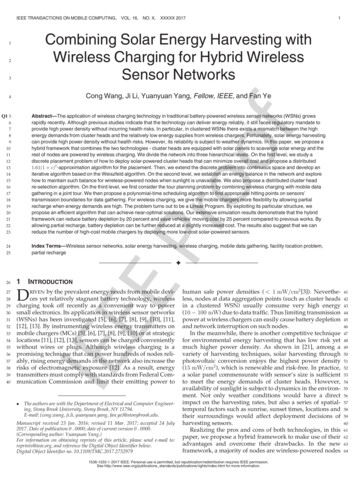
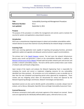
Your Insight into the Technology Behind It›In Brief›The Advantages›The Applications›The System›Technology and Details› ServiceHighly productive fluorescence imagingSpeed, gentle treatment and the optimal wavelength are critical when it comes to multispectral fluorescence imaging. Axioscan 7 employs swift and reproducible LEDillumination, fast filter wheels, and a sophisticated filter concept to efficiently separate a broad range of fluorescence channels.Brilliant illuminationAdvanced filter concept for application flexibilityChoose between Colibri 7, the super-fast 7 wave-The Axioscan 7 filter concept allows the shortest possible exposure times, maximum specimen protec-lengths LED light source from ZEISS, or the whitetion and unparalleled information density without compromising data quality. Three synchronized high-light LED light source X-Cite Xylis. Automatic cali-speed filter wheels for excitation, beam splitting and emission enable fast switching between fluorescencebration guarantees Colibri 7 operates with repro- channels. By using standard filter cubes in a 10-position filter turret, you can capture up to 9 fluorescenceducible output power levels for each wavelengthchannels or even perform polarization light microscopy. When using Colibri 7, you can choose betweenand therefore produces consistent quantitativesingle-band filters for perfect spectral separation or multi-band filters for instant channel switching with-data for all important dyes, fluorescent proteinsout moving any hardware. Newly designed filter sets for multiplexing applications allow for clear spectraland probes. Individual LEDs and integrated delineation without the need for additional software to separate fluorescent channels.excitation filters make an additional filter wheelunnecessary and allow switching times of a fewmilliseconds between the color channels.50 µmThe use of X-Cite Xylis together with a fast excitation filterwheel enables long wavelength sample illumination up to770 nm. In addition, the green gap – typically a problem withLED fluorescent light sources – is overcome and comparable toNon-small cell lung cancer (NSCLC) tissue stained with UltiMapper I/O PD-L1 kit. Nuclear counterstain (blue), CD8 (green), CD68 (orange),PD-L1 (red), panCytoKeratin (magenta). Sample courtesy of Ultivue, Inc. Cambridge, Massachusetts, USAclassic arc lamps in this spectral range.5
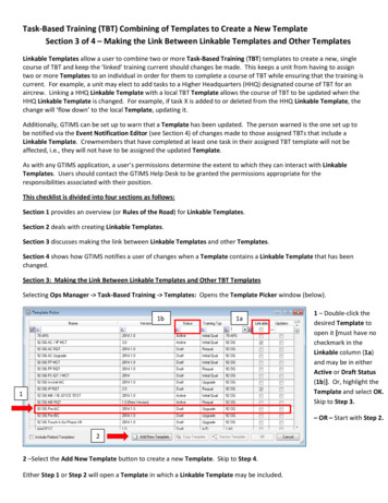
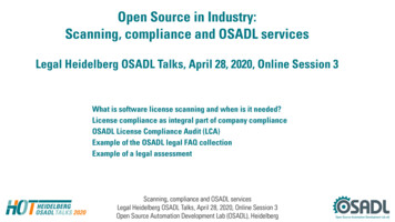
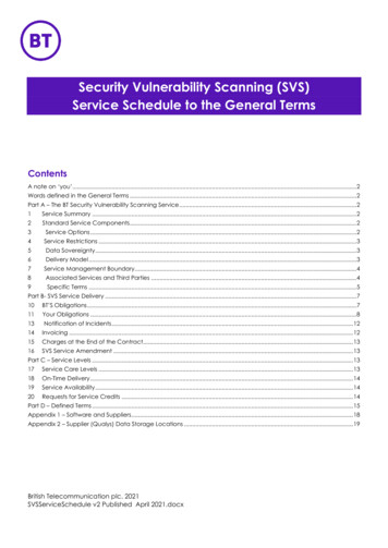
Your Insight into the Technology Behind It›In Brief›The Advantages›The Applications›The System›Technology and Details› ServiceA variety of super-fast brightfieldimaging modesThe newly designed condenser with its motorizedmodulator disk allows automatic switchingbetween different brightfield imaging modes toadapt to the different requirements of your ap-1plications, while maintaining optimal scanningperformance. The motorized aperture adapts to2any selected objective and enables the new TIE3contrast. Circular and linear polarization are now4fully supported, opening a new range of experiments and modality combinations.Axioscan 7 comes with a white light LED lightsource that is now 4 times stronger than itspredecessor. This allows the microscope stageand objective revolver to move continuously whilethe LED flashes in sync with the camera, freezing5movement effects in time by the flash strobe.You benefit from dramatically improved scanspeeds when using standard brightfield imaging,TIE contrast, or the new polarization modes.Perfect color renditionWith its large chip and small pixel size, thenew ZEISS Axiocam 705 color camera perfectlyThe Axioscan 7 brightfield imaging performance is driven by a complements this setup to capture brilliantmotorized condenser and a powerful white light source: images in the many supported imaging modes.1) Motorized modulator disc200 µm2) Circular polarizer3) Motorized linear polarizerMouse kidney wound healing assay, stained with sirius red;4) Motorized aperture diaphragmbrightfield (top) and cross linear polarization.5) White light LED light sourceSample courtesy: Alexander Lomow, Evotec, Germany6
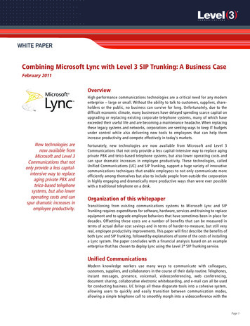
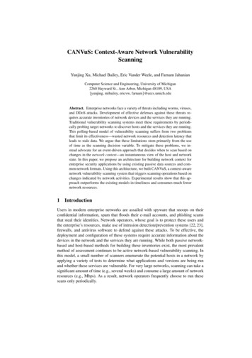
Your Insight into the Technology Behind It›In Brief›The Advantages›The Applications›The System›Technology and Details› ServiceAdvanced cameras for perfect image qualityCameras are crucial components for any automatic imaging system. Axioscan 7 is equipped with the most advanced Peltier-cooled cameras from the ZEISS Axiocamportfolio to support your brightfield and fluorescence applications with state-of-the-art imaging performance.Color camera Axiocam 705 color:Fluorescence camera Axiocam 712 mono:Large chip with perfect color renditionSmall pixels and high-speed imagingWe have implemented the latest color Axiocam,Axiocam 712 mono is the perfect choice for yourthe 705 color CMOS camera. It offers 5-mega-fluorescence imaging applications. It offers smallpixel resolution with a 3.45 µm pixel size and verypixels (3.45 µm), fully capturing the resolutionlow noise. With 55 frames per second acquisition potential of the high numerical aperture optics,speed in Axioscan 7 and a large field of view,and a very low readout noise. Use camera binningAxiocam 705 rapidly accomplishes your brightfieldof 2 2 pixels for increased sensitivity. For theand polarization imaging tasks.most demanding applications with weakest fluorescence signals, the Hamamatsu Orca Flash 4.0 isavailable as an option.1000 µm1000 µmMini pig tongue tissue section stained with masson trichrome.Mouse embryo sagittal cut, embryonic day E13, 12 µm. Sample courtesy: Alexander Lomow, Evotec, GermanySOX2 stained with Alexa488, Pax6 stained with Cy3, Nestinstained with Alexa647, Nuclei in Dapi. Sample courtesy: Ivan Mestres, TU Dresden, Germany7
Your Insight into the Technology Behind It›In Brief›The Advantages›The Applications›The System›Technology and Details› ServiceImproved detection. Better focusing. More context.The new contrast method Transfer of Intensity Equation (TIE contrast) is one of the key advancements in the Axioscan 7.Using this digital method for contrast generationin transparent samples, you record the i nteractionABCof a narrow cone of light with your sample’sstructures in three images: one in focus, and twoout of focus above and below the focal plane.From these three images, the phase informationfor the central plane is automatically extracted.Continuous acquisition in the z dimension, in100 µmcombination with flash illumination and GPUbased fast image processing, enables very fastdelivery of the final contrast images. You canSolanum tuberosum – potatoe starch, 20 Plan-Apochromat 0.8; A) TIE phase contrast, B) TIE relief contrast, C) Brightfieldchoose to present this as either phase contrast orDIC-like relief contrast.TIE contrast is an excellent tool to aid yourABC experiments when working with sensitive fluorescent dyes: Detect transparent tissues with little to no contrast in regular brightfield mode. Speed the subsequent fluorescence imagingprocess with very fast flash-based focusing.100 µm Protect your sensitive dyes from bleaching during focusing by using the lowest light doses. Bring your fluorescent labels into context easilyPleurosigma angulatum – diatomes, 20 Plan-Apochromat 0.8; A) TIE phase contrast, B) TIE relief contrast, C) Brightfieldby applying the additional contrast information.8
Your Insight into the Technology Behind It›In Brief›The Advantages›The Applications›The System›Technology and Details› ServiceZEN Slidescan: Easy to use and versatileAdvanced imaging software brings real benefits to biomedical research, capably handling your wide-ranging and complex tasks while remaining easy to operate.The Axioscan 7 operating software, ZEN Slidescan, is another expression of the Axioscan philosophy: combining the highest scanning performance and simplest operation with application-specific customization options.Smart setup of scanning tasksSeamless imaging and processingEasy-to-create scan profiles form the foundationZEN Slidescan allows you to not only capture virtual slides but also analyze and process the image data—allfor efficient slide scanning. ZEN Slidescanon the one platform. ZEN makes your images sharper and smoother, reinforcing contours, contrasts, bright- supports you with intuitive helpers like the newness and color. Meanwhile deconvolution produces crystal clear images that correspond to optical sections,Smart Profile Wizard and the powerful Advancedfree of out-of-focus light. Easy options for data export enable the analysis with third party tools, in case theProfile Editor. Scan profiles even for complexCZI image format is not natively supported.fluorescence imaging tasks can be created quickly.Just choose the dyes and ZEN Slidescan will calculate all the other settings. Once created, youcan assign and apply scan profiles to numerousslides with ease.Editing of focus pointsPrecise focusing is important for high-qualityscanning results. Axioscan 7 automatically focusesthe various sample areas on your slides. However,for some demanding applications, it may benecessary to check the position of the focus mapsupport points and adjust them manually. Withthe new Edit Focus Points feature, you can addand move focus points to regions that are moresuitable for focusing, even if those regions areoutside the scan area and are not needed forscanning.Editing of focus points9
Your Insight into the Technology Behind It›In Brief›The Advantages›The Applications›The System›Technology and Details› ServiceVirtual slides provide you with a multitude of valuable data so you will need plenty of storage space and agood filing structure to achieve high throughput. ZEISS implemented a central solution that manages yourdata so you can focus on results.ZEN Data Storage: the safe haven for all your imaging dataZEN Data Storage forms the central database that holds not only your digitized slides, but also multidimensional ZEN Connect projects. The server software is easily installed on any Windows-based server hardware.ZEN Data Explorer: permanent access to your research resultsZEN Data Explorer lets you access the stored data from anywhere. Running as a hybrid app on smartphonesand tablets (both iOS and Android) as well as in a web browser, ZEN Data Explorer provides access to yourZEN Data Explorer gallery viewdata storage and handles even large files efficiently. It will be your companion for sharing your findings atconferences and impressing prospective collaboration partners with your data. You can insert annotationsand view images in transmitted light and multichannel fluorescence images with a Z stack.Central Data ManagementCentral storage of images,analysis data, presets,workflows, and reports ZEN Data StorageZEN Data Explorer annotations viewMobile AccessData exploration on tabletcomputers, smartphones,and other mobile devices ZEN Data ExplorerCZI data formatThe ZEISS CZI format provides a wealth of benefits and is supported by a growing number ofother manufacturers. For an updated list, pleasevisit www.zeiss.com/czi.Share data on ZEN Data Storage with students and partners via ZEN Data Explorer.10
Expand Your Possibilities›In Brief›The Advantages›The Applications›The System›Technology and Details› ServiceCore Imaging Facilities: A sound investment that quickly pays for itselfIn core imaging facilities, the demand forhigher throughput and screening capability drivesthe charge towards automated instruments. Automation is convenient, but some platformssuffer from compromises in flexibility or imagequality which will significantly impact the numberof users wanting to make use of it.Axioscan 7 provides automation without sacrificing flexibility or the high quality of imagesyou need to attract a very wide range of usersto your facility. With approaches as varied asmultiplexing in tissue sections to polarizationin rock sections, there is a great opportunity toattract users from departments as diverse as LifeScience and G eology. As well as offering flexibility, Axioscan 7 is designed for 24/7 usage. Thispowerful combination of accommodating a broaduser base with robust design places Axioscan 7Thin section of Karlsbader Sprudelstein, scanned with 10x N-Achroplan 0.45 Pol. A merged image of the cross linear polarized lightas a top performer when it comes to usage hours channels is shown. Sample courtesy of Bernardo Cesare, Universita di Padova, Italyand it quickly pays for itself.Axioscan 7 complements the other instruments inSubsequent higher magnification acquisitionsSupport your users with easy to learn automatedyour facility and conveniently integrates into timeusing other imaging systems in the facility, likescanning that offers great flexibility while requir-saving workflows. Automatic, high quality screen-confocal systems, are easily guided using ZENing minimal training.ing of hundreds of samples for identification ofConnect and as such, previously time-consumingregions or events of interest is fast and efficient.studies are reduced in both time and complexity.11
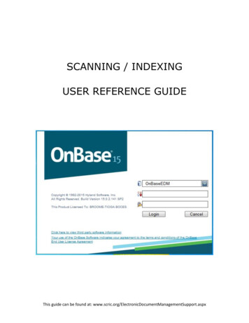
Expand Your Possibilities›In Brief›The Advantages›The Applications›The System›Technology and Details› ServiceZEISS Predictive ServiceMaximizes System UptimeOnce connected to your network and activated,ZEISSEnterprise ServerYour Networkthis advanced technology will automatically trackHTTPSthe health status of your instrument and collectsystem log files in the background to improve remote diagnosis.Relevant technical data such as operating hours,cycle counts or voltages are periodically moni-HTTPStored via a secure connection to our data center.The ZEISS Predictive Service application evaluatesthe performance of your microscope as systemdata can be received and analyzed.Our support engineers will diagnose any issues byanalyzing data on the Enterprise Server – remotelyand without interruption to your operation. Maintain highest system availability Fast and competent supportIncrease your uptime through close monitoringUse secure remote desktop sharing to easily getof the system’s condition as remote supportan expert connected.can often provide immediate solutions. Data security Optimum instrument performanceAs the status of your system is monitored,Ensure highest data security standards using necessary actions can be planned before theywell established technologies like PTC Thing-become urgent.worx and Microsoft Azure Cloud. No personalor image data is uploaded, only machine data.12
Tailored Precisely to Your Applications›In BriefTypical Applications / Typical SamplesTaskZEISS Axioscan 7 Offers›The AdvantagesResearch on Alzheimer pathogenesis and otherage-related diseasesDeveloping analytical models for amyloid deposition (plaques)High-resolution virtual slides in brightfield with image analysis›The ApplicationsCancer researchResearching the basics of cancerExcellent image quality in fluorescence combined with highthroughput thanks to fast filter wheels and sensitive cameras›The SystemMultiplexed imaging Identifying phenotypic fingerprints of different tissue types Optimal spectral separation of up to 7 fluorescent dyes thanksto newly designed filter sets›Technology and DetailsCollagen organization Analyzing fiber orientation using polarization light microscopy Extensive choice of different polarization options (circular polarization, multi-angle cross polarization) ADME / ToxicologyAutomated image analysis workflow and peer reviewswith colleagues at other locationsFindings are reproducible thanks to automated calibration,image analysis, and remote viewing› ServiceFluorescence in-situ hybridization (FISH)Determining the number of single sequence copies in the genomeMultichannel fluorescence, extended depth of fieldTarget identification and characterizationIdentifying and characterizing targets for pharmaceutical activesubstance searchesSensitive fluorescence imaging combined with gentle treatmentof specimens, especially in combination with TIE contrastfocusing options; image analysis functionsImmunological response to allografts and xenograftsIdentifying specific cell phenotypes and developingan understanding of cellular interactions in tissuesHigh dynamic range and zero artifact imagingResearch in the area of neurotraumasQuantifying brain injury measurements, including functional pathwaysof regeneration and microgliaZ stack imaging and robust digitization of the sampleswith high throughputTissue microarrays (TMA)Resource-friendly use of reagents and tissues with increased throughputReliable sample detection and robust scanning processContract research in the biomedical fieldUse of slides that are hard to standardize and various applicationsFlexible and configurable imaging options and tray conceptOrganization of expert networksOrganizing the efficient exchange of information from experts aroundthe world (e.g., cancer centers, tumor databases)Database with integrated platform-independent access to images and documentsInter-study group information managementSustained organization of the data pool in local study groups andevaluation of research findingsMulti-user access to database with specific access rightsExchange of information during spur of the momentdiscussions / meetingsDiscussing findings with peers spontaneously, irrespective of locationExcellent image quality, remote data access with smartphonesand tablet computersPublishing projects onlineMaking own data and images accessible to other peopleProject-based web-centric database system with integratedweb-viewing13
ZEISS Axioscan 7 at Work›In Brief›The Advantages›The Applications›The System›Technology and DetailsZEISS Axioscan 7 offers reliably reproducible image quality, no matter if you repeat your imaging task after aday, a week, a month or on a different machine.› ServiceParaffin-embedded mouse kidneys from healthy wildtype animals (12 weeks). Nephrin stained with Cy3. PCNA APC (FarRed)and DAPI as counterstaining. Imaged with 20 NA 0.8 objective.Sample courtesy: Florian Gembard t, Experimental Nephrology,Department of Internal Medicine III , University Clinic Carl Gustav Carus Dresden , Germany500 µm14
ZEISS Axioscan 7 at Work›In Brief›The Advantages›The Applications›The System›Technology and Details› Service2000 µmColon sample from a patient with Crohn’s disease, imaged with 20 NA 0.8 objective. Green: Cox-1 in Tuft cells in the epithelium – the sensory cells of the gut – and cells in the lamina propria connective tissue. Red: CD 163 – a macrophage marker.Courtesy of Steen Seier Poulsen, Department of Endocrinology and Metabolism, University of Copenhagen, Denmark. The image shown on this page represents research content. ZEISS explicitly excludes the possibility of making a diagnosis or recommending treatment for possibly affected patients on the basis of the informationgenerated with an Axioscan 7 slide scanner.15
ZEISS Axioscan 7 at Work›In Brief›The Advantages›The Applications›The System›Technology and DetailsThe UltiMapper I/O PD-L1 kit from Ultivue addresses whether a tumor is “hot” or “cold” and responsive to immune checkpoint inhibition because of a high immunefiltrate (hot), in contrast to tumors with low immune infiltrates called “cold tumors” or non-T-cell-inflamed cancers – by exploring multiple cell phenotypes such ascytotoxic immune cells (CD8), immunosuppressive macrophages (Markers CD68, PD-L1) or immune evading tumor cells (Markers CK, PD-L1).› Service2000 µmH&E stain of Non-small cell lung cancer (NSCLC) tissue.NSCLC tissue stained with UltiMapper I/O PD-L1 kit. Nuclear counterstain (blue), CD8 (green),Sample courtesy of Ultivue, Inc. Cambridge, Massachusetts, USACD68 (orange), PD-L1 (red), panCytoKeratin (magenta).The images shown on this page represent research content. ZEISS explicitly excludes the possibility of making a diagnosis or recommending treatment for possibly affected patients on the basis of the informationgenerated with an Axioscan 7 slide scanner.16
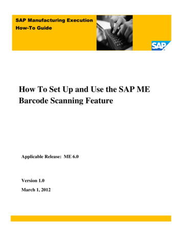
Your Flexible Choice of Components›In Brief›The Advantages›The Applications›The System›Technology and Details31245› Service1 Microscope3 Illumination4 Cameras Axioscan 7 Transmitted light: WL-LED Axiocam 705 color Magazines for 12 or 100 slides Fluorescence: Colibri 7 (385 nm, 430 nm, Axiocam 712 mono Trays for four 26 mm 77 mm slides, two52 mm 77 mm slides or 28 mm 48 mm and106 mm 77 mm slides475 nm, 511 nm, 555 nm, 590 nm, Hamamatsu ORCA-Flash 4.0630 nm, 735 nm) or X-Cite Xylis LT720L(380 nm – 770 nm) Filter wheels:5 Software ZEN Slidescan2 Objectives 10-position ACR for filter cubes or ZEN lite Fluar (5 ) 6-position high-speed excitation ZEN Data Storage N-Achroplan Pol (5 , 10 , 20 ) 6-position high-speed beamsplitter ZEN Data Explorer Plan-Apochromat (10 , 20 , 40 ) 6-position high-speed emission EC Plan-Neofluar Pol (20 , 40 ) EC Epiplan-Neofluar Pol (5 , 10 , 20 , 50 ) Other objectives on request17
System Overview›The Advantages›The Applications›The System›Technology and DetailsmcaxioAIn BriefmcaxioA›271570moronlocoAxiocam 705 color426560-9050-000Axiocam 712 mono426560-9090-000Camera Adapter 60N-C 1" 1.0x426114-0000-000Camera SetHamamatsuOrca Flash 4.0 K3for Axioscan 7426513-9013-000plus: ZEN Software DriverHamamatsu410136-1045-110› ServiceTube module 60N, 90 425524-9000-000Tube module 3x 60N mot., 90 425524-9020-000Filter wheels 2x 6 Pos.Beamsplitter and Emission424953-9012-000or:Reflector turret mot. 10 pos.with ACR for P&C modulesfor Axioscan 7424953-9002-000optional:Multi polarisation D ACR P&C Set424937-9010-000MonitorTFT 27" HP Z27n G2 (68 cm)410350-2701-000Reflected-light illumination mot. FLfor Axioscan 7423670-9000-0001External filter wheel428320-9002-000Illumination adapter423302-0000-0002Mounting frame for 1 slide100 x 76 mm432337-9080-000Mounting frame support for 1 slide28 x 48 mm432337-9090-000Axioscan 7 w / 12-piece magazine430038-9070-000or:Axioscan 7 w / 100-piece magazine430038-9080-000Accessories:Case for transport and storagefor 4 mounting frames432337-9040-000Mounting frame for 2 slides52 x 76 mm432337-9030-000optional:Solid-State Light Source Colibri 7– Type R[G/Y]B-UV423052-9730-000– Type R[G/Y]CBV-UV423052-9741-000– Type FR-R[G/Y]BV-UV423052-9770-000FL light sourceX-Cite Xylis XT720L423006-9000-000Mounting frame for 4 slides26 x 76 mm432337-9020-000Supportfor mounting of 4 slides 26 x 76 mmor mounting of 2 slides 52 x 76 mm432337-9071-00018
In Brief›The Advantages›The Applications›The System›Technology and DetailsAxioscan 7(1)ZEN Slidescan WorkstationDongle / hardware license USB (optional)000000-1066-465Axioscan 7(2)ZEN Data Storage Server› Service(3)Any Devicew / Browser(1)ZEN Slidescan WorkstationDongle / hardware license USB (optional)000000-1066-465(4)(5)Mobile DeviceiOS / Android) Dataw (/2ZENExplorerServerZEN Data StorageZEN DeskWorkstationw / ZEN DataStorage ClientExample configurations, for further details see corresponding product infos:( 3 Workstation,)( 4 10) Gb/s Ethernet connection strongly (recommended,5)Z6 High-EndRev. 2 (410203-9911-000),ZEN (blueAny edition),Device Monitor, Mouse, keyboardMobile DeviceZEN Desk2) ZEN Module Data Storage Server (410135-0031-320), easy installation, with secure user access management options;w/BrowseriOS/AndroidWorkstationserver hardware must be obtained separately: required is a high performance workstation, server system or equivalentw /RAMZENminimum,Data more is better, quad-corew / CPUZENorDatavirtual machine. Minimum requirements: 16 GBbetter, storage:ExplorerStorage ClientRAID system with redundancy recommended, WindowsServer 2012 R2 with update KB2919355or higher,network: 10Gb/s strongly recommended.3) Any device which runs a current browser, Google Chrome (version 78.0.3904.108 or later) recommended can be usedto access data on the ZEN Data Storage server (with appropriate access rights)4) Anymodern iOS (iOS12.4.1or later)or Android (version8.1 orinfos:later) device with ZEN Data Explorer ee correspondingproduct(with appropriate access oduleDataStorageClient(410136-1105-260)for direct access1) Z6 High-End Workstation, Rev. 2 (410203-991
Axioscan 7 is controlled by ZEN Slidescan, which allows you to efficiently create and apply scan profiles, even in complex fluorescence experiments. A wealth of ZEN image analysis tools processes your data accurately afterward. Access your virtual slides anytime, no mat