Transcription
Conductive Keratoplasty for the Correctionof Low to Moderate Hyperopia: U.S.Clinical Trial 1-Year Results on 355 EyesMarguerite B. McDonald, MD,1 Peter S. Hersh, MD,2 Edward E. Manche, MD,3 Robert K. Maloney, MD,4Jonathan Davidorf, MD,5 Moataz Sabry, MD,1 and the Conductive Keratoplasty United States Investigators GroupObjective: To document the 1-year safety, efficacy, and stability results of 355 eyes treated in the multicenter study of conductive keratoplasty (CK) used to correct low to moderate hyperopia.Design: Nonrandomized comparative (self-controlled) trial.Participants: Twenty surgeons at 13 centers performed CK on the eyes of all patients enrolled in amulticenter, 2-year, U.S. phase III clinical trial. Treated eyes had 0.75 to 3.00 diopters (D) of hyperopia andⱕ0.75 D of cylinder. Patients were 40 years of age or older.Intervention: Low-energy, high-frequency current was applied directly into the peripheral corneal stromathrough a delivery tip inserted at 8 to 32 treatment spots. The number of treatment spots was increased forincreasing levels of hyperopia, but the amount of radiofrequency energy remained constant. Emmetropia wasintended. All eyes were treated once (there were no retreatments).Main Outcome Measures: Data from 355 eyes with 1 year of follow-up were analyzed for safety andstability, and data from 318 eyes were analyzed for efficacy and predictability, as well as stability and safety. Allpatients reported on satisfaction and quality of vision after surgery.Results: At 1 year, uncorrected visual acuity was ⱕ20/20 in 56%, ⱕ20/25 in 75%, and ⱕ20/40 in 92% ofeyes. The manifest refractive spherical equivalent refraction was within 0.50 D in 63%, within 1.00 D in 89%,and within 2.00 D in 99%. Seven of 355 eyes lost 2 lines of best spectacle-corrected visual acuity at 1 year,but no eye lost 2 lines. One eye of 355 had induced cylinder of 2.00 D. The cycloplegic refractive sphericalequivalent changed a mean of 0.25 0.50 D between months 3 and 6, 0.11 0.41 D between months 6 and9, and 0.11 0.35 D between months 9 and 12. Refractive stability seemed to be attained by 6 months andremained stable through 12 months. Histology and confocal microscopy showed deep penetration of thetreatment into the stroma. Endothelial cell counts were not changed by the treatment.Conclusions: CK seems to be safe, effective, and stable for correcting low to moderate spherical hyperopiain patients 40 years old or older. Treatment penetration is deep and cylindrical in shape, and it does not damagethe corneal endothelium. Uncorrected visual acuity, predictability, and stability are as good as or better thanthose obtained with other techniques used to correct hyperopia. Ophthalmology 2002;109:1978 –1989 2002by the American Academy of Ophthalmology, Inc.Originally received: November 11, 2001.Accepted: April 11, 2002.Manuscript no. AA 2105831Southern Vision Institute, New Orleans, Louisiana.Cornea and Laser Vision Center, Teaneck, New Jersey.3Stanford University School of Medicine, Palo Alto, California.4Maloney Vision Institute, Los Angeles, California.5Davidorf Eye Group, West Hills, California.The members of the Conductive Keratoplasty United States InvestigatorsGroup are listed in the appendix.Presented in part at the Annual Meeting of the American Academy ofOphthalmology, New Orleans, Louisiana, November, 2001.Drs. Hersh, Manche, Maloney, and Davidorf and the Conductive Keratoplasty Investigators Group participated as clinical investigators in this Foodand Drug Administration phase III study sponsored by Refractec, Inc.(Irvine, CA). These authors participate on Refractec’s Medical AdvisoryBoard and are paid for their time. They otherwise have no proprietary orfinancial interest in the ViewPoint conductive keratoplasty system from21978 2002 by the American Academy of Ophthalmology, Inc.Published by Elsevier Science Inc.Development of thermal techniques to shrink peripheralcorneal collagen and thereby steepen the central cornea haschallenged ophthalmologists for longer than 100 years. Hotwire thermokeratoplasty, used in the 1980s to produce thermal burns that penetrated to 95% of corneal depth in hyperopic eyes, showed a lack of predictability and stability,and further development was abandoned.1– 4 More currentRefractec, Inc. Dr. Moataz Sabry was not an investigator but provided theconfocal microscopy photographs. Dr. Marguerite McDonald is a paidmedical monitor for the phase III study on conductive keratoplasty byRefractec, Inc.The nature of the procedure was explained to all participating patients, andthey all signed informed consent forms before undergoing the procedure.Reprint requests to Marguerite McDonald, MD, Southern Vision Institute,2820 Napoleon Avenue, New Orleans, LA 70115. E-mail: margueritemcdmd@aol.com.ISSN 0161-6420/02/ –see front matterPII S0161-6420(02)01255-1
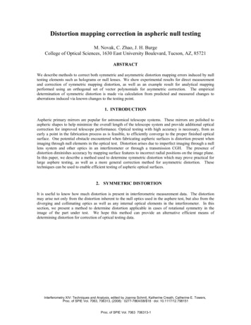
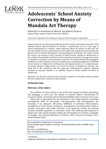
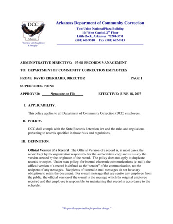
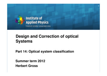
McDonald et al 䡠 Keratoplasty for the Correction of Low to Moderate HyperopiaTable 1. Original and Current NomogramsNumber ofTreatmentSpots8*162432Original Nomogram 0.75 1.00 2.00 3.00DDDDtotototo 1.002.003.004.00Current NomogramDDDD 0.75 D to 0.875 D 1.0 D to 1.625 D 1.75 D to 2.25 D 2.375 D to 3.00 D* None of the first 54 eyes were treated with 8 spots.D diopters.techniques using thermal keratoplasty include noncontactholmium:yttrium–aluminum– garnet laser thermal keratoplasty (Ho:YAG)(LTK; Sunrise Technologies, Fremont,CA),5–9 contact holmium:yttrium–aluminum– garnet LTK(Holmium 25; Technomed, Baesweiler, Germany),9 –12 continuous-wave diode LTK (DTK; Rodenstock, ProLaserMedical Systems, Inc., Dusseldorf, Germany),13,14 and conductive keratoplasty (CK; ViewPoint; Refractec, Inc., Irvine, CA).15 Nonthermal, excimer laser– based techniquesfor correcting hyperopia include photorefractive keratectomy16 –21 and laser in situ keratomileusis.22–28CK is a laserless radiofrequency-based technique for thecorrection of low to moderate hyperopia. The treatmentproduces a homogenous temperature increase that shrinkscollagen in the treated area and forms a cylindrical footprintdeep in the stroma. After a full circle of treatment spots, theperipheral cornea flattens and the central corneal steepens.This article presents the 1-year, single-treatment results of355 available eyes of the 401 enrolled in a U.S. phase IIIFood and Drug Administration (FDA) clinical trial andcompares them with results obtained with other nonexcimerlaser techniques for the correction of hyperopia.Materials and MethodsStudy DesignThis is a report of the 12-month results from the cohort of consecutive enrolled patients whose eyes were treated in a prospective, consecutive-series, multicenter clinical study (FDA phase III)evaluating the safety, efficacy, and stability of the CK procedurefor the correction of 0.75 to 3.00 diopters (D) of hyperopia andⱕ0.75 D of cylinder. The study follow-up will be 24 months.One-year data are available from 318 eyes for the variables thatindicate treatment efficacy (uncorrected visual acuity [UCVA],best spectacle-corrected visual acuity [BSCVA], manifest refractive spherical equivalent refraction [MRSE], and cycloplegic refractive spherical equivalent). For variables that indicate treatmentsafety and stability, data are available for 355 eyes. The discrepancy in number of eyes can be accounted for by the change innomogram after treatment of the first 54 eyes. The original nomogram specifying the number of treatment spots to correct a givenlevel of hyperopia was modified after the first 54 eyes were treatedbecause it had a tendency toward undercorrection (Table 1). Datafrom 24 of those 54 eyes could not be analyzed for efficacyvariables but could be analyzed for safety and stability. Theremaining 30 eyes, however, were found to have been treated withthe number of spots indicated by what later became the revisedFigure 1. The ViewPoint conductive keratoplasty system, including thehandpiece with the Keratoplast tip and a choice of two lid specula that actas the electrical return path. The console weighs 14 pounds.(current) nomogram and were included in efficacy analyses. Forexample, an eye with 1.50 D of hyperopia would be treated with 16spots under both the original and current nomograms, and theefficacy results could be included in the current nomogram eyes.Study DeviceThe ViewPoint CK system (Fig 1) used to perform the CK procedure consists of a radiofrequency energy-generating console; ahandheld, reusable, pen-shaped handpiece attached by a removablecable and connector; a foot pedal that controls the release ofradiofrequency energy; and a speculum that provides a largesurface for an electrical return path. The energy level default is60% of 1 W, and the exposure time default is 0.6 seconds.Attached to the probe is a single-use, disposable, stainless-steelKeratoplast Refractec Inc., Irvine, California tip, 90 m in diameterand 450 m long, that delivers the current directly to the cornealstroma (Fig 2). The tip has a proximal bend of 45 and a distal bendof 90 to allow access to the cornea over the patient’s brow and nasalregions. At the very distal portion of the tip is an insulated stainlesssteel stop (cuff) that ensures correct depth of penetration.Figure 2. Conductive keratoplasty handpiece with Keratoplast tip (90 mwide, 450 m long) and insulated stop at the distal end. Shown next to a7-0 suture.1979
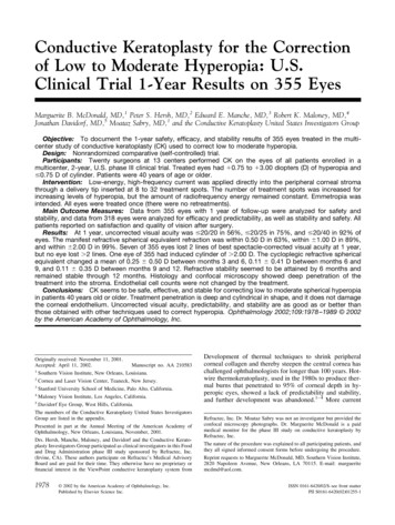
Ophthalmology Volume 109, Number 11, November 2002Figure 3. Treatment application. D diopter; OZ optical zone.PatientsInstitutional review board approval was obtained at each institution. A total of 401 patients at 13 centers in the United States whomet eligibility requirements were enrolled consecutively into thestudy from February 10, 1999, to December 1, 2000, signedinformed consent forms, and had 8 to 32 treatment spots applied tothe cornea during the CK procedure. The intended correction wasemmetropia.Enrolled patients had no existing ocular or chronic systemicdisease, previous ocular surgery, history of herpes infection, steroid-responsive increase in intraocular pressure, intractable keratoconjunctivitis sicca, history of keloid formation, or unstable,progressive hyperopia. Hard contact lens wearers were to discontinue lens use 3 weeks before the final measurements and theprocedure, and soft contact lens wearers were to discontinue lensuse 2 weeks before the final measurements and the procedure.They also had to have clear, undistorted mires on the centralkeratometry examination. Eyes with pachymetry readings of 560 m at the 6-mm optical zone (OZ) and those with a distanceUCVA of better than 20/32 were excluded from study participation.Examination MethodsThe preoperative and postoperative examinations for all eyes included manifest and cycloplegic refractions; uncorrected visualacuity and BSCVA with use of Early Treatment Diabetic Retinopathy Study visual acuity charts or Bailey–Lovie charts (distance)and Jaeger visual acuity charts (near); slit-lamp and funduscopicexamination; and computerized corneal topography. Cycloplegicrefraction was measured 30 minutes after two applications of oneor two drops of 1% cyclopentolate 5 minutes apart. Intraocularpressure was measured with the surgeon’s choice of standardapplanation instruments, including Goldmann, Perkins, or Draegertonometry.The preoperative examinations were performed by the surgeonsor their assistants 1 to 60 days before the CK procedure. Postoperative examinations were performed on days 1 and 7 and monthsTable 2. Study Patient AccountabilityMonth 1Available for safetyanalysisAvailable forefficacy analysisDiscontinuedMissed visitNot yet eligible forintervalLost to follow-up1980Month 3Month 6Month 0.25%0.25%10%1%
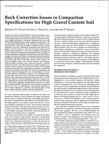
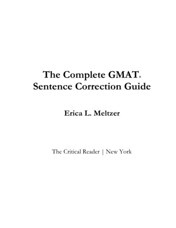
McDonald et al 䡠 Keratoplasty for the Correction of Low to Moderate HyperopiaTable 3. Demographic and Baseline InformationNumber of Patients/Eyes233/401Mean age (SD)RangeMedianRange of Treatment CRSEMean (SD)RangeMedianRange of Treatment MRSEMean (SD)RangeMedian55.3 years (6.4)40 to 7455.6 1.86 D (0.63) 0.75 D to 4.00 D 1.75 D 1.80 D (0.64) 0.38 D* to 3.75 D 1.75 DCRSE – Cycloplegic refractive spherical equivalent; MRSE – Manifestrefractive spherical equivalent.* Includes 2 ineligible eyes with minus CRSE values.SD standard deviation.1, 3, 6, 9, and 12, and results were recorded on standardized dataforms. Patients were also asked to subjectively evaluate the qualityof their postoperative vision and indicate their level of satisfactionon standardized forms.Mesopic contrast sensitivity testing was performed with theOptec 1600X, both with and without a glare source, in a subset of158 study patients before surgery and at 6 and 12 months aftersurgery. Testing without glare was performed with the targetillumination set to 5 cd/m2 and the glare source turned off. A trialframe with the patient’s best correction and test slide 1 wasinserted. The patient was asked to make a forced response to thedirection (left or right) of the point of the line in boxes A1 throughA8. The box number (i.e., A6) of the last correct response wasrecorded. This was repeated with test slide 2. The procedure wasrepeated with lines B1 through B8, C1 through C8, D1 through D8,and E1 through E8. The procedure for testing with glare was thesame except that glare illumination was set to 2 lux.Specular microscopy studies were performed to study the effectof the treatment on the corneal endothelium on 162 eyes at 5 of the13 investigational sites. Measurements were taken with the KonanNonCon Robopachy (Konan Medical, Inc., Hyogo, Japan) at thecentral cornea, the 3-mm OZ, and the 6-mm OZ. Changes frompreoperative levels at these sites were calculated by using paireddifferences statistical testing.Corneal haze was evaluated by slit lamp on a five-level scale ofclear, minimal, trace, mild, moderate, and marked. The protocolfailed to specify that haze only in the central cornea was to bereported and that haze at the treatment site was expected.Confocal microscopy, a noninvasive technique for revealinghistology, was performed 12 months after the procedure by usingthe Nidek Confoscan II (Nidek Ltd., Gamagori, Japan). Thistechnique optically sections living tissues to allow in vivo examination of individual layers.Surgical ProcedureTopical anesthesia was induced with one drop of 0.5% tetracaine,administered three times at 5-minute intervals. Pilocarpine was notadministered. A lid speculum was placed in the eye to be treatedto obtain maximal exposure and to provide the electrical returnpath; the fellow eye was taped closed. Illumination was providedby the operating microscope. While the patient fixated on themicroscope’s light, the cornea was marked with a gentian violet–dampened, eight-intersection CK marker that marks the 7-mm OZand makes radial marks that extend from the 6-mm to the 8-mmOZs (Fig 3). The surface of the cornea was dried with a fiber-freesponge to avoid dissipation of applied energy through a damp surface.The surgeons placed the Keratoplast tip on the cornea at the treatmentmarkings and attempted to place it perpendicular to the cornealsurface. The cuff around the probe, which settles perpendicular to thecornea, helped to achieve perpendicular placement. Light pressurewas applied until the tip penetrated the stroma to its insulator stop.Energy was applied by depressing the foot pedal. All eyes weretreated at the default setting of 350 kHz at 60% power for 0.6 seconds.All eyes were treated with the number of spots indicated by thecurrent nomogram (Table 1; Fig 3). For example, for the lowestamount of correction, 0.75 to 0.875 D, eight spots were placed onthe 7-mm OZ. For correcting 1.00 to 1.625 D, 16 spots wereplaced: 8 on the 6-mm OZ and another 8 on the 7-mm OZ. Forcorrecting 1.75 to 2.25 D, 24 spots were placed: 8 each on the 6-,7-, and 8-mm OZs. For correcting 2.375 to 3.00 D, 24 spots wereFigure 4. Postoperative uncorrected visual acuity (UCVA) over time.1981
Ophthalmology Volume 109, Number 11, November 2002Figure 5. Change over time in uncorrected visual acuity (UCVA).placed: 8 each were placed on the 6-, 7-, and 8-mm OZs, as wereplaced for 1.75 to 2.25 D of correction, and then 8 additional spotswere added between the spots previously made on the 7-mm OZ. Thesequence of spot placement is shown in Fig 3. The probe tip wascleaned of tissue debris with a fiber-free sponge after each treatmentspot. All eyes received a single CK treatment; i.e., no retreatmentswere performed.Postoperative CareAfter treatment, one drop of topical antibiotic solution and onedrop of diclofenac sodium 0.1% (Voltaren; Ciba Vision Ophthalmics, Duluth, Georgia) were administered and continued for 2days. Unpreserved artificial tear solution was the only ocularmedication permitted in the study. The treated eye was notpatched.ResultsAccountability and DemographicsOne eye was enrolled and treated, but no energy was deliveredduring the procedure. Thus the total number of treated eyes was400 of the 401 enrolled (Table 2). Complete follow-up datawere available for 355 eyes for safety variables and for 318 eyesfor efficacy variables. The mean age of enrolled patients was 55 6.4 years (range, 40 –74 years). There were 136 femalepatients and 97 male patients. Most (81%) of the patients werewhite and the others were Black, Asian, or other, or the racewas not recorded. Demographic information is shown in Table3.Figure 6. Predictability of achieved manifest refractive spherical equivalent (MRSE) refraction. D diopter.1982
McDonald et al 䡠 Keratoplasty for the Correction of Low to Moderate HyperopiaFigure 7. Cycloplegic refractive spherical equivalent (CRSE) stability. All visits through 12 months. D diopter.Uncorrected Visual AcuityPredictability and StabilityBefore treatment, 5 (1%) of 363 eyes had 20/20 or better UCVA,96 (26%) of 363 had UCVA of 20/40 or better, and 358 (99%) of363 had 20/200 or better. After surgery, at 1 year, 178 (56%) of318 eyes had UCVA of 20/20 or better, 240 (75%) of 318 had20/25 or better, 294 (92%) of 318 had 20/40 or better, 315 (97%)of 318 had 20/80 or better, and 100% had 20/200 or better. Fig 4shows postoperative UCVA over time. Fig 5 shows UCVAchanges over time for each acuity level. UCVA progressivelyimproved with time for acuity levels of 20/20 or better, 20/25 orbetter, and 20/32 or better and showed no leveling off. For loweracuity levels, however, a leveling-off was apparent within the 12months. The 20/40 acuity leveled off at 6 months, and 20/80leveled off at approximately 1 month.Predictability. At 1 year, 199 (63%) of 318 eyes were within 0.50 D of emmetropia, 282 (89%) of 318 were within 1.00 D,and 316 (99%) of 318 were within 2.00 D (Fig 6).Stability. All eyes (including the early nomogram eyes) wereevaluated for stability. The cycloplegic refractive spherical equivalent refraction changed ⱕ0.50 D in 240 (74%) of 326 eyesbetween the 3- and 6-month visits, in 305 (83%) of 366 betweenthe 6- and 9-month visits, and in 301 (89%) of 340 between the 9and 12-month visits (Fig 7). The MRSE increased a mean of 0.25 0.50 D between 3 and 6 months, 0.11 0.41 D between 6 and9 months, and 0.11 0.35 D between 9 and 12 months. Therefraction seemed to stabilize at 6 months.The plot of the mean MRSE (Fig 8) showed an overshoot ofFigure 8. Mean manifest refractive spherical equivalent (MSRE) refraction (diopters) over time. Twelve-month cohort of 300 eyes treated with thecurrent nomogram. CI confidence interval.1983
Ophthalmology Volume 109, Number 11, November 2002Table 4. Summary Efficacy Variables3 Months 6 Months 12 Months(N ⴝ 358) (N ⴝ 352) (N ⴝ 344)UCVA ⱕ20/20UCVA ⱕ20/25UCVA ⱕ20/40MRSE 0.50 DMRSE 1.00 e 6. Cylinder ChangesCylinder C
1 Southern Vision Institute, New Orleans, Louisiana. 2 Cornea and Laser Vision Center, Teaneck, New Jersey. 3 Stanford University School of Medicine, Palo Alto, California. 4 Maloney Vision Institute, Los Angeles, California. 5 Davidorf Eye Group, West Hills, California. The members o