Transcription
ARTICLEReceived 28 Oct 2011 Accepted 24 Jan 2012 Published 28 Feb 2012DOI: 10.1038/ncomms1701New insights into the Tyrolean Iceman’s originand phenotype as inferred by whole-genomesequencingAndreas Keller1,2,*, Angela Graefen3,*, Markus Ball4,*, Mark Matzas5, Valesca Boisguerin5, Frank Maixner3,Petra Leidinger1, Christina Backes1, Rabab Khairat4, Michael Forster6, Björn Stade6, Andre Franke6, Jens Mayer1,Jessica Spangler7, Stephen McLaughlin7, Minita Shah7, Clarence Lee7, Timothy T. Harkins7, Alexander Sartori7,Andres Moreno-Estrada8, Brenna Henn8, Martin Sikora8, Ornella Semino9, Jacques Chiaroni10, Siiri Rootsi11,Natalie M. Myres12, Vicente M. Cabrera13, Peter A. Underhill8, Carlos D. Bustamante8, Eduard Egarter Vigl14,Marco Samadelli3, Giovanna Cipollini3, Jan Haas15, Hugo Katus15, Brian D. O’Connor16,17, Marc R.J. Carlson18,Benjamin Meder15, Nikolaus Blin4,19, Eckart Meese1, Carsten M. Pusch4 & Albert Zink3The Tyrolean Iceman, a 5,300-year-old Copper age individual, was discovered in 1991 on theTisenjoch Pass in the Italian part of the Ötztal Alps. Here we report the complete genomesequence of the Iceman and show 100% concordance between the previously reportedmitochondrial genome sequence and the consensus sequence generated from our genomicdata. We present indications for recent common ancestry between the Iceman and presentday inhabitants of the Tyrrhenian Sea, that the Iceman probably had brown eyes, belonged toblood group O and was lactose intolerant. His genetic predisposition shows an increased riskfor coronary heart disease and may have contributed to the development of previously reportedvascular calcifications. Sequences corresponding to 60% of the genome of Borrelia burgdorferiare indicative of the earliest human case of infection with the pathogen for Lyme borreliosis.1 Department of Human Genetics, Saarland University, 66421 Homburg, Saar, Germany. 2 Siemens Healthcare, 91052 Erlangen, Germany. 3 Institute forMummies and the Iceman, EURAC research, 39100 Bolzano, Italy. 4 Division of Molecular Genetics, Institute of Human Genetics, University of Tuebingen,72074 Tuebingen, Germany. 5 Febit biomed GmbH, 69120 Heidelberg, Germany. 6 Institute of Clinical Molecular Biology, Christian-Albrechts-UniversityKiel, Kiel, Germany. 7 Genome Sequencing Collaborations Group, Life Technologies, Beverly, Massachusetts 01915, USA. 8 Department of Genetics, StanfordUniversity School of Medicine, Stanford, California 94305, USA. 9 Dipartimento di Genetica e Microbiologia, Università di Pavia, Via Ferrata, 1 27100Pavia, Italy. 10 Unité Mixte de Recherche 6578, Centre National de la RechercheScientifique, and EtablissementFrançais du Sang, Biocultural Anthropology,Medical Faculty, Université de la Méditerranée, 13916 Marseille, France. 11 Department of Evolutionary Biology, University of Tartu and Estonian Biocentre,23 Riia Street, 510101 Tartu, Estonia. 12 Sorenson Molecular Genealogy Foundation, Salt Lake City, Utah 84115, USA. 13 Departamento de Genética, Facultadde Biología, Universidad de La Laguna, Tenerife 38271, Spain. 14 Department of Pathological Anatomy and Histology, General Hospital Bolzano, 39100Bolzano, Italy. 15 Department of Internal Medicine III, University of Heidelberg, 69120 Heidelberg, Germany. 16 The Ontario Institute for Cancer Research,101 College Street, Suite 800, Toronto, Ontario, Canada M5G 0A3. 17 Nimbus Informatics LLC, 104R NC Hwy 54 West, Suite 252, Carrboro, North Carolina27510, USA. 18 Department of Computational Biology, Fred Hutchinson Cancer Research Center, Seattle, Washington 98109, USA. 19 Department ofGenetics, Wroclaw Medical University, 50-368 Wroclaw, Poland. *These authors contributed equally to this work. Correspondence and requests formaterials should be addressed to A.Z. (email: albert.zink@eurac.edu).nature communications 3:698 DOI: 10.1038/ncomms1701 www.nature.com/naturecommunications 2012 Macmillan Publishers Limited. All rights reserved.
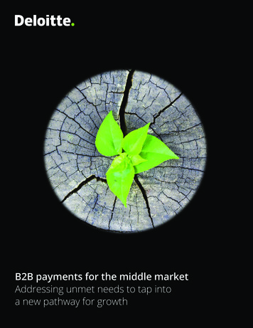
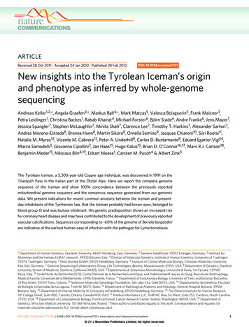
ARTICLEnature communications DOI: 10.1038/ncomms1701In September 1991, two hikers discovered a human corpse (Fig. 1),partially covered by snow and ice, on the Tisenjoch Pass in theItalian part of the Ötztal Alps. The Tyrolean Iceman, a 5,300-yearold Copper age individual, is now conserved at the ArchaeologicalMuseum in Bolzano, Italy, together with an array of accompanying artefacts. An arrowhead lodged within the soft tissue of the leftshoulder, having caused substantial damage to the left subclavianartery, indicated a violent death1. Speculations on his origin, hislife habits and the circumstances surrounding his demise initiateda variety of morphological, biochemical and molecular analyses.Studies of the Iceman’s mitochondrial genome began 3 years afterhis discovery with an analysis of the HVS1 region2 and led to thesequence analysis of the entire mitochondrial genome3,4. Althoughthese mitochondrial DNA (mtDNA) studies yielded conclusivedata, no successful amplification of nuclear DNA from the Icemanhas been reported to date. As cold environments often provide forgood biomolecular preservation and as methods for ancient DNAanalysis have recently improved, a whole-genome sequencing studywas initiated in 2010. A 0.1-g bone biopsy was taken from the Iceman’s left ilium under sterile conditions in the Iceman’s preservationcell at the South Tyrol Archaeological Museum in Bolzano, Italy,DNA was extracted at the Institute of Human Genetics in Tübingen,Germany and a sequencing library was generated at febit GmbH inHeidelberg, Germany. Paired-end high-throughput sequencing onthe SOLiD 4 platform was performed at Life Technologies facilities,Beverly, MA, USA. We found indication for recent common ancestrybetween the Iceman and present-day inhabitants of the TyrrhenianSea (particularly Corsica and Sardinia), that the Iceman probablyhad brown eyes, belonged to blood group O and was probably lactose intolerant. We further found a genetic predisposition for anincreased risk for coronary heart disease (CHD), which may havecontributed to the development of previously reported vascular calcifications. In addition, we found sequences corresponding to 60%of the genome of B. burgdorferi that are indicative of the earliesthuman case of infection with the pathogen for Lyme borreliosis.ResultsOverall sequencing results. We obtained a total of 2.93 109sequencing reads out of which 1.11 109 reads (37.9%) mappeduniquely to the human reference genome (hg18). Paired-end readscovered 96% of the reference genome with small variation amongchromosomes (max 97.74% for chr22, min 89.61% for chrY)(Supplementary Fig. S1a and Supplementary Table S1). The averagedepth of coverage for non-redundant reads excluding duplicates is7.6-fold, ranging from 4.76-fold for chromosome X to 10.8-fold forchromosome Y (Supplementary Fig. S1b). More than 65% of thegenome is covered at least five-fold. Regions with more than 100coverage were excluded from analysis, as these most likely representPCR duplication artefacts.Deamination-based damage, as the most common modificationin ancient DNA5,6, potentially complicates correct identificationof base substitutions that result from evolutionary processes. Thesequencing of a 4,000-year-old palaeo-eskimo’s DNA sequence demonstrated that exclusion of postmortem, damage-based changes hadlittle impact on the high-throughput sequencing result5. However,to estimate the extent of miscoding lesions (that is, postmortemchanges in DNA), we determined the transition and transversionratios (Supplementary Table S2 and Supplementary Table S3) andfound them to be largely consistent with ratios observed for othergenomes recently analysed by high-throughput sequencing.One limitation to the whole-human genome sequencing hasbeen the ability to provide broad access to the underlying sequenceinformation in a useable format such that individuals can readilyquery the genome for their own specific research needs. To enablefull access to the Iceman’s genome to a broader scientific community,we have generated a genome browser (http://IcemanGenome.net)8. Figure 1 The Tyrolean Iceman. The mummy of the Tyrolean Iceman in hispreservation cell at the Archaeological Museum of Bolzano.This browser contains all the aligned sequencing reads to the HumanGenome Reference 19, provides a basic level of annotation including individual single-nucleotide polymorphisms (SNPs), insertionsand deletions, and gene identification. We have also incorporateda query engine for more complicated analysis routines, such as thecomparison of the Iceman’s genome to other contemporary genomes(Methods).Contamination controls. Assessment of the percentage of miscoding lesions and potential human contamination was carried outthrough analysis of the mtDNA. As the mitochondrial genome isnaturally at a very high ratio as compared with the nuclear genome( 1160 coverage versus 7.6 in this specific case), it provides a goodtarget to identify low-frequency variants that can be caused by contamination. Additionally, the Iceman’s mitochondrial genome hadpreviously been sequenced in a different laboratory using a differentsample3, thus serving as an ideal independent comparison. Alleleratios were determined for the previously noted positions at whichthe Iceman differs from the Cambridge Reference Sequence (rCRS,NC 012920). For each read not consistent with the known Icemangenome, the specific exchange type was examined as to whether itis a typical deamination product or whether it constitutes a knownhuman mitochondrial variant (Supplementary Fig. S2, Supplementary Table S4). The average rate of concordance with the known Iceman genome is over 94%, the percentage of exchanges corresponding to miscoding lesions is under 5% and the rate of exchangescorresponding to other known human sequences is 2.5%. Whilethe latter may be caused by modern contamination (either humanor other), it may also suggest heteroplasmy of certain positions ofthe Iceman’s mitochondrial genome. Although this phenomenonwas thought to be rare, recent studies have shown that heteroplasmyis not uncommon in healthy mitochondrial cells6. This analysissuggests a maximum potential contamination level of 2.5%, anamount that would have little impact on the allele calls for thenuclear genome.The next step comprised the analysis of DNA damage patterns.As sequences derived from ancient DNA molecules show characteristic nucleotide composition patterns, those can be used to distinguish them from contaminants of modern DNA fragments. Inparticular, postmortem deamination of cytosine to uracil leads toan increased rate of observed C to T transitions, particularly nearthe ends of the DNA fragments7. Furthermore, postmortem DNAfragmentation occurs at an increased rate at the 3′-end of purinenature communications 3:698 DOI: 10.1038/ncomms1701 www.nature.com/naturecommunications 2012 Macmillan Publishers Limited. All rights reserved.
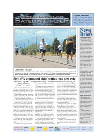
ARTICLEnature communications DOI: 10.1038/ncomms1701SNP analysis. To gain first insight into genetic traits and into physiological and pathological characteristics of the Iceman, we identified 2.2. million SNPs in the Iceman’s genome sequence of which1.7 million SNPs have previously been reported in dbSNP, with 900,000 and 800,000 positions being present in a heterozygous anda homozygous state, respectively (Supplementary Table S5). As the1,000 Genomes Project has demonstrated, using higher sequencecoverage should result in the detection of 3.5 million SNPs perindividual; however, due to the limited amount of ancient DNA,lower sequencing coverage was generated, resulting in the detectionof fewer SNPs. As shown in Supplementary Table S5, we found aconcordance rate of 92% for homozygous SNPs but only of 55%for heterozygous SNPs indicative of an undercalling of heterozygousSNPs as expected for 7.6-fold coverage.We compared the number and frequency of homozygous toheterozygous SNPs known from dbSNP on each chromosome.The highest frequency of homozygous SNPs among autosomes wasdetected for chromosome 13, where 29,400 of the 43,680 SNPs wereshown to be homozygous (67.3%). We detected heterozygous SNPsin regions with high similarity to other genomic regions and pseudoautosomal regions. However, the overall highest rate of homozygousSNPs, namely 96.8% (16,080 of 16,606 SNPs), was detected in the Xchromosome. As the Iceman was a male, these results further demonstrate the low potential degree of contamination, consistent withthe results derived from the mtDNA analysis.We furthermore analysed SNPs, including variants in specificgenomic regions, of potential clinical or functional relevance. Inthe 5,300-year-old mummy, our analysis did not identify variationsin human accelerated regions10, which are apparently 0.15–10–505101520 –20 –15 –10–50105Distance from read endbFrequencynucleotides due to depurination, leading to a characteristic enrichment in purines at the base preceding the sequencing read7.In order to determine whether the Iceman DNA shows thesecharacteristic patterns, we analysed a random subsample of thealigned sequencing reads for their nucleotide composition andmisincorporation patterns. From each of the SAM files containingthe aligned read data of the three sequencing slides, we randomlysampled one out of every 1,000 reads, and combined them into aninitial analysis file containing a total of 1,156,960 reads. To simplifythe subsequent nucleotide substitution analysis, we removed readsthat showed insertions or deletions with respect to the referencesequence, as well as reads that mapped closer than 10 bp from theedges of the respective chromosome. This final read set contained1,148,786 reads, and all subsequent results are based on this set. Foreach read, we then retrieved the corresponding genomic sequencefrom the human genome hg18 build, extended to include sequenceup to 10 bp upstream (downstream) for reads that map to theforward (reverse) strand.The average nucleotide composition around the ends of the analysed reads shows the typical pattern expected for ancient DNAfragmentation (Fig. 2a and b). A sharp increase in the frequency ofpurines (A and G) is observed at the first base upstream of the forward reads, accompanied by a complementary increase of pyrimidines (C and T) just downstream of the reverse reads. Additionally,substitution rates for each of the possible nucleotide mismatchesalong the sequencing read also show the expected pattern. Whilesubstitution rates are generally low ( 0.25%) for most mismatchtypes, C to T transitions show an increased overall rate, in particulartowards the end of the reads (up to 3.5% at the first nucleotide). Asabove, the increase in C to T transitions for the forward reads areaccompanied by the complementary increase in G to A transitionsobserved for the reverse reads. Overall, these results are comparable to other ancient human genomes recently published, such asthe Saqqaq5 and the Australian Aboriginal genome8,9, and therefore provide additional indication that the sequences are 0.005SubstitutionC– TG– AOther05101520 –20–15Distance from read end–10–50Figure 2 Analysis of DNA damage patterns. (a) Average nucleotidecomposition around the ends of the analysed reads. (b) Observed rateof C to T and G to A transitions.sections of the genome showing human-specific evolution over thelast few million years.SNPs of potential clinical relevance with additional direct assessment of further SNPs of phenotypic interest (for example, lactasepersistence, pigmentation and blood group) are summarized in Supplementary Table S6. Not all listed SNPs necessarily infer a phenotypicconsequence or a high predisposition risk. A number of variants supported by morphological or radiological data (such as risk factors foratherosclerosis), or variants that are likely to lead to a specific phenotype (such as lactase nonpersistence) have been resequenced using theSanger method. These are listed together with individual coverage depthin Table 1. Sanger sequencing, carried out using extract from a separatesample, confirmed the next-generation sequencing results in all caseswhere amplification was successfully carried out, further underliningthe low degree of contamination. Additionally, some of the characteristic SNPs of the Iceman’s mitochondrial genome were screened usingmitochondrial primers to confirm authenticity (Table 2).As a next step we analysed the genetic ancestry of the Iceman.As the Iceman’s mtDNA haplotype has not yet been detected amongthousands of sampled contemporary individuals, it has been difficultto assess his genetic ancestry with high accuracy. The first analysiswas to determine if the Iceman’s autosomal DNA shows an affinityto any specific population or if he remains an outlier among contemporary samples. We intersected genotype calls of greater than 6 coverage from his genome with the population reference sampleconsisting of more than 1,300 Europeans genotyped for SNPs on theAffymetrix 500K array11,12, 125 individuals from seven North African populations ranging from Egypt to Morocco on the Affymetrix 6.0 array and 20 Qatari samples from the Arabian Peninsula13.When plotting the Iceman’s genotype along the first two major axesof variation in Principal Component space (PC1 versus PC2), PC1is driven by a north-to-south gradient differentiating North Africans from Europeans, and PC2 aligns individuals along northto-south gradient within Europe (Fig. 3a). The Iceman clusters nearest to southern European samples, suggesting no greater geneticaffinity with the North African or Middle Eastern components ofvariation than present day southern Europeans (Fig. 3a). When considering only European populations, however, we observe that theIceman clusters closest with five outlier contemporary samples fromsouth-western Europe. In particular, the Iceman abuts the Italiansamples originating from geographically isolated regions suchas Sardinia (Fig. 3b). Analysis of a larger set of samples, includingnature communications 3:698 DOI: 10.1038/ncomms1701 www.nature.com/naturecommunications 2012 Macmillan Publishers Limited. All rights reserved.
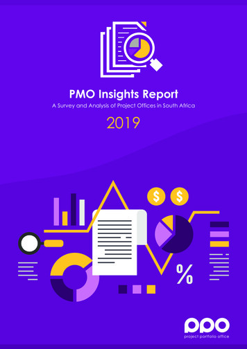
ARTICLEnature communications DOI: 10.1038/ncomms1701Table 1 Verification of nuclear SNPs.dbSNP #(b126)AssociationForwardprimer 5′–3′Reverseprimer 5′–3′rs10757274 Coronary artery CCCCCGTGdiseaseGGTCAAATCTAAGrs2383206 CoronaryTACTATCartery disease CTGGTTGCCCCTTCTGTCrs5351Atherosclerosis TCATCCCTATAGTTTTACAAGACAGCrs4988235 636 Y-hg GCTCAGATCTAATAATCCAGTATCAACTGArs2178500 Y-hg G2aCTATCACCCAGAGACCCCTCArs7892988 Y-hg G2a3TATAACCAAAAATGGCACGATn.n.Y-hg. G2a4TTCTGGAGAGCACTAAGCCACTTCCrs17817449 BMIAAGAAGAGTGATCCCTTTGTGTTTrs1426654 Skin colourCATTTATGTT(light)CAGCCCTTGGATTGTCTCFragment AT IndependentNGSsize (bp) ( C)PCRdatareplication coverageresultsHapMapfrequency ofIceman’s genotype(sample size)aPrimerreferenceCEUTSI8255nsa8G, 1ANANAThis study7855G/G8GG/G 0.246(130)NAThis study7455C/T11153G/G14GG/G 0.088 G/G 0.833(226)(204)Burger2007217259T/T7TT/T TTCAGGAGCTGAACTG8153nsa5CNANAThis study8049G/G16G, 1A,1TG/G CAAT20 T, 14C C/T 0.416(226)C/T 0.567 This study(194)G/G 0.235 This study(204)A/A 1.000 A/A 0.990(126)(204)Graefen200945Abbreviations: NA, not applicable; nsa, no successful amplification/sequencing; n.n., no name (no RefSNP accession ID).aCEU: Utah residents with Northern and Western European ancestry from the CEPH collection; TSI: Tuscan in Italy. HapMap Genome Browser Release #28, (NCBI build 36, dbSNP b126). http://hapmap.ncbi.nlm.nih.gov.Independent verification was
15 Department of Internal Medicine III, University of Heidelberg, 69120 Heidelberg, Germany. 16 The Ontario Institute for Cancer Research, 101 College Street, Suite 800, Toronto, Ontario, Canada M5G 0A3. 17 Nimbus Informatics LLC, 104R NC Hwy 54 West, Suite 2