Transcription
Chen et al. BMC Genomics 2014, 0METHODOLOGY ARTICLEOpen AccessBisQC: an operational pipeline for multiplexedbisulfite sequencingGary G Chen1,2, Alpha B Diallo1,2, Raphaël Poujol1,2, Corina Nagy1,2, Alfredo Staffa3, Kathryn Vaillancourt1,2,Pierre-Eric Lutz1,2, Vanessa K Ota1,2, Deborah C Mash4, Gustavo Turecki1,2,5 and Carl Ernst1,2,5*AbstractBackground: Bisulfite sequencing is the most efficient single nucleotide resolution method for analysis of methylationstatus at whole genome scale, but improved quality control metrics are needed to better standardize experiments.Results: We describe BisQC, a step-by-step method for multiplexed bisulfite-converted DNA library construction, pooling,spike-in content, and bioinformatics. We demonstrate technical improvements for library preparation and bioinformaticanalyses that can be done in standard laboratories. We find that decoupling amplification of bisulfite converted (bis) DNAfrom the indexing reaction is an advantage, specifically in reducing total PCR cycle number and pre-selecting high qualitybis-libraries. We also introduce a progressive PCR method for optimal library amplification and size-selection. Atthe sequencing stage, we thoroughly test the benefits of pooling non-bis DNA library with bis-libraries and findthat BisSeq libraries can be pooled with a high proportion of non-bis DNA libraries with minimal impact on BisSeqoutput. For informatics analysis, we propose a series of optimization steps including the utilization of the mitochondrialgenome as a QC standard, and we assess the validity of using duplicate reads for coverage statistics.Conclusion: We demonstrate several quality control checkpoints at the library preparation, pre-sequencing,post-sequencing, and post-alignment stages, which should prove useful in determining sample and processingquality. We also determine that including a significant portion of non-bisulfite converted DNA with bisulfiteconverted DNA has a minimal impact on usable bisulfite read output.Keywords: DNA methylation, Bisulfite sequencing, BioinformaticsBackgroundDNA methylation has a role in the development ofeukaryotic organisms [1,2] and may represent the interfacebetween genome and environment [3]. Currently, the mostwidely used method for detecting 5-methylcytosine is thetreatment of DNA with sodium bisulfite [4], which resultsin the deamination of all non-methylated cytosine to uracil.Since sodium bisulfite does not convert 5-methylcytosinebases to uracil, this approach allows for the direct interpretation of where methylation has or has not occurredin the genome. This interpretation is complicated by theneed to generate percentage methylation statistics per* Correspondence: carl.ernst@mcgill.ca1Department of Psychiatry, McGill University, Douglas Hospital ResearchInstitute, Montreal, Quebec, Canada2McGill Group for Suicide Studies, Douglas Hospital Research Institute, 6875LaSalle Boulevard, Frank Common Building, Room 2101.2 Verdun, Montreal,QC, H4H 1R3, CanadaFull list of author information is available at the end of the articlecytosine residue because the same locus can have different methylation levels across cells. This complicationunderscores the need to have careful metrics to assess experimental procedures.Bisulfite treatment combined with Next-Generationsequencing (NGS) is the method of choice for the NIHEpigenomics Roadmap [5], a project with a stated goalto map methylation patterns in multiple tissue types.This experimental design underlies the expectation thatthere are different methylation patterns in different tissues,and there is a high likelihood that cells that make up thesetissues themselves have different methylation patterns, evenat the same genomic loci. This variation complicates analysis and can lead to high levels of noise, making data interpretation challenging. One major issue for all bis-DNA 2014 Chen et al.; licensee BioMed Central Ltd. This is an Open Access article distributed under the terms of the CreativeCommons Attribution License (http://creativecommons.org/licenses/by/2.0), which permits unrestricted use, distribution, andreproduction in any medium, provided the original work is properly credited. The Creative Commons Public DomainDedication waiver ) applies to the data made available in this article,unless otherwise stated.
Chen et al. BMC Genomics 2014, 0Next Generation sequencing (BisSeq) experiments is readalignment to a reference genome. This is due to the loss ofunmethylated C residues (observed as T residues after sequencing) leading to decreased complexity. Specifically, thelonger and more diverse a sequenced read, the more likelyit is to align to the genome. Loss of base diversity from thedecrease in C bases means that individual reads may appearto align to multiple regions of the genome. A secondmajor reason for alignment difficulties is the diversity ofmethylation patterns at cytosine loci. In a read containing many C residues, methylation patterns at the samebase could be different between reads. This means thatreads from the same genomic locus could align to different genomic regions.Bisulfite conversion of DNA followed by massivelyparallel sequencing is likely to be the most practicalapproach to map methylation in the coming years,whether in reduced or complete genomic space [6,7].Reduced-Representation bisulfite sequencing (RRBS)uses an MspI digestion (cutting at C VCGG) prior to bisulfite conversion and library preparation to reducegenomic space, resulting in the sequencing of 2.5% ofthe genome; however, a thorough analysis of its limitations, and methods to cope with these inadequacies,has not been fully addressed. Early protocols for bisulfite sequencing [8-10] were described for the IlluminaGenome Analyzer IIx sequencer, where library preparation was singleplexed. Later protocols [11] switched tomultiplexed indexing approaches using Illumina TrueSeqDNA sample preparation. Notably, early protocols focusedon library construction, while recent protocols have explored strategies for successful sequencing and analysis[12]; however, there currently lacks a simple, detailed description of multiplex sequencing using conventional tools;complex, hard-to access strategies are exemplified by theproposal to use ‘Dark’ sequencing [11] in bisulfite sequencing experiments.The purpose of the current work is to provide a simpleBisSeq protocol, with several QC checks throughout thatcan be used in standard laboratories and to test twomain questions: 1) What is the function of pooling nonbisulfite DNA with bisulfite converted DNA in a singlelane, and 2) What is the validity of using duplicate readsto calculate coverage statistics? In this work we carefullyand completely lay out experimental procedures and QCparameters to assess BisSeq experiments and we suggestthat pooling 30% non-bisulfite converted DNA with bisulfite converted DNA has a minimal effect on bisulfiteread output, meaning that sequencing non-bisulfite DNAsamples from unrelated experiments is practical. We findalso that using duplicate reads to calculate coverage is legitimate, but that a simple test to assess whether the total readpool is representative of a read pool with no duplicatesshould be applied first.Page 2 of 22MethodsLibrary preparationAll tissue samples used in this study were provided by theBrain Endowment Bank following protocols approved bythe research ethics board of the University of Miami MillerSchool of Medicine. Brain samples (anterior caudatenucleus) were obtained at autopsy following the principles of the Helsinki declaration and next-of-kin gavewritten informed consent. We used NEBNext IlluminaLibrary Prep Master Mix kit for the library constructionwork. All experiments herein use reduced representationbisulfite sequencing; however, most optimizations canbe applied to standard bisulfite sequencing. Figure 1shows a flow chart of library preparation procedures formultiplexed Reduced-Representation Bisulfite Sequencing (mRRBS), which takes on average 7–10 days. Figure 2shows a molecular level illustration of library preparationafter the adaptor ligation stage.Enzyme digestionAfter DNA purification using the QIAamp genomic DNAmicro isolation kit from human brain, 2-5 μg of genomicDNA was used to carry out the MspI (New England Biolabs) digestion at 37 C for 7 hours using 20 units of enzymeper μg of DNA. Digested DNA was purified by phenol/chloroform (p/c) extraction (49:49:2; phenol:chloroform:isoamyl alcohol). The aqueous top layer containing genomicDNA was precipitated in the presence of NaCl (0.3 M final)and glycogen (25 μg final). The precipitated DNA was pelleted via centrifugation and washed with 500 μl 80% ethanol (rinsing the pellet instead of re-suspending) and thencentrifuged again at 12000 rpm for 20 minutes to solidifythe pellet. The pellet containing MspI fragmented DNAwas resuspended in 100 μl dH20 for the end-repair reaction.Five-ten percent of the purified MspI fragmented DNA wasthen run on a precast 4-20% gradient polyacrylamideTBEx1 gel and stained with ethidium bromide (EtBr; Invitrogen). This is an important step to verify the enzymaticdigestion of DNA; a complete MspI digestion will producevisible satellite bands in a smearing background (Figure 3).A high sensitivity DNA chip to check the completion ofMspI digestion can also be used (Figure 3C).End repair (Filling-in and dA-tailing)MspI recognizes double stranded DNA at 5′-C CGG-3′and cleaves the phosphodiester bonds upstream of CpGdinucleotide. This reaction results in DNA fragments with5′ overhangs, so end repair is necessary to fill-in the 3′ termini of each fragment. This way, all MspI digested libraryfragments should contain a CpG dinucleotide on both endsof the fragment. The NEBNext DNA Library Prep MasterMix Set for Illumina separates the filling-in step fromdA-tailing reaction.
Chen et al. BMC Genomics 2014, 0Page 3 of 22at 20 C for 30 minutes. After incubation, the reactionmixture was diluted to 200 μL with dH2O. Next, weadded 200 μL of p/c at room temperature. After ethanolprecipitation, DNA was resuspended in 50 μL dH2O inpreparation for the dA-tailing reaction.dA-tailing of blunt end MspI fragmentThe enzymatic process that adds an extra adenosine (A)to both the plus and minus strand 3′ termini is referredto as dA-Tailing and is necessary for ligation of the adaptors(which contain a 3′ dT overhang). Following the NEBNextDNA library Prep Master Mix Set for Illumina kit manual,we carried out the dA-Tailing reaction. We used 42 μLblunt-end DNA, 5 μL of NEBNext dA-Tailing ReactionBuffer (10X), and 3 μL Klenow Fragment (3′- 5′ exo-),for a total volume of 50 μL and incubated at 37 C for 30minutes. Next, p/c extraction and ethanol precipitationwere performed following the same steps as outlined above,without the inclusion of glycogen (no glycogen is needed,since previously added glycogen is co- precipitated withgenomic DNA). The final volume of dA-tailed MspI fragment is 30 μl in dH2O.Methylated adaptor designWe used the two-step Illumina adaptor design for theadapters and the PCR indexing primers because it decouples the indexing reaction from library amplification (Figure 2), and allows for more efficient bisulfitetreated DNA library amplification and size selection. Wesynthesized published Illumina paired-end adaptor oligonucleotides, and had all cytosines replaced with 5′methylcytosines in order to prevent the deamination of theadaptor cytosines in the bisulfite conversion reaction. Alladaptors and indexing primers are listed in Table 1.Illumina methylated Y-adaptor annealingFigure 1 Flow chart of library preparation steps.Prior to adaptor ligation, we carried out an adaptor Yfork annealing reaction by combining equal molar ratiosof methylated PE1 and methylated PE2 adaptors. With this,annealed adaptor oligonucleotides can be kept at 20 C formany months before use, provided high temperature andother denaturing conditions are avoided. To perform thisreaction, we mixed 50 μL each of mC-PE1 (25 μM) andmC-PE2 (25 μM) in PCR well-plates and carried out thefollowing denaturing and annealing reaction on the thermalcycler: 95 C, 120 s; 80 C, 60 s; 70 C, 60 s; 60 C, 60 s; 50 C,60 s; 40 C, 60 s; 30 C, 60 s; 4 C indefinitely.Filling- in of MspI rragmented DNALigation of methylated Y-adaptor to dA-tailed DNAfragmentsUsing the MspI fragmented DNA (40–85 μL) describedabove, the NEBNext End Repair Reaction Buffer (10X;10 μL), and the NEBNext End Repair Enzyme Mix (5μL), we incubated the solution (final volume of 100 μl)For the ligation reaction, we used 25 μL dA-tailed DNA,10 μL NEB Quick ligation Reaction Buffer (5X), 10 μLpre-annealed Illumina methylated Y-adaptors, and 5 μLNEB Quick T4 ligase for a total volume of 50 μL. This
Chen et al. BMC Genomics 2014, 0Page 4 of 22Figure 2 Major RRBS library construction steps. This figure demonstrates adaptor ligation (step 1) and barcode indexing (step 2) for Illuminatwo-step library preparation, as well as in between steps including bisulfite treatment. We show how the 2-step procedure affects DNA insertswhen used for RRBS directional sequencing. First, DNA inserts (underlined) with ‘A’ overhangs are ligated to methylated Illumina adaptors (methylatedcytosines are marked in bold), meC-PE1 and meC-PE2. Next, adaptor ligated-DNA inserts are bisulfite treated and amplified using primer indPEPCR1F andindPEPCR2R. All unmethylated cytosines deaminate to uracil. We show two cycles of the PCR reaction toamplify bisulfite fragments to show how DNAinserts change after bisulfite treatment and amplification, as well as to track original top (OT) and original bottom (OB) strands. After an appropriate numberof cycles (appropriate is defined by the visualization of bands shown in this manuscript in the library preparation stage), bisulfite treated libraries can beindexed, then sent for sequencing. Note that for directional sequencing all sequencing reads are either from the original top (OT) or the original bottom(OB) strands. The first three bases of almost all RRBS reads are either CGG or TGG, depending on their genomic methylation state and this applies to readsgenerated from both OT and OB strand. Therefore almost every read in a directional RRBS sequencing experiment that use MspI digestion contains at leastone CpG at the 2nd and 3rd base positions, plus any internal CpGs (provided they are not in CCGG or CCGG sequences). Internal CpGs can be in CCGGsequence where MspI does not cut when the first C is methylated. Abbreviations: C (Bold): methylated C; p: phosphate; s: phosphorothioatebond. Illustrated insert DNA is underlined. P5 (5′ AATGATACGGCGACCACCGA 3′) and P7 (5′ CAAGCAGAAGACGGCATACGA 3′) are flow cellattachment sites.was then incubated for 1 hour at 16 C, followed by asecond incubation for 30 minutes at 20 C.for 50 μL of the ligation mixture. The purified librarieswere then eluted in 100 μL of dH2O.Purification of adaptor- ligated libraryBisulfite conversionWe used the QiaQuick PCR purification kit for the purification of the adaptor-ligated libraries. We used 250 μLof QIA buffer PE to carry out the purification processWe used the Qiagen EpiTect Fast 96 Bisulfite Kit tocarry out the bisulfite conversion of adaptor-ligatedlibrary. This single conversion step is reported to be
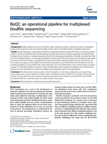
Chen et al. BMC Genomics 2014, 0Page 5 of 22Figure 3 Standards for MspI digestion and progressive PCR. A) MspI digestion of human genomic DNA isolated from human post-mortem braintissues. DNA (200 ng) was digested by MspI and run on a 4–20% precast polyacrylamide gel and stained with EtBr. Arrows show three satellite DNA bandscharacteristic of this enzymatic digestion. B) Agilent 2100 Bioanalyzer chromatogram of MspI digested genomic DNA. C) Bioanalyzer 2100 image of a singlelibrary from an MspI digested DNA sample. Notice that the satellite bands (indicated by arrows) are still visible on the Bioanalyzer image. D)Progressive PCR amplification combined with limited PCR extension time allows for size selection and amplification of six bisulfite convertedlibraries (Lanes 1–5 are distinct RRBS libraries; lane 6 (‘C’) is a negative control). After different progressive PCR cycles (18X, 22X, 24X, or 26X –the same libraries are shown for each cycle number) band intensity increases as cycle number increases. Arrows indicate the three satelliteDNA bands that are still visible in these libraries.sufficient to achieve a 99% conversion rate (discussedin more detail in the results section). We used 50% ofthe purified adaptor ligated library for bisulfite conversion. To purify DNA, the reaction mixture wastransferred into a 96-well plate, with a high affinitymembrane on the bottom of each well (Qiagen). BufferBL, bisulfite conversion mixture and ethanol (96%)were added sequentially, mixed, then left to stand for 2minutes. After spinning, single stranded, bis-convertedDNA was bound to the membrane. We then washedtwice with buffer BW, than twice with buffer BD toachieve a complete on-membrane desulfonation. Thiswas followed by two more washes with BW then a finalelution with buffer EB.PCR amplification of bisulfite converted librariesAgilent Pfu turbo Cx Hotstart DNA polymerase hasthe property of uracil-tolerance and high fidelity DNApolymerization. Amplification of bisulfite-converted libraries was carried out simultaneously with the librarysize selection process by using the progressive PCRmethod. IndPEPCR F (33 nt) and R (32 nt) (Table 1and Figure 2) were used as PCR primers.The detailed PCR reaction mixture (200 μL volume) is asfollows:10X pfu Turbo Cx Rxn Buffer (Agilent)-20 μLdNTP (10 mM each)-4 μLIndPEPCR F (25 M)-1 μL
Chen et al. BMC Genomics 2014, 0Page 6 of 22Table 1 Sequences and specific modifications of oligonucleotides used in the BisQC protocolNameSequence (5′ to 3′)Methylated C adaptor: ted C adaptor: mC-PE2p-GATCGGAAGAGCGGTTCAGCAGGAATGCCGAG-OHPCR primer: IndPEPCR TTCCGATCsTPCR primer: IndPEPCR RGTGACTGGAGTTCAGACGTGTGCTCTTCCGATCsTPE-qPCR F (Phix-PE-qPCR F)AATGATACGGCGACCACCGA-OHPE-qPCR R (Phix-PE-qPCR R)CAAGCAGAAGACGGCATACGA-OHIndex 1R OHIndex 2R OHIndex 3R OHIndex 4R OHIndex 5R OHIndex 6R OHIndex 7R OHIndex 8R OHIndex 9R OHIndex TC-OHIndex TC-OHIndex TC-OHAbbreviations: C (Bold) methylated C, p phosphate, s phosphorothioate bond.IndPEPCR R (25 M)-1 μLPfu Turbo Cx Hot Start DNA Polymerase-2 μLBisulfite converted RRBS library-50 μLdH2O 122 μLThe Pfu Turbo Cx Hot Start DNA Polymerase shouldbe added last. The detailed amplification cycle is asfollowing:1.2.3.4.5.95 C-90 s95 C-30 s60 C-30 s72 C-30 s4 C indefinitely (in 4 C fridge, do not freeze the PCRreaction)Repeat steps 2–4 for 18 cyclesThe final PCR products represent the minimallyamplified and size-selected non-indexed RRBS libraries.We confirmed the correct size amplification by runninga 2.0 % HR Agarose Gel (100 mL 1XTAE 2.0 g of HRAgarose 2.5 μL of 10mg/mL EtBr) or an Invitrogen4-20% gradient polyacrylamide gel (1XTAE), or InvitrogenE-gel 2% with SYBR Safe, all after 18 cycles of PCR.Samples showing faint but visible 150–400 base pair(bp) smearing on the gel have the optimal amplificationPCR cycles. Satellite DNA bands should also be visiblein the smearing background for a well-constructed andoptimally amplified RRBS library (Figure 3). Samplesthat give very faint smearing require additional PCR amplification cycles. Samples requiring extra PCR cycles (usingthe PCR reaction tubes kept at 4 C) can be returned to thethermal cycler for 2–4 more cycles of amplification following the above progressive PCR protocol. Ten μL of these‘additional cycle’ amplified PCR reaction mixtures can berun on a gel and checked for smearing. Thus, finallibrary selection is determined by visual inspection ofgel images for appropriate smearing and satellite bandpatterns (Figure 3). This is required because of the largevariation observed across libraries, even with identical starting DNA concentrations and enzyme digestion times. Finally, we perform a cleanup step with the remaining PCRproducts from all libraries using AMPure XP SPRI beads(Agencourt, Beckman-Coulter) using 60 μl/55 μl (beads/DNA) ratio. After purification, 1 μL of the purified librarywas used for quality control using an Agilent Bioanalyzer2100 High Sensitivity DNA Chips. A good amplified sizeselected and purified library produces a smear covering150–400 bp with visible satellite bands, similar to that seenin Figure 4, with very little primer-dimers.IndexingUsing 50 μL of purified, non-indexed library, we performedthe following PCR indexing reaction for each library. ThePCR primers used for indexing reaction are IndPEPCR F(33 nt) and Index #R primer (43 nt)
Chen et al. BMC Genomics 2014, 0Page 7 of 22Figure 4 Agilent 2100 Bioanalyzer images from final reduced representation bisulfite libraries. A) High Sensitivity DNA Chip from 10 RRBS libraries.Notice that the satellite bands are still visible on the Bioanalyzer gel image B) Chromatogram representation of panel A showing high quality RRBSlibraries.10X pfu Turbo Cx Rxn Buffer (Agilent)-20 μLdNTP (10 mM each)-4 μLIndPEPCR F (25 M)-1 μLIndPEPCR #R (25 M)-1 μLPfu Turbo Cx Hot Start DNA Polymerase-2 μLNon-indexed RRBS library-50 μLdH2O-122 μLThe Pfu Turbo Cx Hot Start DNA Polymeraseshould be added last.We used 60μl/55μl (beads/DNA) ratio. AMPure SPRIbead-purified library was eluted in 60 μL of dH2O. Purifiedlibraries can be screened on an Agilent 2100 BioAnalyzer(Figure 4A and B).Using the following PCR steps:Molecular cloning and sanger sequencing1. 95 C-90 s2. 95 C-30 s5 μl of AMPure bead-purified library (total 60 μl) was usedfor cloning experiments. The single band product wascloned using chemically competent E.coli cells and the3. 65 C-30 s4. 72 C-30 sRepeat 2–4 times5. 4 C indefinitely
Chen et al. BMC Genomics 2014, 0pCR 4-TOPO TA vector (Invitrogen Cat#450030) using 4μl of fresh PCR product, 1 μl of vector, 1 μl of salt solution,and ligated for 5 minutes at room temperature. Four μl ofligated product was added to 50 μl of competent cells andincubated on ice for 30 minutes, 240 μl of S.O.C. medium(Invitrogen Cat#15544-034) was added to the cells incubated in a 37 C shaker for 1 hour. 50 μl of transformed cellswas plated on ampicillin agar plates and incubated for 16hours at 37 C. Prior to sequencing, we assessed some bandsizes by an EcoRI digestion of plasmids (Figure 5A).Sequencing was done using rolling circular amplification, aservice provided by Genewiz, Inc (South Plainfield, NJ).M13F ( 21) sequenced colonies were aligned usingLasergene SeqMan Pro (DNASTAR, Inc. Madison, WI).For methylation assessment of a single, targeted locus,a 250 bp amplicon was isolated from the original RRBSlibrary preparation for sample G12. Two μl of template wasamplified using High Fidelity Platinum Taq (Invitrogen Cat#11304-102) and the PCRx enhancer system to facilitateamplification of CG rich template (Cat# 11495–017), withPage 8 of 22the following thermocycling conditons: 95 for 5 minutes,(95 for 30s, 58 for 30s and 72 for 45 seconds) x 45 cycles,72 for 7 minutes, 4 indefinitely. Bisulfite primers were designed using Methyl Primer Express (Applied Biosystems), forward 5′- GGG AAG AGT TGG TTA GAGAGA -3′ and reverse 5′-AAA ACC CCC TAT AAA AAAACC C-3, corresponding to (HG19) Chromosome 3: 75,718, 452–75, 718, 701.Massively parallel (next generation) sequencingWe used the Illumina HiSeq2000 platform, with single-end50 cycle sequencing. Sequencing was performed by theMcGill University and Génome Québec InnovationCentre in Montreal, Quebec. Data was downloaded ontoour servers in FASTQ format. Illumina de-indexed datawas first processed using FastQC tool v0.10.0 /fastqc/),which provides a user-friendly overview of raw sequencing data. The programs fastx clipper, fastx trimmer and fastx collapser [part of the FASTX-ToolkitFigure 5 Pre-sequencing quality control of bisulfite-converted DNA libraries. A) Cloning and subsequent EcoRI release of library insertsreveals a random size distribution as revealed on an agarose gel. The extra band in some lanes represents enzyme cut sites present in the inserts.B) qPCR dissociation curve of a bisulfite- converted DNA library using primers directed at adaptors to the bisulfite library (blue peak) and PhiXstandard DNA library (green peak). Bisulfite converted DNA libraries have a lower melting temperature due to loss of cytosine residues. The qPCRdissociation curve only serves as a general tool to verify bisufite conversion and is not able to distinguish a minor bisulfite conversion problem.C) Sanger sequencing reaction of a single clone insert from A). Plasmid is detectable by presence of cytosine residues; insert begins at CGGresidue marked by the arrow. Note the lack of cytosine residues in the insert, except at CpG loci (blue ‘C’ peaks under black arrow). Base colorsare: C: Blue, G: Black, T: Red, A: Green.
Chen et al. BMC Genomics 2014, 0(http://hannonlab.cshl.edu/fastx toolkit/)] were usedto remove adaptors, filter low quality reads, and remove reads 20 bp in length.AlignmentThere are many excellent aligners for bisulfite sequencing data including Bismark [13], BSMAP [14,15], andRMAP [16], BS-Seekeer2 [17] and some of these havebeen recently assessed [18]. We opted to use Bismark[14] for its flexibility and compatibility with downstreamprocessing tools such as MethylKit [19], which we useroutinely for post-alignment processing. All user-set features were set to default, except for: -best, -n 2, and –directional. For data visualization, we wrote a customPerl script to convert Bismark output alignment filesinto the SAM (Sequence Alignment/Map) format for thevisualization of the alignment reads into the IntegratedGenomic Viewer [20]. This script is available for download at: www.mcgill.ca/psychiatricgenetics/tools-0. Further description of bioinformatic details can be found inthe results section.ResultsPage 9 of 22Simultaneous amplification and size selection of bisulfitelibraries by progressive PCRProgressive PCR amplification combined with limitedPCR extension time achieves library amplification andsize selection at the same time (Figures 3, 4 and 5). Sizeselection is achieved because standard Taq DNA polymerase extension speed is on the order of 700–800 nucleotides per minute at 72 C. Instead of time-consuminggel-cutting for size selection, optimized progressivePCR can be performed to amplify and size-select libraries. We set the 72 C extension time to 30 secondsfor 150–400 bp libraries. We used 18 cycles of PCRamplification cycles, then transferred plates immediately to 4 C (and never froze samples because extraPCR cycles may be required without adding any freshenzyme or dNTPs). Next, we ran a small aliquot of thePCR product (5 μL) and verified the products on anagarose gel with EtBr. When a smearing range from200–500 bp is nearly visible a minimal amplificationcondition has been achieved (Figure 3D). If there is nosmear visible at all, the complete PCR reaction isreturned to the thermal cycler and amplified for anadditional 2–4 cycles, hence progressive PCR.Post-bisulfite indexing for increased flexibility andimproved library purificationCurrently, most NGS bisulfite DNA protocols take advantage of the ‘one-step’ design, where indexing and adaptorligation occur in one step, but this creates longer products(Adaptors with indexes are 100 nucleotides for the onestep design as opposed to 64 nucleotides in the two-stepdesign) which influences clean-up procedures and allowsless flexibility in library amplification – an important factorin bisulfite DNA projects because of the variability in DNAconcentration after bisulfite treatment. Pre-indexing alsorequires decisions to be made about what samples to pooltogether prior to knowing the quality of each library. To devise a post-library indexing strategy, we used the Illuminatwo-step paired end design (Figure 2) which uses a thirdindexing primer (Table 1 lists the sequences for all oligonucleotides used in these experiments).A major benefit to using the two-step Illumina adaptor/indexing technology beyond the increased flexibility, is thatthey form dimers less than 100 bp, allowing for the use ofspin columns rather than AMPure beads; adaptor dimersusing this strategy are 65 bp in contrast to the 126 bp dimers generated by the TruSeq protocol. Spin columns recover all fragments 100 bps, whereas AMPure beadpurification functions best at high DNA concentrations orwith small volume operations. This means that a greaterproportion of the library is retained after purification.A disadvantage of using the two-step approach is thatfusion products between the adaptor and indexingprimer form, and this product needs to be removed(see Additional file 1
ent methylation levels across cells. This complication underscores the need to have careful metrics to assess ex-perimental procedures. Bisulfite treatment combined with Next-Generation sequencing (NGS) is the method of choice for the NIH Epigenomics Roadmap [5], a project with a stated goal to map methylation patterns in multiple tissue types.