Transcription
Eur Radiol (2010) 20: 450–457DOI 10.1007/s00330-009-1561-9MUSCULO SKELETALMatthieu J. C. M. RuttenGert-Jan SpaargarenTon van LoonMaarten C. de Waal MalefijtLambertus A. L. M. KiemeneyGerrit J. JagerDetection of rotator cuff tears: the valueof MRI following ultrasoundReceived: 13 February 2009Revised: 8 July 2009Accepted: 22 July 2009Published online: 2 September 2009# The Author(s) 2009.This article is published with open access atSpringerlink.comM. C. de Waal MalefijtDepartment of Orthopedic Surgery,Radboud University Nijmegen MedicalCentre,Th. Craanenlaan 7,6500 HB Nijmegen, The Netherlandse-mail: m.dewaalmalefijt@orthop.umcn.nlM. J. C. M. Rutten (*) .G.-J. Spaargaren . G. J. JagerDepartment of Radiology,Jeroen Bosch Ziekenhuis,Nieuwstraat 34,5211NL’s-Hertogenbosch, The Netherlandse-mail: M.Rutten@JBZ.nlTel.: 31-73-6992000Fax: 31-73-6992601G.-J. Spaargarene-mail: g.spaargaren@jbz.nlG. J. Jagere-mail: g.jager@jbz.nlT. van LoonDepartment of Orthopedic Surgery,Jeroen Bosch Ziekenhuis,Nieuwstraat 34,5211NL’s-Hertogenbosch, The Netherlandse-mail: t.v.loon@jbz.nlL. A. L. M. KiemeneyDepartment of Epidemiology andBiostatistics & HTA, RadboudUniversity Nijmegen Medical Centre,Geert Grooteplein Noord 21,P.O. Box 9101, 6500 HBNijmegen, The Netherlandse-mail: b.kiemeney@epib.umcn.nlAbstract Objective: To evaluatethe need for additional magneticresonance imaging (MRI) followingultrasound (US) in patients withshoulder pain and/or disability and tocompare the accuracy of both techniques for the detection of partialthickness and full-thickness rotatorcuff tears (RCT). Methods: In4 years, 5,216 patients underwent USby experienced musculoskeletal radiologists. Retrospectively, patient records were evaluated if MRI andsurgery were performed withinIntroductionBoth ultrasound (US) and magnetic resonance imaging(MRI) can confirm a suspected partial-thickness or fullthickness rotator cuff tear. Both techniques have their5 months of US. US and MRIfindings were classified into intactcuff, partial-thickness and full-thickness RCT, and were correlated withsurgical findings. Results: Additional MR imaging was performed in275 (5.2%) patients. Sixty-eight patients underwent surgery within5 months. US and MRI correctlydepicted 21 (95%) and 22 (100%) ofthe 22 full-thickness tears, and 8(89%) and 6 (67%) of the 9 partialthickness tears, respectively. The differences in performance of US andMRI were not statistically significant(p 0.15). Conclusions: MRI following routine shoulder US wasrequested in only 5.2% of the patients.The additional value of MRI was indetecting intra-articular lesions. Inpatients who underwent surgery, USand MRI yielded comparably highsensitivity for detecting full-thicknessRCT. US performed better in detecting partial-thickness tears, althoughthe difference was not significant.Keywords Musculoskeletal .Shoulder . Rotator cuff tear . US .MRIadvantages and disadvantages, and can be competitive andcomplementary at the same time. Factors of considerationconcerning which technique should be used are availabilityof the test in a timely manner and the skill of the operatorsin carrying out and interpreting a given examination. In the
45147 years, range 18–87 years) also underwent MRI of theshoulder, and 80 of the 275 patients subsequently hadsurgery. Twelve patients were excluded because the timeinterval between US and MRI or surgery exceeded5 months. The remaining 68 patients (37 men and 31women, with a mean age of 48 years, range 24–81 years)formed the study group whose data were analysedretrospectively.Patients were evaluated for the presence of rotator cufftears. Findings were classified into intact cuff, partialthickness and full-thickness rotator cuff tears, andcorrelated with surgical findings, which were consideredthe gold standard. The time interval between the USexamination and MRI ranged from 0 to 98 days (mean41 days). The time interval between US examination andsurgery ranged from 5 to 147 days (mean 99 days) andbetween MRI and surgery 5 to 139 days (mean 59 days).literature the question which test constitutes the mostaccurate, cost effective, expedient or least invasive approach to the diagnosis of rotator cuff tears is stillcontroversial. The question as to which is the best testshould be answered on the basis of clinical experience,availability, and the expected sensitivity and specificity ofthe tests.Although many investigators have evaluated the accuracy of US or MRI separately for the detection of partialthickness (PTT) and full-thickness (FTT) rotator cuff tears,some have directly compared the two tests for the detectionof full-thickness tears [1, 2], partial-thickness tears [3] orpartial-thickness and full-thickness tears [4–10], but mostwith a relatively small number of patients [8–10].In a systematic review of the diagnostic accuracy ofclinical examinations with US and MRI in the detection offull-thickness and partial-thickness rotator cuff tears,Dinnes et al. [11] showed that US and MRI havecomparably high accuracy for the detection of fullthickness tears. Based on the available literature, US isthe method of first choice in the detection of rotator cufftears in our hospital. In the case of unequivocal findings orclinical doubt, additional MRI is requested.The purpose of this study was first to evaluate the needfor additional MRI following US of the shoulder andsecondly to evaluate and compare the accuracy of US andMRI for the detection of partial-thickness and fullthickness rotator cuff tears with surgery as the referencestandard in a selected group of patients.All US examinations were performed by two musculoskeletal radiologists (MR, GJ), using a APLIO device(Toshiba Medical Systems Corporation, Tokyo, Japan)with a 7.5–14-MHz linear array transducer (PLF-805ST).Real-time imaging of the shoulder was performed in astandardised fashion as described in the literature [12].Established criteria were used for the diagnosis of a partialthickness or full-thickness rotator cuff tear [13–15].Materials and methodsMagnetic resonance imagingPatientsAll MRI examinations were performed with an 1.5-T MRsystem (Signa Horizon, GE, ‘s-Hertogenbosch, TheNetherlands). Of the 80 patients, 34 patients underwentMR arthrography (MRA) and 46 patients conventionalMRI. The conventional MRI shoulder protocol consisted ofoblique coronal T2-weighted with fat suppression(2.48 min) and T1-weighted turbo spin echo images(5.58 min), oblique sagittal T2-weighted turbo spin echoimages without fat suppression (2.36 min) and transverseT1-weighted turbo spin echo images (4.52 min). A field ofview of 16 cm was used, the slice thickness was 3 mm, theimaging matrix was 320 224, and three signals wereaveraged for each pulse sequence.The MRA protocol consisted of 3D-gradient T1weighted (SPGR) oblique coronal and axial images(5 min), which were reconstructed, if indicated, in anyother desired plane. SPGR 3D imaging (TR: 51 ms; TE:7 ms; 1 acquisition; 256 256 matrix; FOV: 22 cm, slicethickness 2.8 mm) with 2.8-mm consecutive slices wasused, and coronal T2-weighted turbo spin echo images(2.30 min), sagittal T2-weighted turbo spin echo images(2.40 min) and the ABER view with coronal T1-weightedturbo spin echo images with fat suppression (4.15 min).The inclusion criteria for this retrospective study were: (1)shoulder pain and/or disability for which the patientsunderwent plain radiography and US, (2) an additionalMRI was performed and (3) subsequent surgery wascarried out. The indications to perform MRI following USwere suspicion for having intraarticular pathology (n 94),inconclusive US examination due limited shoulder motion,obesity or undefined findings (n 25), discrepancy betweenclinical and US findings (n 77), and finally as a request formore diagnostic certainty (n 18), and of the remainingpatients (n 61), the indication to perform MRI could notbe determined. Patients were excluded when no surgerywas performed and when the time intervals between USand MRI and/or US and surgery exceeded 5 months.The study was performed in one tertiary teachinghospital. Between January 2004 and December 2007,5,216 patients with shoulder pain and/or disability werereferred by the Department of Orthopaedic Surgery orgeneral practitioners and underwent US. Eighty-one wereoperated upon without MRI. Two hundred seventy-fivepatients (147 men and 128 women, with a mean age ofUltrasound
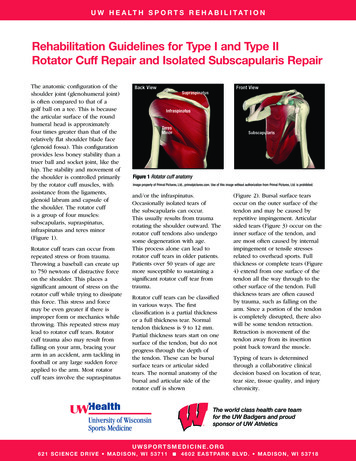
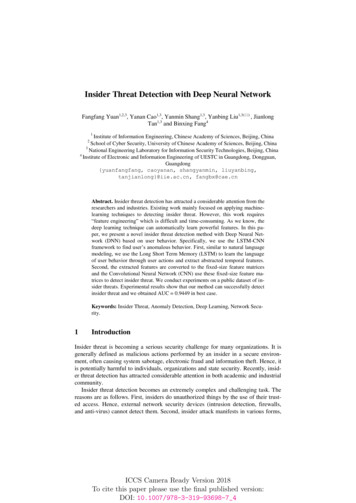
452Fig. 1 Full-thickness rotatorcuff tear. (a) Ultrasound appearance of a full-thickness tear(arrows) at the insertion of thesupraspinatus tendon (SSP).GT greater tuberosity. (b) Thecorresponding oblique coronalgradient T1-weighted MRarthrography image, showingthe same configuration of thefull-thickness tear (arrows) ofthe supraspinatus tendon (SSP).GT greater tuberosityOverall MRA imaging time was 19 min 30 s.The MRI examinations were blinded and retrospectivelyevaluated by the same experienced musculoskeletalradiologists (MR, GJ) who performed the US examinations, at a later date, to avoid any effect on the interpretation of findings at MRI by the recent ultrasoundexamination. This set-up was chosen to exclude thepotential bias of having readers with a different level ofknowledge of anatomy and pathological features of theshoulder.Established criteria were used for the diagnosis of apartial-thickness or full-thickness rotator cuff tear [16–19].An example of a full-thickness rotator cuff tear asdemonstrated by US and MRI is shown in Fig. 1.Statistical analysisUsing cross tabulations, the presence or absence of apartial-thickness or full-thickness rotator cuff tear wascompared among US, MRI and surgery. Based on thetables, sensitivity, specificity, accuracy, the positive predictive value and the negative predictive value werecalculated.The agreement between the results of US and MRI in thedetection of rotator cuff tears was determined by calculating a kappa coefficient. Differences in scoring between USand MRI were tested for statistical significance using amarginal homogeneity test (or McNemar-Bowker test) forrelated samples. All statistical analyses were performedusing SPSS, version 14.0.2 for Windows (Chicago, IL).SurgeryResultsIn all study subjects, surgery (arthroscopy or open) wasperformed by a subspecialty-trained shoulder surgeon, whowas aware of the US and MRI findings. The presence orabsence of a partial-thickness or full-thickness rotator cufftears and intraarticular lesions were recorded.At surgery, 22 full-thickness and 9 partial-thickness rotatorcuff tears were found (Table 1). US correctly depicted 29(94%) of the 31 rotator cuff tears: 21 (95%) of the 22 fullthickness tears and 8 (89%) of the 9 partial-thickness tearsTable 1 Correlation of US and MRI findings with surgical diagnoses of partial-thickness (PTT) and full-thickness (FTT) rotator cuff tearsUltrasoundNo tearMagnetic resonance imaging**PTTSurgical diagnosesNo tear2411PTT08FTT01Total2420Accuracy53/68 (78%)* [66%-87%]Specificity24/37 (65%)* [47%-80%]FTTTotalNo tear21212437922682682600281454/68 (79%)* [68%-88%]26/37 (70%)* [53%-84%]*The exact 95% confidence interval is given between brackets**MRI and MR arthrographyPTTFTTTotal3122263792268
453Table 2 The diagnostic parameters of US and MRI for the diagnosis of partial-thickness (PTT) and full-thickness (FTT) rotator cuff VMagnetic resonance imaging**PTTFTTPTTFTT8/989%* [52%-100%]47/5980% [67%-89%]55/6881% [70%-89%]8/2040% [19%-64%]47/4898% [89%-100%]21/2295% [77%-100%]43/4693% [82%-99%]64/6894% [86%-98%]21/2488 [68%-97%]43/4498% [88%-100%]6/967% [30%-93%]51/5986% [75%-94%]57/6884% [73%-92%]6/1443% [18%-71%]51/5494% [85%-99%]22/22100% [85%-100%]42/4691% [79%-98%]64/6894% [86%-98%]22/2685% [65%-96%]42/42100% [92%-100%]*The 95% confidence interval is given between brackets**MRI and MR arthrographyPPV: positive predictive value (PPV), NPV: negative predictive value (NPV), PTT: partial-thickness rotator cuff tears, FTT: full-thicknessrotator cuff tears(Tables 1 and 2). MRI correctly depicted 28 (90%) of the31 rotator cuff tears: all 22 (100%) full-thickness tears and6 (67%) of the 9 partial-thickness tears (Tables 1 and 2).The overall accuracy of US and MRI in diagnosing fullthickness and partial-thickness tears and intact rotator cuffs(Table 1) was 78% (53/68) and 79% (54/68) with a 95%confidence interval of 66%-87% and 68%-88%, and anoverall specificity of 65% (24/37) and 70% (26/37),respectively.Table 2 lists the sensitivity, specificity, accuracy, andpositive and negative predictive values for both diagnosticimaging techniques in the detection of partial-thickness andfull-thickness rotator cuff tears. The agreement betweenUS and MRI was high: the kappa coefficient was calculatedto be 0.78 (SE 0.06) (Table 3). The differences in thescoring between the two diagnostic tests were notstatistically significant: the marginal homogeneity testwas 0.15, suggesting that the two tests have comparablediagnostic value.For the detection of full-thickness rotator cuff tears withUS and MRI (Fig. 1), there was a high degree of sensitivity,specificity and accuracy (95%, 93%, 94% for US and100%, 91%, 94% for MRI, respectively). The fullthickness rotator cuff tears varied in size from 0.5 to3.0 cm. US and MRI showed three and four false-positivefull-thickness tears, respectively (Table 1). US showed onefalse-negative study (Fig. 2). In this case MRI showed afull-thickness tear, which was proven surgically, whereaswith US it was identified as an extended ( 50%) partialthickness rotator cuff tear (Fig. 2).For the detection of partial-thickness rotator cuff tears,there was a comparable diagnostic value for US and MRIwith a specificity and accuracy of 80%, 81% and 86%,84%, respectively. The sensitivity of US (89%) for thedetection of partial-thickness tears seems to be a little betterthan that of MRI (67%), but these percentages are based ononly nine cases, and the difference is not significant. Thesepartial rotator cuff tears were located superficially, at thearticular side (n 8) and bursal side (n 1). Both US andMRI incorrectly overestimated one partial-thickness tear asa full-thickness rotator cuff tear (Table 1). With conventional MRI, two partial-thickness tears were incorrectlyunderestimated as no tear (Fig. 3).Surgery demonstrated no rotator cuff tears in 37 shoulders(Table 4). US and MRI correctly demonstrated no tears in24 and 26 shoulders, respectively (Table 4). However, USand MRI suggested 2 and 3 full-thickness and 11 and 8partial-thickness rotator cuff tears in the remaining 15 and 13shoulders, respectively, which were not confirmed by surgery.Table 3 Agreement between ultrasound and MRI findings for thediagnosis of rotator cuff tearsMagnetic resonance imaging diagnosis**No tearPTTUltrasound diagnosisNo tear222PTT712FTT00Kappa*: 0.78 (0.06)Marginal homogeneity test †: p 0.15FTT0124*Agreement between US and MRI. The standard error is given inparenthesis**MRI and MR arthrography†Test for significant differences between the two techniquesPTT: partial-thickness rotator cuff tears, FTT: full-thickness rotatorcuff tears
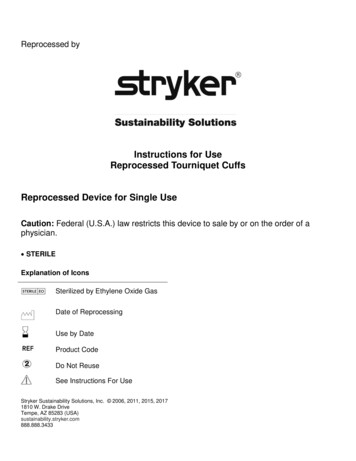
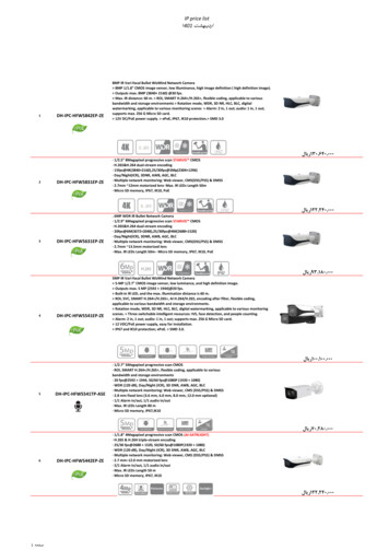
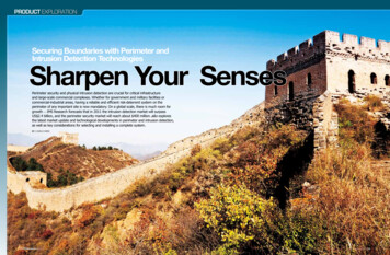
454Fig. 2 Sonographically underestimated full-thickness rotator cufftear. Long (a) and short (b) axis ultrasound section showing anintratendinous partial-thickness tear (arrows) at the insertion of thesupraspinatus tendon (SSP). GT greater tuberosity, H humeralhead. (c) The corresponding oblique coronal gradient T1-weightedMR arthrography image, showing the same intratendinous extendingtear (arrows) of the supraspinatus tendon (SSP), but also leakage ofintraarticularly injected contrast media to the subacromial–subdeltoid bursa (arrowheads), suggestive of a full-thickness SSP tear,which was surgically confirmed. GT greater tuberosityIn 7 cases both US (7/11) and MRI (7/8) demonstrated afalse-positive PTT (Fig. 4). In five of these cases, the PTTwas located intratendinously (Fig. 4) and two at thearticular site near to the insertion of the supraspinatustendon. In four other false-positive PTT cases, US demonstrated a PTT, whereas MRI and surgery did not. In three ofthese cases MRI demonstrated signal distortion in thetendon, which was interpreted as tendinosis. In one of theeight false-positive cases with MRI, US as well as surgerywas negative. Retrospective analyses of the MRI findingsare more suggestive of tendinosis than of PTT.In the two cases in which US identified an FTT andsurgery demonstrated no tear, MRI also identified a FTT. Inone case US and MRI showed an FTT, whereas surgeryshowed a PTT (Table 1). In three cases MRI demonstratedan FTT, while surgery showed no tear; US showed in twocases an FTT as well, but no tear in the other case (Table 3).In seven patients MRI changed the surgical strategybecause in six patients a labral tear and in one patient aglenohumeral ligament tear was found.underwent both imaging techniques. In our institution,US is the method of choice in evaluating patients withshoulder complaints. As in other countries, the use of UShas increased significantly [20]. In our study the use ofUS obviates the need for further imaging in 95% of thecases.The high lifetime prevalence of shoulder pain of 66%[21] and the moderate reliability and reproducibility ofclinical history and clinical examination may be anexplanation for the large number of US examinations. Inthis study we focussed on the presence or absence of rotatorcuff tears. Other diagnoses that are often made with US,e.g., subacromial bursitis and impingement syndrome,were not evaluated. The combination of clinical history,clinical examination and ultrasound fulfil the need fordiagnostic certainty and permit the initiation of therapy inmost cases. The disadvantage of the liberal use of US maybe a large number of negative findings as was an additionalfinding of our study, because the indication to perform USof the shoulder was shoulder pain and/or disability insteadof suspicion for having a rotator cuff tear. Anotherdisadvantage could be a large number of false-positivefindings, as there is a chance of up to 50% of findingabnormalities in an asymptomatic shoulder [22]. Therefore,good clinical examination remains of utmost importance inthe evaluation of patients with shoulder complaints.DiscussionIn the present study we compared the accuracy of US andMRI in the detection of rotator cuff tears in patients whoFig. 3 Partial-thickness rotatorcuff tear in the supraspinatoustendon (SSP) underestimatedwith conventional MRI. (a) Ultrasound showed an intratendinous partial-thickness tear(arrow) in the insertion (i.e., atthe footprint) of the SSP, whichwas confirmed surgically. (b)The corresponding oblique coronal T2-weighted fat saturatedMR image shows high signal inthe SSP, which was wronglyinterpreted as tendinosis. GT greater tuberosity
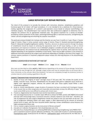
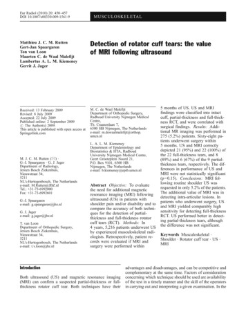
455Fig. 4 False-positive partialthickness rotator cuff tear in thesupraspinatous tendon (SSP).Both ultrasound (a) and thecorresponding oblique coronalT2-weighted fat saturated MRimage (b), show an undersurfacepartial-thickness tear (arrow)in the insertion (i.e., at thefootprint) of the SSP.However, this finding was notconfirmed during surgery.GT greater tuberosityAnother purpose of this retrospective study was toevaluate the diagnostic accuracy of US in cases in whichadditional MRI was requested and to compare these twotechniques.US and MRI findings were compared with surgicalfindings and appeared comparably accurate in diagnosingfull-thickness tears (94% and 94%, respectively) and less,but also comparably accurate for the detection of partialthickness tears (81% and 84%, respectively). Our findingssubstantiate those reported by Dinnes et al. [11] and Teefeyet al. [9], who showed that US and MRI have comparableaccuracy for identifying partial-thickness and full-thickness rotator cuff tears. Although Teefey et al. [9] performeda prospective study, while we retrospectively analysedfindings in a more diverse patient population from dailypractice, both studies show that MRI of the shoulderprovides, with regard to the rotator cuff, little additionalinformation following an US examination. However, in ourselected study group in 7 (10%) of the 68 patients MRIdetected intraarticular pathology, which changed the therapy strategy. Furthermore, for some surgeons MRI mayhave additional value to assess fatty infiltration of therotator cuff; however, we agree with others that US candepict fatty infiltration and atrophy of the rotator cuff asreliably as MRI [23, 24]. Although it is known that US andMRI have comparable accuracy for identifying andmeasuring the size of full-thickness and partial-thicknessrotator cuff tears [9], MRI may be used to define the preciselocation and extent of a rotator cuff tear; however, this wasnot the case in our series.There are several limitations of our study. A potentialdrawback is the operator dependency of US [25–27] andMRI [16, 28]. In an unpublished study we evaluated thelearning curve and the interobserver variability of US in aseries of 200 patients. If US was performed in astandardised manner, the interobserver agreement wasexcellent. The kappa coefficient was calculated to be 0.80(SE 0.05).Furthermore, the study design was prone to bias. Forexample, the study population of the 207 patients whounderwent US and MRI but did not undergo surgeryprobably differs from the 68 patients who were operatedupon (selection bias). On the other hand, the agreementbetween US and MRI in the group of 207 patients was86%, approximately similar to that in the group of 68patients (85%), indicating that the result of our study wasnot biased by this selection.Also, a verification or workup bias was present becauseimaging findings were known by the surgeon andinfluenced the decision whether or not to treat surgicallyand thus influenced patient selection. Preoperative knowledge of the imaging results caused diagnostic review bias,Table 4 Correlation of US and MRI findings with surgical diagnosis of rotator cuff tears overallUltrasound diagnosisNo tearSurgical diagnosisNo tearRC tearTotalAccuracyMagnetic resonance imaging diagnosis**RC tear2413031244455/68 (81%)* [70%-89%]No tearRC tear2611229284055/68 (81%)* [70%-89%]*The 95% confidence interval is given between brackets**MRI and MR arthrographyRC tear: Partial-thickness and full-thickness rotator cuff (RC) tears considered as tearsTotal373168
456as a result of a more thorough exploration of the cuff inorder to find a RCT identified using US or MRI, which ofcourse influences the gold standard.The value of MRI as a follow-up examination isprobably underestimated due to the low threshold torequest US and consequently overuse of US.Finally, there is an imperfect standard bias, which occurswhen the reference standard is not 100% accurate. In ouropinion the so-called gold standard is such a potentialcause of bias. Waldt et al. [29] showed that the diagnosis ofsmall partial-thickness tears are restricted because ofdifficulties in the differentiation among fibre tearing,tendinitis, synovitic changes and superficial fraying attendon margins. Interobserver variability is also introducedby varying definitions and/or synonyms used by bothsonologists and surgeons. Kuhn et al. [30] showed that sixcurrently described rotator cuff classification systems havedemonstrated little interobserver agreement among experienced shoulder surgeons. In our experience in these studiesthe ‘gold standard’ was more a ‘silver handicap’, especiallywith regard to the detection of partial-thickness rotator cufftears (Fig. 4). When we assume that the seven cases inwhich both US and MRI showed a non-surgically provenPTT were true-positives, then the sensitivity, specificity,accuracy, PPV and NPV of US would increase to 94%,90%, 91%, 75%, 98%, and those of MRI to 81%, 98%,94%, 93%, 94%, respectively, which is almost as good asthe accuracy for diagnosing full-thickness rotator cufftears. The imperfect standard bias may cause an underestimation of the reported accuracy for diagnosing partialthickness rotator cuff tears with US and MRI.At the RSNA meeting of 2008 a special focus sessionwas dedicated to the question “Musculoskeletal US: Hasthe Time Come?” We have demonstrated that diagnosticUS of the shoulder in patients with periarticular complaintsin our institution performed by a radiologist fulfils theclinical need for diagnosis and further management. We areof the opinion that if musculoskeletal radiologists ignoreincreasing requests for US of the shoulder, these examinations will soon be performed by rheumatologists [27, 31],orthopaedic surgeons [32–34], physiotherapists or familyphysicians who have been reported to be able toperforming US of the shoulder equally well.In summary, in patients with periarticular shoulder pain,US is a reliable diagnostic tool that obviates the need forfurther imaging in most cases. Our study established thatUS and MRI yield comparably high sensitivity, diagnosticaccuracy and positive predictive value in detecting fullthickness rotator cuff tears. In detecting partial-thicknessrotator cuff tears both tests are less accurate; however, USappears to be more sensitive than MRI.Finally, following US of the shoulder performed by adedicated radiologist, MRI offers little additional value, withregard to the detection of rotator cuff tears. Of course localsetting and other factors such as equipment availability,personal expertise and preference, patient preference [35]and cost effectiveness [36] may play a role in choosing whichimaging technique will be used.Open Access This article is distributed under the terms of theCreative Commons Attribution Noncommercial License whichpermits any noncommercial use, distribution, and reproduction inany medium, provided the original author(s) and source are credited.References1. Chang CY, Wang SF, Chiou HJ, MaHL, Sun YC, Wu HD (2002) Comparison of shoulder ultrasound and MRimaging in diagnosing full-thicknessrotator cuff tears. Clin Imaging 26:50–542. Swen WA, Jacobs JW, Algra PR et al(1999) Sonography and magnetic resonance imaging equivalent for theassessment of full-thickness rotator cufftears. Arthritis Rheum 42:2231–22383. Vlychou M, Dailiana Z, Fotiadou A,Papanagiotou M, Fezoulidis IV, Malizos K (2009) Symptomatic partialrotator cuff tears: diagnostic performance of ultrasound and magneticresonance imaging with surgical correlation. Acta Radiol 50:101–1054. Nelson MC, Leather GP, Nirschl RP,Pettrone FA, Freedman MT (1991)Evaluation of the painful shoulder. Aprospective comparison of magneticresonance imaging, computerized tomographic arthrography, ultrasonography and operative findings. J BoneJoint Surg Am 73-A:707–7165. Bachmann GF, Melzer C, HeinrichsCM, Mohring B, Rominger MB (1997)Diagnosis of rotator cuff lesions: comparison of US and MRI on 38 jointspecimens. Eur Radiol 7:192–1976. Kenn W, Hufnagel P, Muller T et al(2000) Arthrography, ultrasound andMRI in rotator cuff lesions: a comparison of methods in partial and smallcomplete ruptures. Fortschr Roentgenstr 172:260–2667. Ferrari FS, Governi S, Burresi F, VigniF, Stefani P (2002) Supraspinatus tendon tears: comparison of US and MRIarthrography with surgical correlation.Eur Radiol 12:1211–12178. Martin-Hervas C, Romero J, NavasAcien A, Reboiras JJ, Munuera L(2001) Ultrasonographic and magneticresonance images of rotator cuff lesionscompared with arthroscopy or opensurgery. J Shoulder Elbow Surg10:410–4159. Teefey SA, Rubin DA, Middleton WD,Hildebolt CF, Leibold RA, YamaguchiK (2004) Detection and quantificationof rotator cuff tears. Comparison ofultrasonographic, magnetic resonanceimaging, and arthroscopic findings inseventy-one consecutive cases. J BoneJoint Surg Am 86-A:708–71610. Fotiadou A, Vlychou M, PapadopoulosP, Karataglis DS, Palladas P, FezoulidisIV (2008) Ultrasonography of symptomatic rotator cuff tears comparedwith MR imaging and surgery. Eur JRadiol 68:174–179
45711. Dinnes J, Loveman E, McIntyre L,Waugh N (2003) The effectiveness ofdiagnostic tests for the assessment ofshoulder pain due to soft tissue disorders: a systematic review. HealthTechnol Assess 7:1–16612. Rutten MJ, Maresch BJ, Jager GJ,Blickman JG, van Holsbeeck MT(2007) Ultrasound of the rotator cuffwith MRI and anatomic correlation.Eur J Radiol 62:427–43613. van Holsbeeck MT, Kolowich PA,Eyler WR et al (1995) US depiction ofpartial-thickness tear of the rotator cuff.Radiology 197:443–44614. Teefey SA, Hasan SA, Middleton WD,Patel M, Wright RW, Yamaguchi K(2000) Ultrasonography of the rotatorcuff. A comparison of ultrasonographicand arthroscopic findings in one hundred consecutive cases. J Bone JointSurg Am 82-A:498–50415. Rutten MJ, Jager GJ, Blickman JG(2006) Ultrasound of the rotator cuff:pitfalls, limitations and artifacts.Radiographics 26:589–60416. Balich SM, Sheley RC, Brown TR,Sauser DD, Quinn SF (1997) MRimaging of the rotator cuff tendon:interobserver agreement and analysis ofinterpretive errors. Radiology 204:191–19417. Jbara M, Chen Q, Marten P, Morcos M,Beltran J (2005) Shoulder MR arthrography: how, why, when. RadiolClin North Am 43:683–69218. Kassarjian A, Bencardino JT, PalmerWE (2006) MR imaging of the rotatorcuff. Radiol Clin North Am 44:503–52319. Seibold CJ, Mallisee TA, Erickson SJ,Boynton MD, Raasch WG, Timins ME(1999) Rotator cuff: Evaluation withUS and MR Imaging. Radiographics19:685–70520. Awerbuch MS (2008) The clinicalutility of ultrasonography for rotatorcuff disease, shoulder impingementsyndrome and subacromial bursitis.Med J Aust 188:50–5321. Luime JJ, Koes BW, Hendriksen IJ et al(2004) Prevalence and incidence ofshoulder pain in the general population;a systematic review. Scand J Rheumatol 33:73–8122. Milgrom C, Schaffler M, Gilbert S, vanHolsbeeck M (1995) Rotator cuffchanges in asymptomatic adults: theeffect of age, hand dominance, andgender. J Bone Joint Surg [Br] 77:296–29823. Khoury V, Cardinal E, Brassard P(2008) Atrophy and fatty infiltration ofthe supraspinatus muscle: sonographyversus MRI. Am J Roentgenol190:1105–111124.
using SPSS, version 14.0.2 for Windows (Chicago, IL). Results At surgery, 22 full-thickness and 9 partial-thickness rotator cuff tears were found (Table 1). US correctly depicted 29 (94%) of the 31 rotator cuff tears: 21 (95%) of the 22 full-thickness tears and 8 (89%) of the 9 partial-thickness tears Fig. 1 Full-thickness rotator cuff tear.