Transcription
Journal of Behavioral and Brain Science, 2018, 8, 599-614http://www.scirp.org/journal/jbbsISSN Online: 2160-5874ISSN Print: 2160-5866Electroencephalographic Correlates of SexualArousal Induced by Sexually-Explicit Reading inHuman FemalesMiguel Angel Guevara1, Carolina Gómez-Navarro1, Claudia Amezcua-Gutiérrez1*,Marisela Hernández-González1, Anders Ågmo2Institute of Neuroscience, CUCBA, University of Guadalajara, Guadalajara, Jalisco, MéxicoPsychology Department, Tromsø University, Tromsø, Norway12How to cite this paper: Guevara, M.A.,Gómez-Navarro, C., Amezcua-Gutiérrez,C., Hernández-González, M. and Ågmo, A.(2018) Electroencephalographic Correlates ofSexual Arousal Induced by Sexually-ExplicitReading in Human Females. Journal ofBehavioral and Brain Science, 8, ceived: October 6, 2018Accepted: November 4, 2018Published: November 7, 2018Copyright 2018 by authors andScientific Research Publishing Inc.This work is licensed under the CreativeCommons Attribution InternationalLicense (CC BY en AccessAbstractThis study explored the cortical functionality in young women while readinga sexually-explicit text. Electroencephalographic activity (EEG) was recordedin heterosexual women while reading either a sexually-explicit text or onewith neutral content. Absolute power (AP) and the degree of EEG synchronization among the prefrontal, temporal and parietal cortices were analyzedfor the different EEG bands. To evaluate sexual arousal, valence and generalactivation, Likert-type and Manikin scales were applied. Subjects scored thesexually-explicit text as more pleasurable, and reported greater general activation and sexual arousal than while reading the neutral one. During reading ofthe sexually-explicit text only, they showed a higher AP in the beta and gamma bands in temporal areas in both hemispheres, and in the theta band in parietal ones, with a lower degree of EEG synchronization between prefronto-temporal areas in the fast bands. Results show that both hemispheres wereactivated in these sexually-aroused women. The low bilateral synchronizationbetween prefrontal and temporal regions indicates the independent functionality of these cortices, which could be a requirement for inducing and maintaining sexual arousal during reading of sexually-explicit texts in women.These data should broaden our knowledge of the cortical mechanisms thatunderlie sexual arousal in women.KeywordsSexual Arousal, EEG, Sexually-Explicit Reading, Erotic Reading, Women1. IntroductionIn research with humans, sexual arousal (SA) is commonly induced in laboratoryDOI: 10.4236/jbbs.2018.811037Nov. 7, 2018599Journal of Behavioral and Brain Science
M. A. Guevara et al.conditions by exposure to such sensory stimuli as videos, photos, audio tapesand sexually-explicit texts [1] [2] [3] [4]. Effectiveness in inducing SA varieswith the type of sexual stimuli utilized and, although few studies have compareddifferent stimuli, they have shown that sexually-explicit videos generate thiscondition more efficiently than photographic prints [5], slides [6] or audio tapes[1] in both men and women. This may explain why most laboratory studies haveused sexual videos. However, there are some studies in which it was shown thatreading erotic texts also induce sexual arousal.Erotic, or sexually-explicit, texts (SET) have been defined as written narrativesthat describe and recount sexual experiences with stimulation during oral or vaginal sex and orgasm [7]. Back in the 1950s, Kinsey and colleagues [8] describedthat erotic reading generates SA and, more specifically, that women reportedfeeling sexually-aroused by reading such materials. Similarly, Osborn & Pollak[3] measured SA in young female volunteers (aged 22 - 31 years) both subjectively—using self-report scales—and physiologically by means of a vaginal photoplethysmograph, while they read a SET. Their results showed that the high ASvalues self-reported by those women coincided with a higher vaginal pressurepulse.Several functional magnetic resonance imaging (fMRI) and positron emissiontomography studies have demonstrated the activation of subcortical and corticalareas during SA states and orgasm in men and women, highlighting the activation of the prefrontal, parietal and temporal cortical areas [4] [9] [10] [11]. Theprefrontal cortex (PFC) is involved in evaluating and integrating neural information, in “cold” executive functions, such as planning and working memory[12] [13], and in self-regulation of sexual impulses [14] and SA [11], while thetemporal lobe, where the amygdala is located, plays a pivotal role in recognizingemotions from visual stimuli [15], in emotional regulation, and in SA [4] [16].Finally, the parietal and prefrontal cortices participate in attentional processesand the processing of relevant stimuli [17] [18], two requirements for inducingsexual arousal.It is well-known that the perception and processing of stimuli with emotionalcontent (such as sexual stimuli) require the activation and functional synchronization of different cortical and subcortical structures [4] [10] [19]. Thoughseveral brain structures have been associated with the detection and processingof sexual stimuli, much more information is needed concerning the neural activity and connectivity of these regions. The recording of electroencephalographicactivity (EEG) is a non-invasive procedure with high temporal resolution thatmakes it possible to obtain information on brain functioning during differentbehavioral and physiological states. In humans, the frequency spectrum of EEGis divided into six bands, according to frequency: delta (δ, 1.5 - 3.5 Hz), theta (θ,3.5 - 7.5 Hz), alpha1 (α1, 7.5 - 10.5 Hz), alpha2 (α2, 10.5 - 13.5 Hz), beta 1 (β1,13.5-19.5 Hz), beta 2 (β2, 19.5-30 Hz) and gamma (γ, 31-50 Hz). Low frequencies, such as delta, are recorded in the deeper stages of sleep and during states ofmotivational urges triggered by biological rewards, attention and salience detecDOI: 10.4236/jbbs.2018.811037600Journal of Behavioral and Brain Science
M. A. Guevara et al.tion [20]. Theta is normally seen in drowsiness and lighter stages of sleep, as wellas in pleasant [21], relaxed [22], and positive emotional states [23]. Alpha isnormally recorded in awake individuals with eyes closed. A decrease in alpha isan index of increased brain activation associated with the processing of emotional responses to relevant stimuli [24]. Changes in the alpha range have beenassociated with attentional processes and the inhibition of irrelevant information[25], while Beta is seen under conditions of arousal [26] and increased alertness[27], or as a response to optimal sensory stimuli. The gamma band, finally, hasbeen associated with cognitive processing and perceptual experience [28].EEG absolute power (AP) is an index of the number of neurons that dischargesynchronously; thus, it reflects the capacity for, or performance of, cortical information processing [29], whereas EEG correlations estimate the functionalconnectivity, or degree of synchronization, between EEG signals recorded in twodifferent cerebral areas [30]. Studies using EEG recording have reported a lowerdegree of synchronization, or correlation (rEEG) between the prefrontal andposterior cortices (parietal and temporal) during sexual activation induced inyoung men by looking at photographs that depict sexual intercourse [19]. Todate, however, few studies have examined brain functioning during SA in women [9] [10] and, as far as we know, none have recorded EEG activity during SAin women induced by erotic reading.Thus, considering that reading SET induces a state of sexual arousal [3] [7] [8][31], and that this state is associated with the activation of the prefrontal, parietaland temporal cortices, the aim of the present study was to characterize corticalfunctionality by means of EEG recording in young women while reading a SET.We hypothesized that, compared to a neutral text, while reading a SET, womenwould present a prevalence of the theta and fast bands in the cortical areas involved in attentional and emotional processes, including the temporal and parietal areas. Moreover, given that the prefrontal cortex plays a critical role in the“cold” executive functions and self-regulation of SA [11], we further hypothesized that the women would present a lower EEG synchronization of the fastbands between the prefrontal and posterior cortices during SA induced by reading a SET.2. Methods2.1. ParticipantsThe sample consisted of 18 young, healthy, heterosexual women aged 25 - 30(mean 23.50; SE 0.80), all of them right handed and with regular menstrualcycles. All women decided participate volunteered and without compensation ina single experimental session which lasted approximately one hour. All participants were assured that confidentiality would be maintained and that they werefree to withdraw from the experiment at any time without penalty. None of thewomen reported any previous brain disease, psychopathology, sexual dysfunctionor neural injury, and none were under any type of medication or drug known toDOI: 10.4236/jbbs.2018.811037601Journal of Behavioral and Brain Science
M. A. Guevara et al.influence EEG recording. Subjects were asked to refrain from drinking caffeineor alcohol during the 12 h prior to the recording sessions. All recordings weremade between post-menstrual days 4 - 8. The study was approved by the Institute’s Committee on Research Ethics, and complied with all APA ethical standards. Subjects were only allowed to participate after signing an informed consent form which stated that they would read erotic texts as part of the study.2.2. StimuliTwo types of texts were used, one classified as neutral, the other as sexually-explicit, based on a previous pilot study applied to 14 young heterosexualwomen (mean age 23.6 years).The sexually-explicit text (SET) was made up of excerpts from the Fifty Shadestrilogy, which was written by a woman: Fifty Shades of Gray [32], Fifty ShadesDarker [33], and Fifty Shades Freed [34]. Considering that the most appealingand arousing films for women are those that depict heterosexual vaginal intercourse [35], the excerpts from these books described this sexual act performedby a young heterosexual couple, including anatomical sexual references, passionate kissing, fondling, sexual activity and sexually-explicit descriptions. TheNeutral Texts (NT) were descriptions of places like cities or the interior of houses and buildings, taken from: Anne Frank, Tales from the Secret Annex [36],The Neverending Story [37], Men Who Don’t Love Women [38] and Love in theTime of Cholera [39]. The excerpts from both texts had similar characteristics interms of writing, and were connected in a way that followed a sequence so thatthey did not seem disjointed. Each text had a total of approximately 700 words,divided into 7 paragraphs of about 70 - 100 words each. The paragraphs werepresented (one-by-one) in white letters on a black computer screen at a size of480 640 pixels. The font was Times New Roman, size 22. All participants had30 seconds to read each paragraph, so a total reading time of 3 min per text wasconsidered.2.3. Questionnaires and Scales UtilizedTo ensure that the women studied were heterosexual, they answered the KinseyScale [40] at the beginning of the experiment. This scale ranges from 0 (for thosewho identified themselves as exclusively heterosexual) to 6 (for those whoself-identify as exclusively homosexual). Ranges of 1 - 5 thus indicate womenwho recognize varying levels of desire for sexual activity with either sex, including “incidental” or “occasional” desires for sexual activity with the same sex.This study included only heterosexual women who reported scores of 0 or 1.The sexual functioning in women was evaluated by means of the “FemaleSexual Function Index” (FSFI) [41], a 19-item questionnaire, that has been developed as a brief, multidimensional self-report instrument for assessing sixdifferent dimensions of sexual function in women (desire, arousal, lubrication,orgasm, satisfaction, and pain) as well as a total score. The scores quantifies theDOI: 10.4236/jbbs.2018.811037602Journal of Behavioral and Brain Science
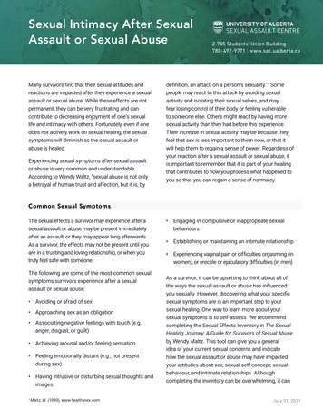
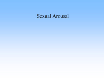
M. A. Guevara et al.sexual experience over the past 4 weeks, on a scale of 1 to 6, where 1 is themaximum value, and 6 the minimum or null. A score 26.55 is classified as female sexual disfunction [42].To obtain the measures of valence and the degree of both general and sexualarousal, each participant answered two scales immediately after finishing eachtext. The first was the Manikin Self-Assessment Scale (SAM) [43], a non-verbal,pictorial assessment technique that directly measures pleasure (positive andnegative valence) and general arousal. For valence, images ranging from a smiling, happy figure to a frowning, unhappy one represent the pleasure dimension(stimuli rated 1 - 3 were considered “unpleasant”; 4 - 6, “neutral”; and 7 - 9,“pleasant”). General arousal was rated using images that ranged from an excited,wide-eyed figure to a relaxed, sleepy one. Here, ratings of 1 - 5 were considered“not activated”, and 6 - 9 “activated”. The second instrument was the SexualArousal Scale (SAS), which is based on the principles of the ManikinSelf-Assessment Scale (a Likert-type scale). It was used to indicate the degree ofsubjective experience of vaginal lubrication (or sexual arousal) subsequent toreading the SET. It consists of a series of 5 drawings of smiling faces of differentintensities that represent vaginal lubrication, where 1 no lubrication (no sexualactivation) and 9 very high lubrication (high sexual activation). This allowedus to obtain a quantitative measure of the subjective experience of sexual arousal(see Figure 1). The utility of this scale has been demonstrated in previous studies [44].2.4. EEG Recordings and ProcedureAll recordings were made in a sound-attenuated room at 22 C - 23 C betweenFigure 1. Statistical data and graphic representation of the subjective scores of valence(a), general arousal (b), and sexual arousal (c) reported by participants using the ManikinSelf-Assessment and Sexual Arousal Scales. Wilcoxon test. *p (W) 0.01 as compared toNT.DOI: 10.4236/jbbs.2018.811037603Journal of Behavioral and Brain Science
M. A. Guevara et al.10:00 and 15:00 hours and without artificial light. Electrodes were placed on theright and left prefrontal (F3, F4), temporal (T3, T4) and parietal (P3, P4) corticesaccording to the 10 - 20 international system [45], and referred to linked earswith impedances maintained below 10 Kohms. Linked ears were chosen as thereference to avoid, as far as possible, reference electrode and volume conductioncontributions to the EEG correlations. In addition, electrooculograms were recorded to detect eye movement artifacts using a bipolar montage with electrodesplaced at the outer canthi of both eyes. As a measure of peripheral activation,heart rate was simultaneously recorded by means of an electrode placed on thefront of the left wrist.EEG data were recorded using a Grass P-7 polygraph(Grass Technologies, Co., USA) with filters set at 1 and 60 Hz, digitalized at asampling rate of 512 Hz, and stored on a PC using CAPTUSEN acquisitionsoftware [46]. Subjects were seated in a comfortable chair in front of a laptopcomputer in the room with door closed. All EEG recordings were made in twoconditions, each lasting 3 minutes: first while reading the neutral text and afterwhile reading the sexually-explicit text.EEG signals were analyzed off-line with CHECASEN software [47], whichdisplays the EEG epochs on a computer screen. By moving 2 cursors along theEEG signals it is possible to select and store artifact-free EEG segments (causedby eye movements, muscle activity or heart beat) that correspond to the specificperiods of interest. In this study, 60 epochs were selected from each text for eachparticipant. Fast Fourier Transform (FFTs) analyses were applied to the artifact-free data in 1-s epoch samples, with the spectral graph ranging from 1 - 60Hz at 1 Hz-resolution. FFTs were performed using EEG magic software [48].Absolute power (AP), defined as the power density of each frequency band expressed in microvolts squared (mV2/Hz), was calculated for each condition andtraditional EEG band: delta, δ (1 - 3 Hz), theta, θ (4 - 7 Hz), alpha1, α1 (8 - 10Hz), alpha2, α2 (11 - 13 Hz), beta 1, β1 (14 - 19 Hz), beta 2, β2 (20 - 30 Hz), andgamma, γ (31 - 50 Hz). As mentioned above, EEG synchronization or correlation is a mathematical index that makes it possible to calculate the degree of similarity between two EEG signals in different frequency bands, or at specificfrequencies. It is sensitive to both phase and polarity, regardless of amplitude[30]. Therefore, the correlation between cortices in the same hemisphere (intrahemispheric correlation, or rINTRA) was also calculated for each of the six frequency bands by means of Pearson Product-moment correlation analysis.2.5. Statistical AnalysesThe parameters of valence, general arousal and sexual arousal between conditions (NT vs. SET) were compared by Wilcox on tests.A Student t test for correlated groups was used to compare the absolute powerand rINTRA of the six traditional EEG bands for the prefrontal, temporal andparietal areas in the two conditions (reading SET vs. NT). For statistical purposes,AP values were transformed into logarithms: ln(x), where x AP value in microvolts2 [49]. Correlation values were averaged over all epochs of the same conDOI: 10.4236/jbbs.2018.811037604Journal of Behavioral and Brain Science
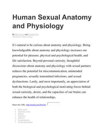
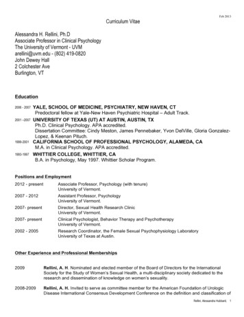
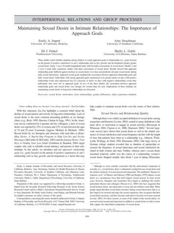
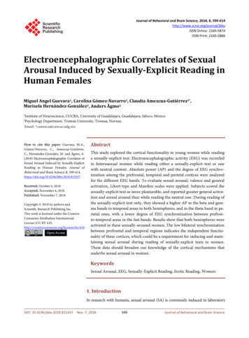
M. A. Guevara et al.dition, for each participant and pair of derivations. To approximate a normaldistribution, correlation values were transformed into Fisher’s Z scores.3. Results3.1. ScalesAs mentioned previously, only women who answered 0 or 1 on the Kinseyscale—i.e., exclusively heterosexual–were included. Regarding the evaluation ofthe texts, the results of the Manikin Self-Assessment Scale indicated significantbetween-condition differences (SET vs. NT) in valence (W 15.00 p (W) 0.0189), which showed that reading the SET produced more pleasure than reading the NT (Figure 1(a)). A significant difference was found for general arousal(W 0.00 p (W) 0.0004) with the SET producing greater arousal than the NT(Figure 1(b)). The results of the sexual arousal scale showed that only readingthe SET induced a state of SA (W 0.00 p (W) 0.0002) (Figure 1(c)).3.2. EEG3.2.1. Absolute PowerStatistical analyses revealed significant differences between conditions (readingSET vs. NT) in the AP of the different EEG bands. During reading of SET, higher APs of the beta 1 (t 2.47 p 0.024), beta 2 (t 2.88 p 0.016), and gamma (t 2.88 p 0.010) bands in the left (T3) temporal area, and of the beta 2(t 2.52 p 0.021) and gamma (t 2.41 p 0.027) bands in the right temporal area (T4) were observed, compared to NT (Figure 2).In the parietal cortex, in contrast, a higher AP was obtained only duringreading of the SET in both the left (P3, t 2.573 p 0.019) and right (P4, t 3.078 p 0.006) hemispheres, specifically in the theta band, compared to theNT condition (Figure 3).Figure 2. Absolute Power (Mean 2 SE) transformed into logarithms of the EEG frequency bands recorded in temporal (T3 and T4) regions of the women while reading theneutral (NT) and sexually-explicit (SET) texts. MD Mean of differences. Students t testfor correlated groups. *p 0.05 as compared to NT.DOI: 10.4236/jbbs.2018.811037605Journal of Behavioral and Brain Science
M. A. Guevara et al.Figure 3. Absolute Power (Mean 2 SE) transformed into logarithms of the EEG frequency bands recorded in parietal (P3 and P4) regions of women while reading the neutral (NT) and sexually-explicit (SET) texts. MD Mean of differences. Students t test forcorrelated groups. *p 0.05 as compared to NT.Figure 4. Intrahemispheric correlation (Mean 2 SE) transformed into Fisher’s Z scoresof the EEG frequency bands in the left (F3-T3) and right (F4-T4)) fronto-temporal regionsof the women while reading the neutral (NT) and sexually-explicit (SET) text. MD Mean of differences. Students t test for correlated groups. *p 0.05 as compared to NT.No significant differences were obtained in the AP of the EEG bands in theprefrontal areas.3.2.2. Intra-Hemispheric CorrelationCompared to reading the NT, the SET condition was associated with a lower degree of synchronization, or rINTRA, between the prefrontal and temporal (F3-T3)cortices. In the left hemisphere, this decreased correlation was found in beta 1 (t 2.462 p 0.024), whereas in the right hemisphere it was observed in the fasterbands (alpha1, t 2.948 p 0.008; alpha2, t 3.114 p 0.006; beta 1, t 2666p 0.036; beta 2, t 3.912 p 0.001; and gamma, t 2.235 p 0.039) (Figure 4).DOI: 10.4236/jbbs.2018.811037606Journal of Behavioral and Brain Science
M. A. Guevara et al.4. DiscussionTo the best of our knowledge, this is the first study to characterize the corticalEEG activity of young women during sexual arousal induced by reading sexually-explicit texts. According to participants’ self-reports on the Manikin and sexualarousal scales, they scored the SET as more pleasurable, and indicated that itgenerated greater general activation and greater sexual arousal than the NT. Thisresult confirms reports in other studies [3] [7] [8] [31] that demonstrated the effectiveness of SET in generating a state of SA in women. Though we used an indirect, or subjective, measure—namely, vaginal lubrication self-reported on aLikert-type scale—our data are consistent with the results of other studies thathave shown a direct correlation between increased vaginal pressure pulse (usingvaginal photoplethysmography) and the high SA values self-reported by participants [3].It was only while reading the SET that these women presented a greater AP inthe fast bands (beta 1, beta 2 and gamma) in the temporal areas of both hemispheres. An increased AP in beta 2 and gamma has been associated with positiveemotional states induced by reading [50]. In particular, the gamma band hasbeen associated with the processing of emotional stimuli, especially in temporalareas [51]. Also, studies using imaging techniques (fMRI) have reported activation of temporal areas during states of sexual arousal induced by visual stimulation (photos, videos) in both sexes [10] [14] [52]. These findings suggest that theincreased AP of the fast bands in bilateral temporal regions could be associatedwith the state of sexual arousal and the positive affect (greater pleasure) that thewomen reported after reading the sexually-explicit texts.This study also showed a greater bilateral AP of the theta band in the parietalareas during SA. The parietal lobe has been related to attention, the integrationof somatosensory and visual information [17] [18], and, through fMRI studies,to SA induced by erotic stimuli [14]. The prevalence of the theta band has beenreported in the posterior cortical areas in response to emotional stimuli [53] andduring pleasant and relaxed states [22]. Therefore, its prevalence in parietalareas, together with the higher AP of the fast bands in temporal areas, representa state of cortical activation that could be associated with the affective (pleasant)evaluation and SA induced by reading sexual texts.Contrary to our expectations, no significant differences were found in the APof the prefrontal areas during the reading of SET compared to NT. The prefrontal cortex participates in the syntactic processing of reading; i.e., the correctcombining and ordering of words in a discourse [54] and in other cognitiveprocesses, such as working memory, concept formation [55] [56], and visualimagery [57]. All these processes are required to understand narrative texts; thatis, those that combine statements to form a coherent story [58]. The stimuli usedin this study were of a narrative nature, so understanding and processing the information they contained required using similar cognitive resources, ones thatare modulated primarily by the prefrontal cortex. Thus, a possible explanationDOI: 10.4236/jbbs.2018.811037607Journal of Behavioral and Brain Science
M. A. Guevara et al.for the absence of prefrontal activation in these subjects while reading the SETcould be that the processing that takes place in the prefrontal cortex while reading is similar regardless of the emotional content involved; hence, the functionality observed did not vary. A second possible interpretation is that the level ofsexual arousal generated by reading the SET was insufficient to produce changesin the functionality of the prefrontal cortex, so no significant differences werefound between the reading of the two texts.Regarding the degree of EEG synchronization, or correlation between corticalareas, a lower rINTRA of the fast bands (alpha, beta) between the prefrontal andtemporal cortices in both hemispheres was found only while the women read theSET. This decreased rINTRA agrees with the findings of a previous study, whichfound a lower prefronto-temporal correlation in beta 1 and a lower prefronto-parietal correlation in the theta and alpha2 bands in heterosexual men whileobserving photographs with sexual content [19]. To explain this lower intrahemispheric synchronization, several aspects must be considered. F3 and F4 specifically constitute the so-called dorsolateral region of the prefrontal cortex [59],an area related to goal-directed behavior, the evaluation and integration of neural information, and “cold” executive functions, such as planning, working memory and inhibitory control [12]. The temporal cortex, in contrast, has beenwidely-associated with recognizing emotions from visual stimuli [15] and severalaspects of the sex drive or sexual desire [16] [60]. Moreover, it has been suggested that the inhibitory processes that are active between periods of SA, andthat act to prevent its emergence, are exerted by regions in the temporal lobes[4]. On the other hand, the fast bands—beta 1, beta 2, gamma—have been related to the inter-neuronal communication of inhibitory networks [61] and toinformation transfer between regions [62]. Thus, it is possible that the functionaldissociation between the prefrontal and temporal areas observed in our work isrequired to suppress any timidity or mental tasks that might interfere with theinduction of the positive emotional sexual arousal associated with reading erotictexts.Interestingly, our study did not find a lower EEG correlation between the prefrontal and parietal cortices, perhaps as a result of the cognitive processes involved in reading the texts presented. As mentioned above, understanding anarrative passage (i.e., a text with statements that form a coherent story) requireshigher-order processes, such as attention and working memory [63] in whichthe prefrontal [7] and parietal [17] cortices play pivotal roles. In addition, ananatomical circuit described between the parietal and prefrontal areas has beenwidely-associated with the attentional processes responsible for achieving andmaintaining sensitivity to incoming stimuli [18]. Because both texts used in thisstudy were of a narrative nature, their reading, regardless of content, requiredattentional and memory processes and the integration of concepts. The fact thatthe prefrontal and parietal cortices participate in these processes leads us to suggest that they work together; thus explaining why a similar degree of correlationDOI: 10.4236/jbbs.2018.811037608Journal of Behavioral and Brain Science
M. A. Guevara et al.between them was obtained during reading of both texts.Our results contrast with the lower prefronto-parietal correlation observed inmen and women during sexual arousal induced by observing visual stimuli withsexual content (photos and videos) [19]. However, it is important to point outthat visual stimulation does not require dealing with spelling, punctuation or thevisual images characteristic of written language; hence, these different types ofstimuli are processed distinctly. Moreover, following Anokhin et al. [9], from anevolutionary perspective, rapid assessment of a visual scene is critical for an organism’s adaptation and survival, quite unlike reading, which is a reconstructionof messages represented by graphic symbols. Thus, it is not surprising to find adifferent functionality and degree of cortical EEG coupling during sexual arousalinduced by observing visual stimuli compared to sexual arousal induced byreading texts with sexual content.One particularly interesting result of our work is that the activation of thetemporal and parietal cortices, as well as the lower prefronto-temporal correlation, were observed in both hemispheres during reading of the SET. Althoughnumerous fRMI and EEG studies have reported distinct patterns of hemisphericlateralization during sexual activation and orgasm, mainly in men [64] [65], it isprobable that a lower pattern of lateralization will be observed in women duringsexual arousal, as in the case of linguistic demands. Indeed, Pugh et al. [66] andShaywitz et al. [67] demonstrated gender-based differences in the brain regionsactivated by grammatical and reading tasks that indicated a stronger pattern ofleft-lateralization in men than women. Other works provide additional evidenceof the bilateral activation of several brain structures in women during suchprocesses as spatial orientation or language use [67] [68].These EEG data show that during sexual arousal induced by reading erotictexts the prefrontal and parietal cortices function similarly. This is likely required for subjects to attend to cognitive demands involved in understandingand then, reasoning about, of the texts presented. The bilateral activation of theparie
The sexually-explicit text (SET) was made up of excerpts from the Fifty Shades trilogy, which was written by a woman: Fifty Shades of Gray [32], Fifty Shades Darker [33], and Fifty Shades Freed [34]. Considering that the most appealing and arousing films for women are those that depict heterosexual vaginal inter-