Transcription
Lateral Release Can Increase Range of Flexion duringComputer Assisted Total Knee Arthroplasty withPatellar MaltrackingPornpavit Sriphirom MD*,Chaiyaporn Siramanakul MD**, Boonyawat Chanopas MD**** Department of Orthopedic Surgery, Rajavithi Hospital, College of Medicine, Rangsit University, Bangkok, Thailand** Department of Orthopedic Surgery, Banphaeo Hospital (Prommitr Branch), Bangkok, Thailand*** Department of Orthopedic Surgery, Medical Development Clinic, Bangkok, ThailandObjective: The patellar tracking is an important factor that affect range of motion after total knee arthroplasty (TKA).Intraoperative patellar maltracking during TKA can be improved by performing lateral release. We hypothesized that TKAwith patellar maltracking after undergoing lateral release can increase intraoperative range of flexion.Material and Method: A prospective study was conducted on 110 knees that underwent computer assisted TKA. The patellartracking was assessed with no thumb test technique. Fifty-two knees were classified into negative no thumb test group, and58 knees were classified into positive no thumb test group. The positive no thumb test group further received lateral releasewith outside to inside technique. The range of flexion was recorded before and after final implantation in both groups, andrecorded after lateral release in positive no thumb test group.Results: After final implantation, the negative no thumb test group had significant greater flexion angle than the positiveno thumb test group (128.20 and 123.90 ). The range of flexion after performing the lateral release in positive no thumbtest group increased the flexion up to 127.60 . Thus, there was no significant difference from the negative no thumb testgroup (128.20 ). After the lateral release was performed, the flexion angle had significantly increased by 3.70 .Conclusion: The results indicated that intraoperative lateral release in patellar maltracking can improve range of flexionin computer assisted TKA.Keywords: Lateral release, Patellar maltracking, Computer assisted total knee arthroplastyJ Med Assoc Thai 2017; 100 (3): 295-300Full text. e-Journal: http://www.jmatonline.comIn Asian culture, squatting and sittingcross-legged is very common in religious activities.Therefore, total knee arthroplasty (TKA) requiresincreased knee flexion. There are many intraoperativefactors that affect the range of flexion after TKAincluding flexion and extension gap imbalance, jointline elevation, retention of posterior osteophyte offemoral condyle, femoral component malposition, andpatellar tilt and shift. Those have been considered tobe important factors that affect postoperative range offlexion(1-7). Intraoperative patellar tilt or maltrackingduring TKA can be managed by performing a lateralrelease procedure(8). The biomechanical effect oflateral release has been examined and it could decreasepressure on the lateral patellar facet in flexion(9). TheCorrespondence to:Siramanakul C, Department of Orthopedic Surgery, BanphaeoHospital (Prommitr Branch), Sukhumvit 39, Wattana, Bangkok10110, Thailand.Phone: 66-2-2590333, Fax: 66-2-2584751E-mail: chaisira@hotmail.comJ Med Assoc Thai Vol. 100 No. 3 2017incidence of some degree of lateral release has beenreported to be as high as 40%(10). Various studies havereported that lateral release does not compromise theclinical outcomes or increase the complication rateof TKA(11-14). However, effects of lateral release inimproving the range of flexion during TKA have notbeen studied. We hypothesized that the intraoperativelateral release will not only improve patellar trackingbut will increase flexion angle during TKA navigationas well.Material and MethodThis prospective study was performedbetween January 2009 and January 2010. The studyprotocol was approved by our institutional ethicscommittee. All 110 osteoarthritis knees underwentprimary TKA with mobile posterior stabilize prosthesis(emotion PS, Aesculap, Tuttlingen, Germany). Allcases underwent TKA under the navigation system(Orthopilot version 4.3 tibia cut first) with gapbalancing technique. The exclusion criterion was TKA295
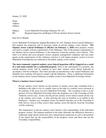
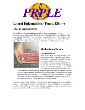
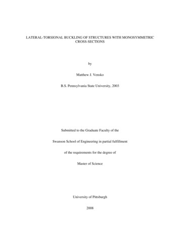
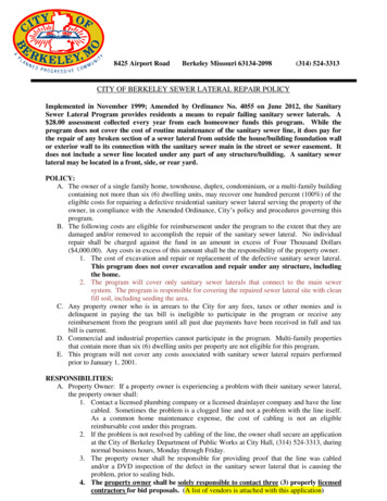
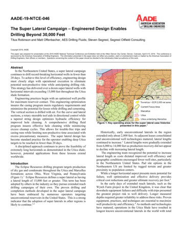
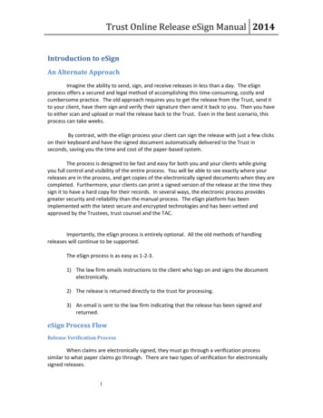
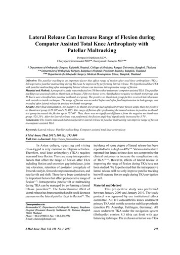
with resurfacing patella. When the operation wasbegun, the tourniquet was inflated with knee maximallyflexed and mini-midvastus arthrotomy was used in allcases. The operation was performed step by step undernavigation. The deformity and range of flexion bygravity were recorded after each registration step andat final implantation. After cementing of the finalprostheses, patellar tracking was evaluated with “nothumb test”(15,16) from knee flexion 30 to 80 and thetourniquet was not released. If the absolute no degreeof patellar opening or tilting and perfectly tracking ofpatella within the trochlear groove of the femoralprosthesis was found, it was classified in the “negativeno thumb test” group. If any degree of patellar openingor tilting during range of motion trialing was found,the lateral release was performed and classified inthe “positive no thumb test” group (Fig. 1). The rangeof motion was recorded again after lateral release(Fig. 2). The lateral release was performed in a stagedfashion with outside to inside technique. The lateralretinaculum was sequentially released in three stageswith reassessment of patellar tracking between eachstage until no opening or tilting of the patella could bedetected. The outside to inside technique was begunwith open the lateral skin flap over the patella deep toFig. 1Photograph showed positive no thumb test withmedial opening of patella.Fig. 2Photograph showed the range of the flexion bygravity without released tourniquet before and afterthe lateral release was recorded from navigation.296deep fascia to expose the lateral retinaculum. The firststage of lateral release started from the level of thesuperior pole of the patella, beginning 5 mm lateral tothe lateral border of the patella to the inferior pole ofthe patella (Fig. 3). The second stage was extendedfrom the level of the lower pole of the patella to theGerdy’s tubercle (Fig. 4). The line of incision wasparallel to the patellar tendon. The third stage, theincision was made upward from the superior pole ofthe patella to the superficial layer of vastus lateralisobliquus tendon (Fig. 5, 6). The superior lateralgenicular artery was not identified during release.The statistical analysis used to comparepreoperative deformity between two groups wasindependent sample t-test. The statistical analyses forcomparison flexion angle, final implantation, andpostoperative flexion angle between two groups andincluding a comparison between flexion angle of finalimplantation and after lateral release in lateral releaseFig. 3Photograph showed the first stage of lateral release.The lateral patellofemoral ligament and patellomeniscal ligament were cut.Fig. 4Photograph showed the second stage of lateralrelease. The incision extended from the lowerpole patella toward Gerdy’s tubercle.J Med Assoc Thai Vol. 100 No. 3 2017
Fig. 5Photograph showed the third stage of lateral release.The incision extended from upper pole patellatoward superficial layer of vastus lateralis obliquustendon.group were paired two-tailed Student’s t-test. Statisticalsignificance was set at p-value 0.05. Sample sizecalculated for one-tailed t-test study with 0.05probability level, 0.5 anticipated effect and 80%statistical power level was 51 knees in each group.SPSS for windows (Version 9.0, SPSS, Chicago,Illinois, USA) was used for the statistical analysis.ResultsThe 110 TKAs were classified into two groupsaccording to no thumb test after final implantation.The negative no thumb test group consisted of52 knees. The average age of patients in this groupwas 69.2 years. The deformity consisted of 46 varusknees had average 9.28 varus, and six valgus kneeshad average 3.16 valgus. The positive no thumb testgroup had 58 knees. The average age of patients was69.0 years. The positive no thumb test group consistedFig. 6Photograph showed the absent patellar openingafter the lateral release was performed.of 52 varus knees, with an average degree of deformityof 9.38 varus, and six valgus knees had average 3.00 valgus. There was no significant difference betweentwo groups in term of preoperative deformity (Table 1).The preoperative flexion angle and the angle afterthe final implantation were analyzed in both groups(Table 2). The results revealed no statistical significantdifference in preoperative flexion angle between thetwo groups. However, it was noted that after finalimplantation, the negative no thumb test group hadsignificant greater flexion angle than the positive nothumb test group.After the lateral release was performed, theflexion angle had significantly increased by 3.70 compared with the final implantation (p 0.00).Finally, postoperative flexion angle was comparablein each group, with no significant difference in flexionangle after operation (Table 2).Table 1. Preoperative deformity between the negative no thumb test and the positive no thumb test groupDeformityNegative no thumb test groupPositive no thumb test groupp-value*Varus deformity9.28 9.38 0.93Valgus deformity3.16 3.00 0.87* Independent sample t-testTable 2. Preoperative, final implantation, and postoperative flexion angle of the negative no thumb test and the positiveno thumb test groupNegative no thumb test groupPositive no thumb test groupp-value*Preoperative flexion angle124.00 122.00 0.91Final implantation flexion angle128.20 123.90 0.00Postoperative flexion angle128.20 127.60 0.437* Paired two-tailed Student’s t-testJ Med Assoc Thai Vol. 100 No. 3 2017297
DiscussionAccording to the present study’s results, thenegative no thumb test group had significant greaterflexion angle after the final implantation than thepositive no thumb test group. This indicated thatpatellar tracking is one of the factors affecting rangeof motion after TKA. Kawamura and Bourne had asimilar conclusion(7). The lateral release is a procedureto correct patellar maltracking. The lateral release canbe performed using either of two techniques: “insideout” and “outside-in”. The inside-out technique wasperformed in the knee joint and release throughsynovial lining and joint capsule, which exposed thejoint to subcutaneous plane under the skin. Thistechnique has associated risks of hematoma under theskin and spread of infection into the joint(17). Strachanet al reported six stages of outside-in lateral releaseproximal to distal manner(11). Maniar et al describedthree steps of outside-in technique with: step-1 releasewas from the midpatella to the upper tibial border,step-2 release was from the midpatella to the proximalpole of the patella, and step-3 release was proximal tothe superior pole of the patella; and reported aspreserving the superior lateral genicular artery in 76%of patients and no complications were seen at followup with functional and radiographic examinations(18).The technique we used was to release from the upperpole of the patella to the lower pole of the patella, inthe first step release the lateral patellofemoral andpatellomeniscal ligament(19). In the second step, fromthe lower pole of the patella to the Gerdy’s tubercle werelease the patellotibial ligament. The ligament wasattached to the lateral tibial condyle between the tibialtuberosity and Gerdy’s tubercle(19). In the third step,the incision was made upward from the superior poleof the patella to the superficial layer of vastus lateralisobliquus tendon. The role of lateral release forimproving range of motion is rarely mentioned,especially in TKA navigation. Kawamura and Bournefound the tilt angle of patella had a negative correlationwith postoperative range of flexion(7).According to our results, after performingthe lateral release for the correction of the patellar tilt,the flexion angle was increased 3.7 (from 123.9 to127.6 ) and postoperative flexion angle in positive nothumb test group had no significant difference fromthe negative no thumb test group. The results indicatedthat patellar tracking effects on range of motion duringTKA and lateral release can improve range of flexionin TKA with patellar maltracking. Laskin(20) andLombardi et al(21) had recommended releasing the298tourniquet before performing lateral release andfound that it could reduce the incidence of lateralrelease between 6% and 22%. However, the perfectintraoperative patellar tracking is the goal of TKAand lateral release does not associate with negativeoutcomes. The short- and long-term improvement ofthe range of motion should be followed in the future.The postoperative follow-up is an important factor thatrequires the cooperation of the patients, if we determineto have the long-term postoperative range of motion.Therefore, the efforts of the patients in conscientiouslyparticipating in the postoperative physical therapyprogram are required.ConclusionBased on the data of the present study, we canconclude that lateral release can increase intraoperativerange of flexion in TKA with patellar maltracking.What is already known on this topic?The intra-capsular factors influencing rangeof flexion after TKA were implant design, ligamentbalance, flexion-extension gap balance, height of jointline, and patellar tracking.What this study adds?Patellar tracking is a significant factor ofintraoperative range of flexion. Lateral release inpatellar maltracking has also been improvingintraoperative range of flexion during TKA.Potential conflicts of interestNone.References1. Dennis DA, Komistek RD, Scuderi GR, ZingdeS. Factors affecting flexion after total kneearthroplasty. Clin Orthop Relat Res 2007; 464:53-60.2. Lo CS, Wang SJ, Wu SS. Knee stiffness on extensioncaused by an oversized femoral component aftertotal knee arthroplasty: a report of two cases anda review of the literature. J Arthroplasty 2003; 18:804-8.3. Dennis DA. A stepwise approach to revision totalknee arthroplasty. J Arthroplasty 2007; 22: 32-8.4. Figgie HE 3rd, Goldberg VM, Heiple KG, MollerHS 3rd, Gordon NH. The influence of tibialpatellofemoral location on function of the kneein patients with the posterior stabilized condylarknee prosthesis. J Bone Joint Surg Am 1986; 68:J Med Assoc Thai Vol. 100 No. 3 2017
1035-40.Goldstein WM, Raab DJ, Gleason TF, Branson JJ,Berland K. Why posterior cruciate-retaining andsubstituting total knee replacements have similarranges of motion. The importance of posteriorcondylar offset and cleanout of posterior condylarspace. J Bone Joint Surg Am 2006; 88 (Suppl 4):182-8.6. Massin P, Gournay A. Optimization of theposterior condylar offset, tibial slope, and condylarroll-back in total knee arthroplasty. J Arthroplasty2006; 21: 889-96.7. Kawamura H, Bourne RB. Factors affecting rangeof flexion after total knee arthroplasty. J OrthopSci 2001; 6: 248-52.8. Sodha S, Kim J, McGuire KJ, Lonner JH,Lotke PA. Lateral retinacular release as a functionof femoral component rotation in total kneearthroplasty. J Arthroplasty 2004; 19: 459-63.9. Ostermeier S, Holst M, Hurschler C, WindhagenH, Stukenborg-Colsman C. Dynamic measurementof patellofemoral kinematics and contact pressureafter lateral retinacular release: an in vitro study.Knee Surg Sports Traumatol Arthrosc 2007; 15:547-54.10. Engh GA, Parks NL, Ammeen DJ. Influence ofsurgical approach on lateral retinacular releasesin total knee arthroplasty. Clin Orthop Relat Res1996; (331): 56-63.11. Strachan RK, Merican AM, Devadasan B,Maheshwari R, Amis AA. A technique of stagedlateral release to correct patellar tracking in totalknee arthroplasty. J Arthroplasty 2009; 24: 735-42.12. Ritter MA, Campbell ED. Postoperative patellarcomplications with or without lateral release5.J Med Assoc Thai Vol. 100 No. 3 201713.14.15.16.17.18.19.20.21.during total knee arthroplasty. Clin Orthop RelatRes 1987; (219): 163-8.Ritter MA, Herbst SA, Keating EM, Faris PM,Meding JB. Patellofemoral complications followingtotal knee arthroplasty. Effect of a lateral releaseand sacrifice of the superior lateral geniculateartery. J Arthroplasty 1996; 11: 368-72.Kusuma SK, Puri N, Lotke PA. Lateral retinacularrelease during primary total knee arthroplasty: effecton outcomes and complications. J Arthroplasty2009; 24: 383-90.Dugal SS, Scott RD. Extensor mechanismcomplications. In: Rand JA, editor. Total kneearthroplasty. New York: Raven Press; 1993: 394.Insall J. Surgical techniques and instrumentationin total knee arthroplasty. In: Windsor RE, ScottWN, editors. Surgery of the knee. 2nd ed. NewYork: Churchill Livingstone; 1998: 768.Johnson DP, Eastwood DM. Lateral patellarrelease in knee arthroplasty. Effect on woundhealing. J Arthroplasty 1992; 7 (Suppl): 427-31.Maniar RN, Singhi T, Rathi SS, Baviskar JV,Nayak RM. Surgical technique: Lateral retinaculumrelease in knee arthroplasty using a stepwise,outside-in technique. Clin Orthop Relat Res 2012;470: 2854-63.Merican AM, Amis AA. Anatomy of the lateralretinaculum of the knee. J Bone Joint Surg Br2008; 90: 527-34.Laskin RS. Lateral release rates after total kneearthroplasty. Clin Orthop Relat Res 2001; 88-93.Lombardi AV Jr, Berend KR, Mallory TH, DoddsKL, Adams JB. The relationship of lateral releaseand tourniquet deflation in total knee arthroplasty.J Knee Surg 2003; 16: 209-14.299
�ื่อด านข างลูกสะบ าช �ข าขณะการทําผ าตัดเปลี่ยนข อเข าเทียมด วยคอมพิวเตอร นําวิถีในผู ป วยที่มีตําแหน งลูกสะบ าผิดปกติพรภวิษญ ศรีภิรมย , ชัยพร ศิระมานะกุล, บุญวัฒน �ค : ตําแหน งของลูกสะบ าเป นป �ญต อองศาการงอเข าหลังการผ าตัดเปลี่ยนข อเทียม ตําแหน งลูกสะบ าที่ผิดปกติขณะผ าตัดสามารถแก ไขด � เยือ่ ข างลูกสะบ า �ษานีค้ อื การตัดเลาะเนือ้ เยือ่ ข างลูกสะบ าในผู ป วยที่มีตําแหน � าขณะผ าตัดเปลี่ยนข �องศาการงอเข าได ด �: เป นการศึกษาไปข างหน าในผู ป วยที่ผ าตัดเปลี่ยนข อเทียมด วยคอมพิวเตอร นําวิถีจํานวน 110 เข า ตําแหน งของลูกสะบ าประเมินด �หน งของลูกสะบ า พบว า 52 เข า จัดในกลุ มตําแหน งลูกสะบ าปกติ และ 58 เข า จัดในกลุ �ลูกสะบ า โดยกลุ �ลูกสะบ าจะได �อเยื่อด านข างลูกสะบ าโดยวิธีตัดด านนอกเข าสู ด านในเข า องศาการงอเข าจะได รับการบันทึกก อนและหลังใส ข �ุ มรวมทั้งได � �ื้อเยื่อด านข างลูกสะบ าด วยผลการศึกษา: หลังจากใส ข อเข าเทียม กลุ มที่ตําแหน งลูกสะบ �ข ามากกว ากลุ �ลูกสะบ าอย างมีนัยสําคัญ (128.20 และ 123.90 ) �เนือ้ เยือ่ ข างลูกสะบ าในกลุม ทีม่ กี ารเอียงของลูกสะบ า พบว าองศาการงอเข �ถงอเข าได ถึง 127.60 และพบว าสุดท ายองศาการงอเข าของทั้งสองกลุ มไม แตกต างกัน �ื่อข างลูกสะบ �ารงอเข าได 3.70 อย �: การตัดเลาะเนือ้ เยือ่ ข างลูกสะบ าในผูป ว ยทีม่ ตี าํ แหน งของลูกสะบ าผิดปกติขณะผ าตัดสามารถเพิม่ องศาการงอเข าในการผ าตัดข อเข าเทียมด �อร นําวิถี300J Med Assoc Thai Vol. 100 No. 3 2017
Therefore, total knee arthroplasty (TKA) requires increased knee flexion. There are many intraoperative factors that affect the range of flexion after TKA including flexion and extension gap imbalance, joint line elevation, retention of posterior osteophyte of femoral condyle, femoral component malposition, and patellar tilt and shift. Those have been considered to be important factors that .