Transcription
Updates in RIGINAL ARTICLEEvolving knowledge in surgical oncology of pancreatic cancer:from theory to clinical practice—a fifteen‑year journey at a tertiaryreferral centreRiccardo Casadei1,2 · Claudio Ricci1,2 · Carlo Ingaldi1,2 · Laura Alberici1,2 · Emilio De Raffele1,2 · Bianca Barcia1,2 ·Cristina Mosconi3 · Margherita Diegoli3 · Mariacristina Di Marco5 · Giovanni Brandi5 · Rocco Maurizio Zagari4 ·Nico Pagano4 · Leonardo Henry Eusebi4 · Carla Serra2 · Marina Migliori2 · Alessandra Guido2 · Donatella Santini6 ·Francesca Rosini6 · Deborah Malvi6 · Francesco Minni1,2Received: 3 March 2022 / Accepted: 22 July 2022 The Author(s) 2022AbstractPancreatic ductal adenocarcinoma (PDAC) is an increasing disease having a poor prognosis. The aim of the present study wasto evaluate the effect of different models of care for pancreatic cancer in a tertiary referral centre in the period 2006–2020.Retrospective study of patients with PDAC observed from January 2006 to December 2020. The demographic and clinicaldata, and data regarding the imaging techniques used, preoperative staging, management, survival and multidisciplinarytumour board (MDTB) evaluation were collected and compared in three different periods characterised by different organisation of pancreatic cancer services: period A (2006–2010); period B (2011–2015) and period C (2016–2020). One thousandfour hundred seven patients were analysed: 441(31.3%) in period A; 413 (29.4%) in B and 553 (39.3%) in C. The proportion ofpatients increased significantly, from 31.3% to 39.3% (P 0.032). Body mass index (P 0.033), comorbidity rate (P 0.002)and Karnofsky performance status (P 0.001) showed significant differences. Computed tomography scans (P 0.001),endoscopic ultrasound (P 0.001), fine needle aspiration, fine needle biopsy (P 0.001), and fluorodeoxyglucose-positronemission tomography/computed tomography (P 0.001) increased; contrast-enhanced ultrasound (P 0.028) decreased. ThecTNM was significantly different (P 0.001). The MDTB evaluation increased significantly (P 0.001). Up-front surgeryand exploratory laparotomy decreased (P 0.001), neoadjuvant treatment increased (P 0.001). The present study showedthe evolving knowledge in surgical oncology of pancreatic cancer at a tertiary referral centre over the time. The differentmodels of care of pancreatic cancer, in particular the introduction of the MDTB and the institution of a pancreas unit to thedecision-making process seemed to be influential.Keywords Pancreatic ductal adenocarcinoma · Incidence · Diagnosis · Treatment · SurvivalIntroductionRiccardo Casadei and Claudio Ricci shared the first authorship.* Riccardo Casadeiriccardo.casadei@unibo.it12Division of Pancreatic Surgery, IRCCS, AziendaOspedaliero Universitaria di Bologna, S.Orsola-MalpighiHospital, Via Massarenti n.9, 40138 Bologna, ItalyDepartment of Internal Medicine and Surgery (DIMEC),Alma Mater Studiorum, University of Bologna,S.Orsola-Malpighi Hospital, Bologna, ItalyApproximately 60,430 and 14,263 new diagnoses of pancreatic cancer were estimated in the U.S. and Italy in 2021,respectively, with an increasing incidence rate of 0.5–1%3Department of Radiology, IRCCS AziendaOspedaliero-Universitaria di Bologna, Bologna, Italy4Division of Gastroenterology, IRCCS AziendaOspedaliero-Universitaria di Bologna, Bologna, Italy5Medical Oncology, IRCCS AziendaOspedaliero-Universitaria di Bologna, Bologna, Italy6Division of Pathologhy, Azienda Ospedaliero-Universitariadi Bologna, Bologna, Italy13Vol.:(0123456789)
Updates in Surgeryper year [1, 2]. Based on GLOBOCAN 2018 estimates, pancreatic cancer accounted for 458,918 new cases and caused432,242 deaths (4.5% of all deaths caused by cancer) in2018 [3]. The 5-year survival rate was approximately 10%for the first time in 2020, and pancreatic cancer is projectedto become the second-leading cause of cancer death in theU.S. by 2030 [4, 5]. Much effort is being made to resolve thisalarming situation. Imaging resolution has been improved byenhanced computed tomography with a high-quality pancreatic protocol and by the introduction of endoscopic ultrasound [6]. The accurate assessment of surgical indications,techniques, and perioperative care have been very usefulregarding better short-term results. Furthermore, adjuvantand neoadjuvant radiochemotherapy, and immunotherapieswere more frequently incorporated into multimodal treatment [7, 8] to obtain better long-term results and, finally,consensus recommendations for improving pancreatic cancercare were adopted [9]. Nationwide trends regarding the incidence, treatment and survival of pancreatic cancer patientsto adequately measure the impact of these improvements inthe management of pancreatic cancer have been reportedin only a few studies [10–18]. To the Authors’ knowledge,there are no studies which evaluate the changes in trendsregarding pancreatic ductal adenocarcinoma in a tertiaryreferral centre over the time periods. The aim of the presentstudy was to evaluate the effect of different models of carefor pancreatic cancer by analysing the changes in trends ofthe demographic and clinical data, the imaging techniquesused, the management adopted and long-term survival in atertiary referral centre in the period 2006–2020.MethodsStudy designThis was a retrospective study of patients affected by PDACobserved at S. Orsola-Malpighi Hospital, Bologna, Italy,from January 2006 to December 2020. The study wasapproved by the Ethical Committee of S. Orsola-MalpighiHospital (code: 642017 U/Oss), and patient informed consent was obtained from all the participants enrolled in thestudy. Patients were included in this study if they provided aconsent to include their anonymized data in future research.All patients with an ICD-9-CM code diagnosis of 157.0,157.1, 157.2, 157.8, and 157.9, and ICD-9-CM procedures52.51, 52.52, 52.53, 52.6, and 52.7 were extracted andentered into the database for pancreatic diseases (PANBO,code: 064/2017/U/OSS). Patients were included in this studyonly when admitted to S. Orsola-Malpighi hospital. Patientsseen only in outpatient clinics were not included. The following data were collected for each patient: (1) demographicand clinical; gender, age, co-morbidities, body mass index13(BMI), symptoms (back pain, diabetes, weight loss, jaundice, stenting), Ca 19–9 serum value and Karnofsky performance status (KPS); (2) imaging techniques used: thoracoabdominal computed tomography (CT) scan, magneticresonance (MR), endoscopic ultrasound (EUS), contrastenhanced ultrasound (CEUS), fluorodeoxyglucose-positronemission tomography/computed tomography (FDG-PET/CT), and fine needle aspiration (FNA) or fine needle biopsy(FNB), and 3) preoperative staging, management and survival; site of the tumour (head/uncinate process, body/tailand multiple), cTNM according to the American Joint Committee on Cancer AJCC 8 edition [19], National Comprehensive Cancer Network (NCCN) classification [20] (resectable, borderline resectable, locally advanced and metastatic),multidisciplinary tumour board (MDTB) evaluation, type ofmanagement (up-front surgery, palliative care, neoadjuvantapproach), surgical resection, unnecessary surgery and overall survival. Using these data, three different periods wereidentified and compared: period A (2006–2010); period B(2011–2015) and period C (2016–2020). The three different periods were characterised by the different organisationof pancreatic cancer services. In the period 2006–2010, thediagnostic and therapeutic decision-making process wasrelated only to common evidence-based recommendationsregarding pancreatic cancer. The decisions were usuallymade by a single medical doctor or a single team of specialists (for example, surgeons). However, in this periodthere was a multidisciplinary tumour board but it played amodest role in the decision-making process because it wasnot recognized by our Hospital. In the period 2011–2015integrated healthcare policies promoted specialisation andput a multidisciplinary tumour board (MDTB) at the centreof the decision-making process. The MDTB consisted ofseveral specialists with expertise in the field of pancreaticcancer (surgeons, gastroenterologists, radiologists, oncologists and radiation oncologists, pathologists, endoscopistsand diabetologists). In this period, the diagnostic and therapeutic decision-making process was entrusted to the MDTB.All decisions were made by the MDTB, and a report wasissued. Finally, in the third period, from 2016 to 2020,healthcare policies included the MDTB and a Pancreas Unit.The Authors’ Pancreas Unit was equipped with a dedicatedsurgical team and a hospital ward, and was able to guaranteethe different specialties necessary for performing pancreaticsurgery: gastroenterology, interventional radiology, digestiveendoscopy, diagnostic radiology, nuclear medicine, diabetology, intensive care unit (ICU), pain medication, nutritionalmedicine, psychosocial oncology and pathology units withproven experience in pancreatic diseases.
Updates in SurgeryStatistical analysisAll the categorical variables were described as frequencies and percentages while the continuous variables werereported as median or means, interquartile range (IQR) orstandard deviation (SD). Comparison of the three groupswas carried out using the Fischer’s exact test, Student’s t testand Pearson chi square test. Two-tailed P values less than0.05 were considered statistically significant. All statisticalanalyses were carried out by running the Statistical Packagefor the Social Sciences (SPSS, Chicago, IL), version 13 ona personal computer.ResultsFrom January 2006 to December 2020, a total of 2178patients with a suspected diagnosis of pancreatic cancerwere enrolled. Of the latter, 771 were excluded: 184 werewithout a diagnosis of PDAC, 450 had insufficient documentation (lack of key imaging studies, in particular for patientsfrom other countries), 81 were eliminated at diagnosis(patients who did not receive any treatment or who were lostto follow-up soon after diagnosis) and 56 had been treatedin another hospital. In total, the patients having a diagnosisof PDAC were 1407: 441(31.3%) in period A (2006–2010).413 (29.4%) in period B (2011–2015), and 553 (39.3%) inC (2016–2020) (Fig. 1).The demographic and clinical characteristics of thepatients affected by PDAC in the three periods analysed aresummarised in Table 1. The proportion of patiens affectedby PDAC admitted to our Hospital increased significantlyover the time periods, from 31.3% (2006–2010) to 39.3%(2016–2020) (P 0.032). There were no statistically significant differences regarding gender, age, symptoms,stenting and Ca 19–9 serum value. On the contrary, theBMI (period A 23.9 kg/m2; period B 24.3 kg/m2 andperiod C 24.4 kg/m2; P 0.033), comorbidity rate (periodA 75.5%; period B 85.0% and C 80.3%; P 0.002)and KPS (period A 78.2%, period B 74.5%, and periodC 79.2%; P 0.001). showed significant differences in thethree periods.Fig. 1 Flowchart of the selection process of the patientsenrolled by pancreatic ductaladenocarcinoma (PDAC)observed from 2006 to 2020.PDAC pancreatic ductal adenocarcinoma13
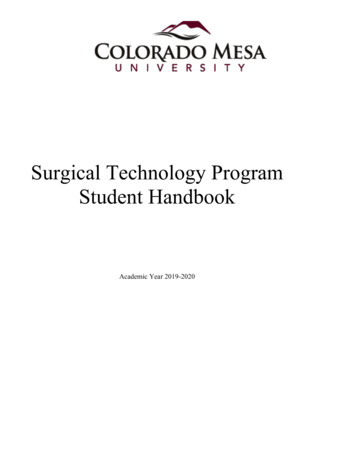
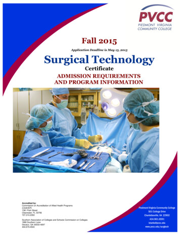
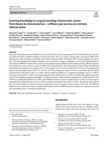
Updates in SurgeryTable 1 Demographic andclinical characteristics of thepatients affected by pancreaticductal adenocarcinoma(n 1407) in the three periodsanalysedParametersGenderMaleFemaleAge, (years)BMI (Kg/m2)*ComorbidityNoYesSymptomsNoYesBack painNoYesDiabetesNoYesWeight lossNoYesJaundiceNoYesStentNoYesCa 19.9 (U/mL) KPSTotalTotal N (%)Mean (SD)Group A2006–2010Group B2011–2015Group C2016–2020738 (52.5)669 (47.5)72.1 (11.5)24.2 (4.2)244 (55.3)197 (44.7)70.7 (10.9)23.9 (4.1)206 (49.9)207 (50.1)71.3 (11.7)24.3 (4.2)288 (52.1)265 (47.9)72.1 (11.7)24.4 (4.5)279 (19.8)1128 (80.2)108 (24.5)333 (75.5)62 (15)351 (85)109 (19.7)444 (80.3)509 (36.2)898 (63.8)158 (35.8)283 (64.2)141 (34.1)272 (65.9)210 (37.9)343 (62.1)1172 (83.3)235 (16.7)368 (83.5)73 (16.5)331 (80.2)82 (19.8)473 (85.5)80 (14.5)1071 (76.2)336 (23.8)323 (73.2)118 (26.8)312 (75.5)101 (24.5)436 (78.8)117 (21.2)968 (68.8)439 (31.2)304 (68.9)137 (31.1)287 (69.5)126 (30.5)377 (68.2)176 (31.8)777 (55.2)630 (44.8)232 (52.6)209 (47.4)219 (53)194 (47)326 (58.9)227 (41.1)864 (61.4)543 (38.6)500 (90–3142)77.5 (16.6)1407258 (58.5)183 (41.5)493 (91–3110)78.2 (16.7)441 (31.3)251 (60.7)162 (39.3)559 (83–3412)74.5 (17.9)413 (29.4)355 (64.2)198 (35.8)468 (92–2960)79.2 (14.9)553 (39.3)P 0.1780.931 0.0010.032The bold values represent the significant valuesSD Standard Deviation, BMI Body Mass Index, KPS Karnofsky Performance StatusTable 2 and Fig. 2 described the percentages of the imaging techniques used in the three periods analysed. A CT scan(from 89.4% in period A to 95.4% in period B to 95.1% inperiod C (P 0.001), EUS (from 11.1% in period A to 32.2%in period B to 49.4% in period C (P 0.001), FNA/FNB(from 39.0% in period A to 50.6% in period B to 57.7% inperiod C (P 0.001), FDG-PET/CT (from 9.7% in periodA to 19.4% in period B to 16.5% in period C (P 0.001)showed a significantly increasing rate over the time periods.On the contrary, CEUS (from 16.3% in period A to 12.3% inperiod B to 10.7% in period C (P 0.028) showed a significantly decreasing rate over the time periods.Table 3 reports the differences regarding preoperativestaging, type of management and overall survival. Figure 3shows only the differences regarding preoperative staging.The cTNM was significantly different in the three periods13(P 0.001). In particular, stage IA increased from period A(6.8%) to period C (11.6%).The MDTB evaluation increased significantly fromperiod A (32.6%) to period C (58.3%) (P 0.001). First,therapeutic decision making changed significantly overthe time periods (P 0.001). In particular, up-front surgery decreased from 39.2% in period A to 26.6% in periodC while the neoadjuvant approach increased from 6.4%in period A to 9.9% in period C. The resection rate ofthe entire sample was 25.6% with only slight differencesamong the three periods (28.2% in period A, 24.5% inperiod B and 24.4% in period C; P 0.339). R statuschanged significantly over the time (P 0.001). In particular in the last period, the R0 status decreased significantly respect to the two previous period (34.1% versus66.1 and 64.1%, respectively). Mini-invasive laparoscopicapproach increased significantly over the time from 0% of
Updates in SurgeryTable 2 Trends of the imagingtechniques in the three periodsanalysedImaging techniquesCT A/FNBNoYesTotalTotal N (%)Median (IQR)Group A2006–2010Group B2011–2015Group C2016–202093 (6.6)1314 (93.4)47 (10.6)394 (89.4)19 (4.6)394 (95.4)27 (4.9)526 (95.1)1196 (85.0)211 (15.0)372 (84.4)69 (15.6)335 (85.9)58 (14.1)469 (84.8)84 (15.2)952 (67.6)455 (32.4)392 (88.9)49 (11.1)280 (67.8)133 (32.2)280 (50.6)273 (49.4)1125 (87.1)182 (12.9)369 (83.7)72 (16.3)362 (87.7)51 (12.3)494 (89.3)59 (10.7)1193 (84.8)214 (15.2)398 (90.3)43 (9.7)333 (80.6)80 (19.4)462 (83.5)91 (16.5)707 (50.3)700 (49.7)1407269 (61)172 (39)441 (31.3)204 (49.4)209 (50.6)413 (29.4)234 (42.3)319 (57.7)553 (39.3)P value 0.0010.796 0.0010.028 0.001 0.001The bold values represent the significant valuesIQR interquartile range, CT Computed Tomography, MR Magnetic Resonance, EUS Endoscopic UltraSound, CEUS Contrast Enhanced UltraSound, FDG-PET/CT FluoroDeoxyglucose-Positron EmissionTomography/Computed Tomography, FNA/FNB Fine Needle Aspiration/BiopsyFig. 2 Changes in trends of theimaging techniques in the threeperiods analysed. CT ComputedTomography, FNA/FNB FineNeedle Aspiration/Biopsy, EUCEndoscopic ultrasound, FDGPET/CT Fluoro DeoxyGlucosePositron Emission Tomography/Computed Tomography, MRMagnetic Resonance, CEUSContrast Enhanced Ultrasoundthe period 2006–2010 to 11.9% of the period 2016–2020(P 0.001). Exploratory laparotomy decreased significantly from period A (12.0%) to the period C (4.7%)(P 0.001). Median overall survival was 8.3 months(3–19) and it did not significantly change over the timeperiods (period A 7 months, period B 7 months andperiod C 10 months; P 0.097). Table 4 reported thepostoperative outcomes after pancreatic resection forPDAC. Overall morbidity, 30-days mortality, 90-daysmortality, length of stay (LOS) were not significantly different in the three periods. CR-POPF resulted significantlydifferent over the time. In particular, CR-POPF increasedfrom period A to period C (P 0.001) (grade B, from 10.6to 22.7%; grade C, from 0 to 3.0%, respectively).13
Updates in SurgeryTable 3 The staging,management and survival ofpatients affected by pancreaticductal adenocarcinoma(n 1407) in the three lMultiplecTNM*IAIBIIAIIBIIIIVNCCN classificationResectableBorderlineLocally advancedMetastaticMDTB evaluationNoYesInitial therapeutic decision makingUp-front surgeryNeoadjuvantPalliative careSurgical resectionNoYesR statusR0R1R2Mini-invasive approachNoYesExploratory laparotomyNoYesOverall Survival (months)TotalTotal N (%)Median (IQR)Group A2006–2010Group B2011–2015Group C2016–2020P value852 (60.6)457 (32.5)98 (6.9)279 (63.3)134 (30.4)28 (6.3)238 (57.6)138 (33.4)37 (9)335 (60.5)185 (33.5)33 (6)115 (8.2)266 (18.9)103 (7.3)104 (7.4)291 (20.7)528 (37.5)30 (6.8)108 (24.5)39 (8.9)30 (6.8)65 (14.7)169 (38.3)21 (5.1)62 (15)29 (7)34 (8.2)108 (26.2)159 (38.5)64 (11.6)96 (17.4)35 (6.3)40 (7.2)118 (21.3)200 (36.2)520 (36.9)202 (14.4)157 (11.2)528 (37.5)161 (36.5)74 (16.8)37 (8.4)169 (38.3)149 (36.1)51 (12.4)54 (13)159 (38.5)210 (37.9)77 (13.9)66 (11.9)200 (36.3)787 (57.5)581 (42.5)297 (67.4)144(32.6)273 (66.1)134 (33.9)226 (41.7)316 (58.3)442 (31.4)107 (7.6)858 (61.0)173 (39.2)28 (6.4)240 (54.4)122 (29.5)24 (5.8)267 (64.7)147 (26.6)55 (9.9)351 (63.5)1047 (74.4)360 (25.6)317 (71.9)124 (28.2)312 (75.5)101 (24.5)418 (75.6)135 8)65(64.4)34(33.7)2(1.9)46(34.1)87(64.4)2(1.5) 19(88.1)16(11.9) 0.0011296 (92.1)111 (7.9)8.3 (3–19)1407388 (88)53 (12)7 (3–17)441 (31.3)381 (92.3)32 (7.7)7 (3–17)413 (29.4)527 (95.3)26 (4.7)10 (4–20)553 (39.3)0.251 0.001 0.001 0.0010.339 0.0010.097The bold values represent the significant valuesIQR interquartile range, NCCN National Comprehensive Cancer Network, MDTB MultiDisciplinaryTumour Board*cTNM according to the AJCC 8th edition [19]DiscussionIn the present study, a total of 1407 patients with PDACwere analysed, and were compared in relation to the threeperiods characterised by different models of care for pancreatic cancer in a tertiary referral centre during the period132006–2020. In the literature, there are only a few nationwide studies [10–18] which have analysed the changesin trends regarding PDAC over time. On the contrary, tothe Authors’ knowledge, the present study represents thefirst study which analysed the changes in trends regarding PDAC together with the associated models of carefor pancreatic cancer in a tertiary referral centre over a
Updates in SurgeryFig. 3 Incidence of cTNM staging of patients affected by pancreatic ductal adenocarcinomain the three period analysedTable 4 Postoperativeoutcomes after pancreaticresection for pancreatic ductaladenocarcinoma over the timePostoperative dataMorbidity according toCDC classificationNoGrade IGrade IIGrade IIIa–IIIbGrade Iva–IVbGrade VCR-POPFNot a risk *No/BLBCLOS (in day)30-days mortalityNoYes90-days mortalityNoYesTotalN (%) orMedian (IQR)Group A2006–2010Group B2011–2015Group C2016–2020P value153 (42.5)60 (16.7)73 (20.3)41 (11.4)22 (6.1)11 (3.1)53 (42.7)21 (16.9)24 (19.4)15 (12.1)10 (8.1)1 (0.8)40 (39.6)18 (17.8)19 (18.8)12 (11.9)7 (6.9)5 (5)60 (44.4)21 (15.6)30 (22.2)14 (10.4)5 (3.7)5 (3.7)0.75353 (14.8)243 (67.5)57 (15.9)7 (2)12 (10–19)0 (0.0)111 (89.4)13 (10.6)0 (0.0)13 (10–18)24 (23.5)67 (65.9)9 (9.4)1 (1.2)12 (10–19)23 (16.7)78 (57.6)30 (22.7)4 (3.0)12 (9–20) 0.001350 (97.2)11 (3.1)123 (99.2)1 (0.8)96 (95.1)5 (4.9)130 (96.3)5 (3.7)0.171345 (95.8)15 (4.2)360121 (97.6)3 (2.4)124(34.4)95 (94)6 (5.9)101(28.1)129 (96.6) 6 (4.4)0.4130.968135(37.5)The bold value represents the significant valueIQR InterQuartile Range, CDC Clavien Dindo Classification, CR-POPF Clinically Relevant PostOperativePancreatic Fistula, * total pancreatectomy, BL Biochemical Leak, LOS Length of Stayfifteen-year time period. However, there is a recent studyreporting on 3000 pancreaticoduodenectomy performedat a single centre over 20 years that provided time-relatedchanges in management of pancreatic cancer [21].Many differences were found among the three periodsin which different models of care were adopted. The epidemiological and clinical characteristics of the population,the incidence rate, the BMI, the comorbidity rate and KFSwere significantly different in each of the time periods. Theincreasing incidence of PDAC has been reported by onlya few nationwide studies [10–18]. The data from the Danish Cancer Registry [13], covering a sixty-one year periodfrom 1943 to 2003, reported a relatively steady incidence ofPDAC. On the contrary, the Dutch population-based study[10], covering only a twenty-year period from 1997 to 2016,found an ever-increasing incidence of PDAC from 12.1to15.3 per 100,000 individuals, and the annual incidence ofpancreatic cancer in Finland increased from 19 per 100,000in 2000 to 24 per 100,000 in 2016 [21]. The present studyshowed an ever-increasing number of patients affected13
Updates in Surgeryby PDAC from the period 2006–2010 (31.3%) to periodC (39.3%) (P 0.032). This finding indicated that betterdetection (in particular of, small lesions) and classificationof tumours by means of improved diagnostic procedures waspossible. Secondly, the major number of patients affected bypancreatic cancer in a tertiary referral centre could be theresult of a centralisation policy. The body mass index, thecomorbidity rate and KFS showed a decreasing incidencein periods B and C with respect to the period A. This indicated that, when the tumour was diagnosed in periods B andC, the patients were in better general health with respectto period A, probably because the symptoms related to thetumour were recognized early and an early diagnosis waspossible. In addition, this finding could be related to the factthat a majority of the patients were fit for pancreatic resection. Finally, the proportion of patients receiving a biliarystent remained stable over the time. Indeed, our preferredapproach to palliation of biliary obstruction is endoscopicstenting.Regarding the imaging techniques, the present studyshowed that, the trends of all the diagnostic procedures,except for MR, changed over the time periods. First, a CTscan was the most frequent procedure performed over thetime periods (more than 90% of the patients underwent a CTscan) and its incidence significantly increased from periodA (89.4%) to period C (95.1%) (P 0.001). These data indicated that a CT scan represented the best imaging techniquefor the diagnosis and staging of the disease, in particularowing to its reliability in assessing the involvement of thepancreatic vessels due to the pancreatic cancer. Nevertheless, of all the diagnostic procedures, EUS was the imagingtechnique which was found to be the most increased overthe time periods (from 11.1% in period A to 49.4% in periodC, P 0.001), even if it remained a second level diagnostictool as shown by its low incidence rate (range 10–50%). Theincreasing use of EUS was probably related to its usefulnessin detecting small lesions better than a CT scan and in performing FNA/FNB (these procedures increased from 39.0%in period A to 57.7% in period C; P 0.001). An FDG-PET/CT scan was performed in a few cases (approximately 10%of cases), confirming its value as a third level diagnostic procedure. However, its use significantly increased from 9.7%in period A to 16.5% in period C. This finding could bejustified by the ever-increasing use of neoadjuvant therapyand by the need to compare the morphological and functional findings of the disease before and after the neoadjuvant therapy with the aim of properly defining the surgicalindication. Finally, the use of CEUS decreased significantlyfrom 15.6% in period A to 10.7% in period C (P 0.028),probably because its usefulness was limited to only thoseselected cases in which there was a “CT scan doubt” regarding distant metastases to the liver.13Regarding staging, it should be noted that, in the totalsample, 34.4% (stages IA, IB, IIA) were limited to the pancreas, 28.1% (Stages IIB, III) presented lymph node involvement and 37.5% (Stage IV) were metastatic. Over the timeperiods, a significant increase in early stages (stage 1A from6.8% in period A to 11.6% in period C) was noted, probablyrelated to better knowledge of the disease and improvementin diagnostic tools, capable of recognising small tumours.Regarding the treatment of pancreatic cancer, throughoutthe entire study period, the majority of patients received onlypalliative treatment (61.0%), including mainly chemo- andradiotherapy, with an increasing incidence of palliative careover the time periods. This finding indicated that pancreatic cancer was most often diagnosed at an advanced stage,partially due to late presenting symptoms, and it has poorbiological behaviour which could explain the negligibleimprovement in overall survival in the entire population. Itis to underline that chemo and radiotherapy changed overthe time. In particular, in the period 2006–2010, monochemotherapy was used (gemcitabine alone). Subsequently,polichemotherapy schedules as well as gemcitabine plusNAB-Paclitaxel, gemcitabine plus capecitabine and, finally,modified FOLFIRINOX (Leucovorin, 5-Fluorouracil, Irinotecan and Oxaliplatin) were adopted.The use of up-front surgery, as a first therapeuticapproach, decreased significantly over the time periods(from 39.2% in period A to 26.6% in period C; P 0.001),possibly indicating (1) better preoperative evaluation andimproved detection of non-resectable disease and (2)changes in trends of early therapeutic decision makingwith a decreasing rate of up-front surgery, probably infavour of neoadjuvant treatment which increased over thetime periods from 6.4% in period A to 9.9% in periodC. This finding confirmed the results reported by somenationwide studies [22, 23], suggesting that neoadjuvanttreatment for pancreatic adenocarcinoma is only utilised ina minority of cases; however, its use appears to be increasing over time in both borderline and resectable tumours ashas been recommended by various guidelines [2, 20]. Theincreasing use of neoadjuvant chemotherapy seems to berelated with the major effectiveness of the new chemotherapeutic schedules, in particular those including morethan one chemotherapic agent. R status changed over thetime. In particular R0 status decreased from the first period(66.1%) to the last period (34.1%) and, at the same time,the R1 status increased from 33.1 to 64.4%. These dataseem to be related with a more accurate study of the specimen by pathologists because surgical technique improvedover the time. Mini-invasive approach increased over thetime from 0 to 11.9%. This datum is clearly related tothe learning curve of each surgeon. Exploratory laparotomy significantly decreased from 12.0% in period A to4.7% in period C as, in recent years, the indications for
Updates in Surgerysurgery have been more carefully evaluated regarding bothimprovements in diagnostic procedures and the importantsupport of the MDTB which includes radiologists, gastroenterologists, and oncologists with expertise in thepancreatic field. Finally, the MDTB evaluation of patientsaffected by PDAC increased significantly over the timeperiods from 32.6% in period A to 58.3% in period C. Thisfinding indicated that the MDTB represented an essentialinstrument of effective cancer policy which allowed providing the highest quality care the patients needed anddeserved. The MDTB could be considered to be the corecomponent in organising cancer care and setting down theelements for guiding changes in trends over time. Postoperative outcomes were similar in the three periods. Theincreasing rate of CR-POPF could be related with a moreaccurate definition of a pancreatic fistula over the time.Even with all these changes in trends regarding pancreaticcancer, patient survival has not improved significantly inthe past 15 years, even if the slight improvement in survival could be said to represent a trend (P 0.097). However, a survival benefit of three months between period A(7 months)and period C (10 months) was not negligibleas the majority of patients only received palliativecare,confirming not only the delayed diagnosis of the diseaseand its poor biological behaviour, but also the increasingeffectiveness of the chemotherapeutic agents.The present study had some limitations, namely the retrospective design from large databases which could potentiallylead to coding error. In addition, the results of the study mayhave been affected by the different effectiveness of the imaging techniques over the time periods, by the improvementin pancreatic surgery, and the efficacy of chemo- and radiotherapy. The main strength of the study was the analysis ofall patients affected by PDAC observed at S.Orsola-MalpighiHospital, a tertiary referral centre.In conclusion, despite its limitations, the present studyshowed the evolving knowledge in surgical oncology of pancreatic cancer at a tertiary referral centre over the time. Theincreasing incidence, the greater use of EUS and FNA/FNB,a decreasing rate of exploratory laparotomy and, finally, animportant modification in management in favour of neoadjuvant treatments should be noted. All these changes wererelated to improvement in the diagnostics and therapeutictools used to manage pancreatic cancer, and
pancreatic cancer in Denmark from 1943 to 2003: a nationwide study. Pancreas 38:374-378 14. Søreide K, Aagnes B, Møller B et al (2010) Epidemiology of pan-creatic cancer in Norway: trends in incidence, basis of diagnosis and survival 1965-2007. Scand J Gastroenterol 45:82-92 15. Zhou J, Enewold L, Stojadinovic A et al (2010) Incidence .