Transcription
This material is protected by U.S. copyright law. Unauthorized reproduction is prohibited. To purchase quantity reprints,please e-mail reprints@ons.org or to request permission to reproduce multiple copies, please e-mail pubpermissions@ons.org.Demystifying Lymphedema:Development of the LymphedemaPutting Evidence Into Practice CardEllen Poage, MSN, FNP-C, MPH, CLT-LANA,Marybeth Singer, MS, APN-BC, AOCN , ACHPN, Jane Armer, PhD, RN, FAAN,Melanie Poundall, RN, and M. Jeanne Shellabarger, RN, MSNCancer treatment is the leading cause of lymphedema in developed countries. Development and severity of lymphedemahave a significant impact on comfort, psychological distress, and overall quality of life. Incidence statistics have rangedfrom 5%–60%, with onset of symptoms ranging from immediately after treatment to 30 years after treatment. Oncologynurses caring for patients throughout the cancer trajectory have a critical role to play in early assessment of risk, promptidentification of lymphedema, and implementation of evidence-based, individualized treatment plans in collaboration withtherapists. As part of an Oncology Nursing Society (ONS) project team, the authors of this article undertook a review ofcurrent literature to identify effective interventions for the treatment of secondary lymphedema. Following the guidelinesestablished by the ONS Evidence-Based Practice Resource Team, the authors evaluated current clinical practice guidelines,systematic reviews, and research studies conducted since 1998. The team reviewed and synthesized the literature anddeveloped evidence tables and a Putting Evidence Into Practice (PEP) card. The data were reviewed by experts in thefield of lymphedema management. The lymphedema ONS PEP card, a user-friendly, succinct summary of interventions, wasreleased at the 33rd Annual ONS Congress in May 2008.Lymphedema is caused by a disruption or malformationof the lymphatic system that results in high-proteinswelling of the affected body part. Although it maybe acute or chronic, transient or progressive, lymphedema often is seen clinically as a chronic, progressive condition starting with seemingly innocuous superficialswelling that waxes and wanes. If left untreated, lymphedemamay evolve into a permanent, disfiguring condition which ismanageable but no longer reversible or curable.Primary lymphedema has no known acquired causes and develops from an insufficiency in structure and/or function of thelymphatic system. The insufficiency is characterized by a failureof the lymph system to keep up with the lymph load demandsof the affected body part. Secondary lymphedema is more common and, in developed countries, often is caused by surgicalremoval of lymph nodes or the use of radiation on lymph nodesduring breast cancer treatment. The staging and treatment ofcancers of the head and neck, ovaries, vulva, prostate, and anyother cancer that may involve removal and/or irradiation oflymph nodes for managing disease also can trigger secondarylymphedema. Lymphedema is the result of hydrophilic proteincongestion of the interstitial spaces in the tissues of the limb(s)or trunk (Mortimer, 1998), causing swelling of the affectedarea (see Figure 1). Primary and secondary lymphedema can betreated with similar approaches (Foldi, 1998).At a GlanceF Although lymphedema may be a prevalent, debilitatingoutcome of cancer therapy, knowledge of lymphedema, itstreatment, and how to reduce risk is increasingly available.F Evidence supports early lymphedema diagnosis and referralfor therapies to reduce patient burden.F A ready-to-use synthesis of evidence-based information assistsnurses in answering patient questions about lymphedema.Ellen Poage, MSN, FNP-C, MPH, CLT-LANA, is a certified lymphedematherapist and nurse practitioner at the Rehabilitation Associates ofNaples in Fort Myers, FL; Marybeth Singer, MS, APN-BC, AOCN , ACHPN,is a nurse practitioner at the Massachusetts General Hospital GilletteCenter for Breast Cancer in Boston; Jane Armer, PhD, RN, FAAN, is aprofessor in the Sinclair School of Nursing at the University of Missouriand director of nursing research at the Ellis Fischel Cancer Center, both inColumbia; at the time this article was written, Melanie Poundall, RN, wasa staff nurse in the infusion unit at the Massachusetts General HospitalGillette Center for Breast Cancer; and M. Jeanne Shellabarger, RN, MSN,is a coordinator of the breast clinic at the Ellis Fischel Cancer Center. Nofinancial relationships to disclose. (Submitted June 2008. Accepted forpublication August 24, 2008.)Clinical Journal of Oncology Nursing Volume 12, Number 6 Demystifying LymphedemaDigital Object Identifier: 10.1188/08.CJON.951-964951
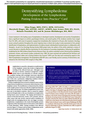
Lymphedema may cause debilitating, distressing, and disfiguring changes (Armer, Radina, Porock, & Culbertson, 2003; Foldi,1998; Ridner, 2002) at every stage (see Table 1). Early recognition and treatment of lymphedema provide optimal outcomesand may alleviate or minimize the physical and emotionalburden of the condition. More than 2.47 million breast cancersurvivors living in the United States (Ries et al., 2007) are at riskof developing lymphedema after treatment. The occurrence oflymphedema after breast cancer treatment has been estimatedto be 5%–60%, with some onset as late as 30 years after treatment (Armer & Stewart, 2005; Petrek & Heelan, 1998). If theestimate of lymphedema incidence is set at 25%, more than600,000 breast cancer survivors would be affected.Most of the research to date has focused on upper-extremitylymphedema following breast cancer treatment. Relatively fewresearch-supported interventions exist for lymphedema in otherareas of the body. Despite the pervasive risk of lymphedemafollowing treatment, research is lacking to support many ofthe clinical recommendations proposed to reduce the risk oflymphedema or prevent its progression to more advanced andpermanent stages (Ridner, 2002).Lymphedema may initially be dismissed as swelling, discomfort, and inflammation after surgery. Other early indications oflymphedema include self-reported sensations of heaviness, swelling, tingling, fatigue, or aching (Armer et al., 2003). In addition,axillary paresthesia and pain in the breast, chest, and arm havebeen reported as symptoms of lymphedema (Bani et al., 2007),Table 1. Stages of LymphedemaStageDescription0Subclinical or pre-lymphedema. Typically includes all patientswho have had lymph node dissection. Swelling is not evident,although impaired lymph flow is present. The stage may lastfor a long time.IAccumulation of fluid and protein in tissue is present. Elevationmay influence the limb; pitting may be present.IIIncludes swelling that does not reduce with elevation; pittingis present with fibrosis.IIIFibrotic tissue has indiscernible pitting; includes skin thickeningand large limb volume known as elephantiasis, a morbid condition where lymphstasis and chronic inflammation develop intofibrosclerosis and additional tissue swelling (Foldi, 1998).Note. From “The Diagnosis and Treatment of Lymphoedema. ConsensusDocument of the International Society of Lymphology,” by the International Society of Lymphology, 2003, Lymphology, 36(2), pp. 85–86.Copyright 2003 by the International Society of Lymphology. Adaptedwith permission.although they also may be associated with other after-treatmenteffects. Given the common and seemingly transient nature ofearly-onset lymphedema, patients and providers often ignore theearly warning signs and deny their significance.This article was developed out of a need to have nurses takenotice of complaints often regarded as minor and to offer appropriate evidence-based interventions for reducing risk of andtreating lymphedema.In a study of 263 patients with lymphedema, Jeffs (2006)found that 156 (59%) patients developed symptoms within thefirst year after surgery, but only 92 (35%) of the patients soughtassistance from a specialist within three months of symptomonset. Twenty-nine patients (17%) delayed treatment longer than12 months from onset because of a lack of awareness of theircondition or therapy options. Increasing awareness and accessto trained therapists have the potential to reverse the deleterious effects of delayed treatment.The authors of this article critically reviewed the literature toidentify and evaluate evidence-based interventions for cancerrelated secondary lymphedema. The goal was to accuratelydescribe lymphedema, its myriad consequences, and evidencebased interventions so oncology nurses can confidently identifypatients who are at risk for or who are experiencing early-stagelymphedema and recommend prompt and effective interventions.MethodsFigure 1. Legs of Caucasian Male With Right LegAffected by LymphedemaNote. Copyright by Bart’s Medical Library/Phototake. All rights reserved.Used with permission.952The Oncology Nursing Society’s (ONS’s) Lymphedema PuttingEvidence Into Practice (PEP) team consisted of two advancedpractice nurses, two staff nurses, and a nurse researcher. Theteam undertook a thorough review of current literature toidentify effective interventions for the treatment of secondarylymphedema. The evidence-based review consisted of evaluationof current clinical practice guidelines, systematic reviews, andresearch studies reported from 1997–2007. Search engines usedDecember 2008 Volume 12, Number 6 Clinical Journal of Oncology Nursing
included MEDLINE , the National Library of Medicine’s database,CINAHL , CancerLit, and the EBM-Cochrane database. Some 218articles were extracted, reviewed, and categorized with the ONSWeight of the Evidence classification system (see Table 2).Detailed evidence tables were created, reviewed, and weightedfollowing PEP team reviews and conference calls from August toNovember 2007. An ONS PEP card was developed based on thisreview, externally reviewed by experts in the field of lymphedemamanagement, revised, and finalized in May 2008. A detailed listof defined terms is available at www.ons.org/outcomes/volume4/anxiety.shtml (see Table 3).Many studies investigating interventions for the treatment oflymphedema are limited by small sample size, lack of controlgroups, and limited follow-ups. The rigors of current therapies alsomay pose considerable challenges for patients and clinicians alikeas they are dependent on access to lymphedema therapists.Effective Interventions for Reducingthe Risk for and Promoting Treatmentof Secondary LymphedemaRecommended for PracticeComplete decongestive therapy: Complete decongestive therapy (CDT), also recognized in the literature as complexdecongestive physiotherapy and complex physical therapy, is therecommended treatment for lymphedema. CDT combines multiple modalities with the purpose of achieving the maximum possible swelling reduction in a limb or affected body area. Severalstudies support the use of CDT for the treatment of lymphedema(Browning, 1997; Moseley, Carati, & Piller, 2007). In addition,rigorously developed practice guidelines support the use of CDT(Lymphoedema Framework, 2006; National Lymphedema Network Medical Advisory Committee, [NLN MAC], 2006).CDT is a two-phase therapy which initially includes an intensive phase when the limb volume is reduced during treatment bya therapist (NLN MAC, 2006) and a maintenance phase when thepatient is instructed in self-management.The intensive phase is comprised of five components or modalities: manual lymph drainage (MLD); compression appliedthrough short-stretch compression bandages and compressiongarments; meticulous skin and nail care; remedial exercise; andeducation in self-care. The maintenance phase consists of simplelymphatic drainage, nightly compression bandaging (CB), daytime use of compression garments, skin care, and exercise.CDT reduces congested interstitial lymphatic fluid and excesslimb volume and has been shown to improve shoulder range ofmotion (Didem, Ufuk, Serdar, & Zumre, 2005; Szuba, Achalu, &Rockson, 2002) and decrease pain (Hamner & Fleming, 2007;Moseley et al., 2007). Hamner and Fleming reported that 76 of135 patients (56%) had pain associated with their lymphedema.After CDT, 56 of the 76 patients (74%) were pain free. However, the study was retrospective and did not distinguish stageTable 2. Putting Evidence Into Practice Weight-of-Evidence Classification SchemaDescriptionExamplesRecommended for practiceEffectiveness is demonstrated by strong evidence from rigorously designed studies, metaanalyses, or systematic reviews. Expectedbenefit exceeds expected harms.At least two multisite, well-conducted, randomized, controlledtrials (RCTs) with at least 100 subjectsPanel of expert recommendation derived from explicit literaturesearch strategy; includes thorough analysis, quality rating,and synthesis of evidenceLikely to be effectiveEffectiveness has been demonstrated by supportive evidence from a single rigorously conducted controlled trial, consistent supportiveevidence from well-designed controlled trialsusing small samples, or guidelines developedfrom evidence and supported by expert opinion.One well-conducted RCT with fewer than 100 patients or atone or more study sitesGuidelines developed by consensus or expert opinion withoutsynthesis or quality ratingBenefits balanced with harmsClinicians and patients should weigh thebeneficial and harmful effects according toindividual circumstances and priorities.RCTs, meta-analyses, or systematic reviews with documentedadverse effects in certain populationsEffectiveness not establishedData currently are insufficient or are of inadequate quality.Well-conducted case control study or poorly controlled RCTConflicting evidence or statistically insignificant resultsEffectiveness unlikelyLack of effectiveness is less well establishedthan those listed under not recommended forpractice.Single RCT with at least 100 subjects that showed no benefitNo benefit and unacceptable toxicities found in observationalor experimental studiesNot recommended for practiceIneffectiveness or harm clearly is demonstrated, or cost or burden exceeds potentialbenefit.No benefit or excess costs or burden from at least two multisite, well-conducted RCTs with at least 100 subjectsDiscouraged by expert recommendation derived from explicitliterature search strategy; includes thorough analysis, qualityrating, and synthesis of evidenceWeight-of-Evidence CategoryNote. Based on information from Mitchell & Friese, n.d.Clinical Journal of Oncology Nursing Volume 12, Number 6 Demystifying Lymphedema953
Table 3. Definitions of Lymphedema TermsTermDefinitionBioimpedenceMeasures tissue resistance to an electrical current to determine extracellular fluid volumeBody mass index (BMI)A number calculated from a person’s weight and height. BMI provides a reliable indicator of fat content for mostpeople and is used to screen for weight categories that may lead to health issues. Elevated BMI may affect risk fordevelopment for lymphedema following cancer treatment and impact progression and management of lymphedema(Centers for Disease Control and Prevention, 2008).CellulitisAn infection that spreads freely, quickly, and uncontrollably within the deeper tissues of the skin. Cellulitis becomes a lifethreatening emergency when it spreads through the lymphatic or circulatory systems and can reach vital organs and otherbody parts (lymphangitis), requiring prompt treatment with antibiotics. Cellulitis usually is caused by the bacteria Staphylococcus aureus (Lymphatic Research Foundation, 2006).Complete decongestivetherapy (CDT)The system of lymphedema treatment that includes manual lymph drainage (MLD), compression techniques, exercise,skin care, and self-care training (Lymphatic Research Foundation, 2006). CDT is comprised of an initial reductive phase(phase I), followed by an ongoing, individualized maintenance phase (phase II). Components include MLD; multilayer,short-stretch compression bandaging (CB); remedial exercise; skin care; education in self-management; and elasticcompression garments (National Lymphedema Network Medical Advisory Committee [NLN MAC], 2006).Compression garmentA knit, two-way stretch sleeve or stocking that is worn to assist in controlling swelling and to aid in moving lymphfrom the affected area. A compression garment is worn only while the patient is awake and active (Lymphatic ResearchFoundation, 2006). The garment should be individualized for each patient.ErysipelasA painful skin infection that affects the skin plus the subcutaneous tissues and lymphatic structures that are locatedjust under the skin (cellulitis affects the deeper tissues). Erysipelas also requires prompt treatment with antibiotics andis caused by streptococci bacteria. Erysipelas rapidly invades and spreads through the lymphatic vessels, damaging thelymph vessels and increasing the formation of fibrosis in the affected tissues. Erysipelas is one of the most commoncomplications of lymphedema and tends to recur; correlated with stage of lymphedema (Lymphatic Research Foundation, 2006).Exercise (low intensity)Although activity and exercise may temporarily increase fluid load, appropriate exercises may enable the patient withlymphedema to resume exercise and activity while minimizing the risk of swelling exacerbation. Compression garmentsor CB must be used during exercise to counterbalance the excessive formation and stasis of interstitial fluid (NLN MAC,2006). Exercise plans must be individualized for each patient. See NLN MAC (2008b) guidelines for specific suggestions. Lymphedema exercises (decongestive) are a standard and integral part of phase I and phase II CDT programs forindividuals with lymphedema (NLN MAC, 2006).Infrared perometryPerometry using infrared light beams to measure the outline of the limb, which then can be used to estimate limb volumeLymphangitisA potentially life-threatening bacterial infection involving the lymphatic vessels that may spread to the bloodstream;sometimes associated with cellulitis (Lymphatic Research Foundation, 2006).LymphedemaA progressive, chronic condition that may appear as swelling of one or more limbs and may include the correspondingquadrant of the trunk. Swelling also may affect other areas, such as the head and neck, breast, or genitalia. Swellingoccurs from an accumulation of fluid and other elements (e.g., subcutaneous fat, protein) in the tissue spaces becauseof an imbalance between interstitial fluid production and transport (usually low output failure) (International Society ofLymphology, 2003). Lymphedema arises from congenital malformation of the lymphatic system or damage to lymphaticvessels and/or lymph nodes (Lymphoedema Framework, 2006). The leading cause of lymphedema in the United Statesis cancer and its treatment (NLN MAC, 2006).MLDA treatment technique that uses a series of rhythmic, light strokes to reduce swelling and improve the return of lymph tothe circulatory system (Lymphatic Research Foundation, 2006). The technique encourages fluid away from congested areasby increasing activity of normal lymphatics and bypassing ineffective or obliterated lymph vessels. MLD is widely advocated,but little research data conclusively support its use (Badger, Preston, Seers, & Mortimer, 2004; McNeely et al., 2004; Williams, Vadgama, Franks, & Mortimer, 2002; Woods, 2003). The most appropriate techniques, optimal frequency, and indications for MLD, as well as the benefits of treatment, have not been clarified. MLD is a specialist’s skill that requires regularpractice to maintain competence. Deep, heavy-handed massage should be avoided because it may damage tissues andexacerbate edema by increasing capillary filtration (Lymphoedema Framework, 2006).Multilayer or CBA specialized form of compression used in the treatment of lymphedema. Bandages are the most effective and flexibleform of compression, particularly in the early stages of treatment. Bandages provide proper compression when thepatient is active or when the patient is resting. They can also be easily adjusted to fit changing limb size and compression needs (Lymphatic Research Foundation, 2006). Multiple layers of short-stretch bandages are applied to the(Continued on next page)954December 2008 Volume 12, Number 6 Clinical Journal of Oncology Nursing
Table 3. Definitions of Lymphedema Terms (Continued)TermDefinitionMultilayer or CB(continued)lymphedematous area(s). Short-stretch bandages have limited extensibility under tension (50%), in contrast to Ace (Becton, Dickinson and Co.) bandages (300%). To achieve an effective compression gradient, bandages must be strategicallyapplied with low to moderate tension using more layers in the distal portions of the affected limb(s). Pressure within theshort-stretch bandages is low when the patient is inactive (resting pressure). Muscle contractions increase interstitial pressure (working pressure) as muscles expand within the limited volume of the semi-rigid bandages. Interstitial cycling between low-resting and high-working pressures creates an internal pump that encourages movement of congested lymphalong the distal to proximal gradient created by the bandaging. The non-elastic bandage sheath also counters refilling offluid and reduces tissue fibrosis, adding more volume reduction (Lymphoedema Framework, 2006; NLN MAC, 2006).Pneumatic compressionpumpThis is the basic component of intermittent pneumatic compression, a widely used technique where an electrical aircompression pump is attached to an inflatable plastic garment that is placed over the affected limb. The garment isinflated and deflated cyclically for a set period, usually about 30–120 minutes. The pressure produced by the garmentcan be varied. Garments may be single chambered or contain multiple chambers (usually 3, 5, or 10) that are inflatedsequentially to provide a peristaltic massaging effect along the length of the limb (Lymphoedema Framework, 2006).Simple lymphatic drainageSimplified self-massaging techniques that normally take about 20 minutes and are done daily. Physical therapist orother specialist also can teach a partner, friend, or relative how to perform the massage (Activa Health Care, n.d.). Thetechnique incorporates simplified hand movements in a set sequence, which work across lymphatic watersheds towardfunctioning lymphatics. Treatment is mainly to the neck and trunk area, although the limb may be treated dependingon patient need and the condition of the limb. No oils or creams are used (British Lymphology Society, 1999).Note. Based on information from Oncology Nursing Society, 2008.of lymphedema, timing of referral, or factors that may maketherapy less effective or pain more significant. Moseley et al.reviewed three studies that reported subjective symptom improvement after CDT, but none reported long-term follow-up. Inall, Moseley et al. reviewed nine studies reporting lymphedemaimprovement after about one month of CDT.Positive therapeutic outcomes have routinely been documented when trained therapists provided CDT (LymphoedemaFramework, 2006; McNeely et al., 2004). Experts typically recommend comprehensive CDT when lymphedema is moderate tosevere (Jeffs, 2006; Koul et al., 2007; Lymphoedema Framework;Moseley et al., 2007). When lymphedema is detected early orsymptoms are mild, CDT may be modified to exclude one or twoof the five modalities (Koul et al.; Lymphoedema Framework;Moseley et al.). Modifying CDT also may be appropriate whena patient is unwilling or unable to participate in comprehensiveCDT (Jeffs). The lymphedema therapist will be knowledgeable inways to appropriately modify the therapy (NLN MAC, 2006).Early treatment of lymphedema with CDT is less costly andburdensome to the patient and yields far better outcomes (Hamner & Flemming, 2007; Jeffs, 2006; McNeely et al., 2004; Moseley et al., 2007). In one well-designed, randomized, controlledtrial by McNeely et al., when MLD with CB was compared to CBalone, a significantly larger relative reduction in arm volume wasseen in patients with mild lymphedema versus chronic lymphedema, regardless of therapeutic intervention. Because mostresearch has focused on breast cancer–related lymphedema,additional research is needed to assess the benefits of CDT fordifferent oncology populations and to tailor the therapy to theirunique needs.Compression bandaging: CB is a systematic applicationof short-stretch bandages with various types of padding. Wrapsare applied with moderate tension at the distal portions of theaffected limb(s), gradually decreasing to low tension in the moreproximal portions. CB is distinguished from elastic bandagingbecause of the relative inelasticity of the special bandaging material used in CDT. The special bandages are known as short- orlow-stretch versus the high-stretch material of the more familiarelastic bandages. This type of bandaging is physiologically correct for the purpose of reducing volume of lymphedematouslimbs and is less likely to cause injury from excessive pressure.CB is used 24 hours per day during intensive therapy. Bandagesare used nightly in combination with a daytime compressiongarment during maintenance therapy.Research supports the use of CB alone to reduce swelling (Lymphoedema Framework, 2006). In a study by Jeffs (2006), patientswho had received CB and MLD achieved a 40% reduction, whereaspatients who had only CB achieved a 25% reduction.In a well-designed, prospective, randomized, controlled trialby McNeely et al. (2004), patients who had four weeks of CB,with (n 21) or without MLD (n 24), experienced a significantreduction in limb volume. A major finding was the significantlylarger reduction in the MLD with CB group for subjects withmild lymphedema (n 7) compared to subjects in all other subgroups (n 38). In addition, the study found that the greatestbenefits occurred after two weeks of daily treatment, supporting the use of CB as an intervention for lymphedema, potentiallyconserving time, energy, and resources. The few studies thatexamined CB with and without MLD validate the contributionof CB to managing lymphedema and provide evidence of valueadded with MLD.In a prospective cohort by Vignes, Porcher, Arrault, andDupuy (2007) (N 537), nonadherence with low-stretch bandaging and elastic sleeves were risk factors for progression oflymphedema after one year of maintenance treatment. Continued use of low-stretch self-bandages allowed additional volumereduction during maintenance therapy, compared to no use ofbandages. Unfortunately, patients often resist bandaging andthe wearing of elastic compression garments. Bani et al. (2007)provided patients (N 742) with information about compressionClinical Journal of Oncology Nursing Volume 12, Number 6 Demystifying Lymphedema955
sleeves or garments, but use did not increase. Adherence witheffective self-care interventions is an area ripe for additionalresearch to help improve outcomes and quality of life in cancersurvivors.Infection treatment: Patients with lymphedema are atincreased risk for infection. Cellulitis, an acute infection of theskin and underlying tissue that is characterized by painful swelling, erythema, and heat, often is caused by normal skin flora entering through a break in the skin (Braunwald et al., 2001). Themost common cause of infection in the lymphedematous limbis group A hemolytic streptococcus bacterium or Streptococcuspyogenes, although the emergence of Staphylococcus aureusco-infection, particularly with concerns for methicillin-resistantS. aureus, and co-infection with numerous other organisms,must be considered in at-risk populations (Bernard, 2008; Lymphoedema Framework, 2006). Erysipelas is an acute superficialnon-necrotizing dermal infection caused by S. pyogenes that ischaracterized by rapid onset of fiery red edema of the affectedextremity with well-defined indurated borders (Bernard; Braunwald et al.). Despite effective treatment, generally with penicillin-based therapies, swelling may persist. Immediate attention tosigns of infection and prompt initiation of antibiotic therapy arecritical to preventing sepsis. Antibiotic coverage should includecoverage for strep and staph species. Careful history of traumaand injury is important, although, in many cases, no injury isapparent. The presence of comorbidities, age, neutropenia, andallergies will determine antibiotic choices.Antibiotics for first-line treatment include penicillin-basedtherapies (if no history of allergy exists), either orally (if no signsof systemic infection are seen) or by IV. Oral penicillins, such asamoxicillin and dicloxacillin, often are used and continued for aperiod of no less than 14 days or until inflammation has resolved.For patients with penicillin allergy, clindamycin or clarithromycin may be used (Bernard, 2008; Lymphoedema Framework,2006). Consult with infectious disease colleagues regardingantibiotic choices, particularly in the case of recurrent infection.One of the most common errors made when treating an infectionin the lymphedematous limb is too short of a treatment course. Atleast a 14-day course of antibiotic therapy after an acute episodehas responded clinically is recommended (Lymphoedema Framework); it may take one to two months of therapy for symptoms tocompletely resolve in some patients.Antibiotics should begin as soon as possible. Criteria for hospital admission include presence of Fevers, hypotension, tachycardia, confusion, or vomiting Continuing symptoms despite oral antibiotics for 48 hours Unresolved local symptoms despite the use of first- andsecond-line oral antibiotics.Simple lymph drainage and MLD should be avoided duringacute infection with fever. If tolerated, reduced-tension compression garments or CB may be applied. Prolonged periodswithout compression should be avoided (Lymphoedema Framework, 2006), and Bernard (2008) suggested aggressively initiating treatment after infection to decrease edema. Recurrentinfections occur in up to 20% of patients (Bernard); the mostf
groups, and limited follow-ups. The rigors of current therapies also may pose considerable challenges for patients and clinicians alike as they are dependent on access to lymphedema therapists. effective interventions for reducing the risk for and promoting Treatment of Secondary Lymphedema. recommended for practice