Transcription
(2021) 19:335Zhao et al. J 1080-3Journal of NanobiotechnologyOpen AccessREVIEWRecent advances in selective photothermaltherapy of tumorLiping Zhao1†, Xu Zhang2†, Xiaoxia Wang1, Xiuwen Guan1,3,4, Weifeng Zhang1,3,4* and Jinlong Ma1,3,4*AbstractPhotothermal therapy (PTT), which converts light energy to heat energy, has become a new research hotspot incancer treatment. Although researchers have investigated various ways to improve the efficiency of tumor heat ablation to treat cancer, PTT may cause severe damage to normal tissue due to the systemic distribution of photothermalagents (PTAs) in the body and inaccurate laser exposure during treatment. To further improve the survival rate ofcancer patients and reduce possible side effects on other parts of the body, it is still necessary to explore PTAs withhigh selectivity and precise treatment. In this review, we summarized strategies to improve the treatment selectivityof PTT, such as increasing the accumulation of PTAs at tumor sites and endowing PTAs with a self-regulating photothermal conversion function. The views and challenges of selective PTT were discussed, especially the prospects andchallenges of their clinical applications.Keywords: Photothermal therapy, Selective killing, Targeted enrichment, Self-regulatingIntroductionCancer therapy is one of the most significant challengesfacing the health care industry today [1]. Accordingto a recent survey, in 2020, the number of new cancerpatients globally is approximately 19.29 million, and thenumber of deaths has reached 9.6 million. Cancer’s highincidence and mortality have led researchers worldwideto work hard to develop more accurate and rapid diagnostic strategies and effective anticancer methods [2,3]. As an effective treatment, traditional treatments(chemotherapy, radiotherapy, and surgery) are the mostcommonly employed clinical treatment methods. However, patients may have a high risk of treatment failureor posttreatment side effects during or after traditionaltreatment [4, 5]. Among the emerging cancer therapies,photothermal therapy (PTT) utilizes the photothermal*Correspondence: zhangwf@wfmc.edu.cn; majinlong99@hotmail.com†Liping Zhao and Xu Zhang contributed equally to this work1College of Pharmacy, Weifang Medical University, Weifang 261053,ChinaFull list of author information is available at the end of the articleeffect of photothermal agents (PTAs), which convertsabsorbed light energy to heat to cause thermal burns onthe tumor. PTT has high research value because of itssimple operation, short treatment time, and rapid recovery [6, 7]. More importantly, PTT is a highly effective andnoninvasive therapy that can eliminate various types ofcancer. It is well known that the ultimate goal of cancertreatment is to kill cancer cells without damaging normal cells [8–10]. The greatest problem of PTT is the systemic distribution of PTAs in the body and non-precisionexposure of lasers, which can cause serious side effectson normal tissues around tumors when using existingPTAs for PTT [1].Increasingly, researchers have recognized the advantages of selective killing of tumor cells in PTT anddeveloped a variety of strategies to achieve selectivekilling by PTT (Scheme 1), such as increasing the concentration of PTAs at the tumor site and giving PTAsa self-regulating photothermal conversion capability [1,11–14]. The simplest and most universal solution is toincrease the enrichment amount of PTAs at the tumor The Author(s) 2021. Open Access This article is licensed under a Creative Commons Attribution 4.0 International License, whichpermits use, sharing, adaptation, distribution and reproduction in any medium or format, as long as you give appropriate credit to theoriginal author(s) and the source, provide a link to the Creative Commons licence, and indicate if changes were made. The images orother third party material in this article are included in the article’s Creative Commons licence, unless indicated otherwise in a credit lineto the material. If material is not included in the article’s Creative Commons licence and your intended use is not permitted by statutoryregulation or exceeds the permitted use, you will need to obtain permission directly from the copyright holder. To view a copy of thislicence, visit http:// creat iveco mmons. org/ licen ses/ by/4. 0/. The Creative Commons Public Domain Dedication waiver (http:// creat iveco mmons. org/ publi cdoma in/ zero/1. 0/) applies to the data made available in this article, unless otherwise stated in a credit line to the data.
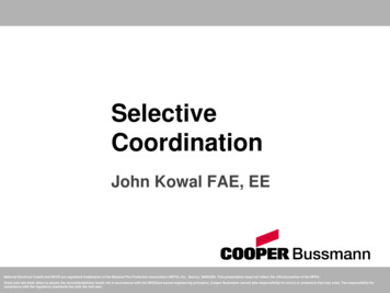
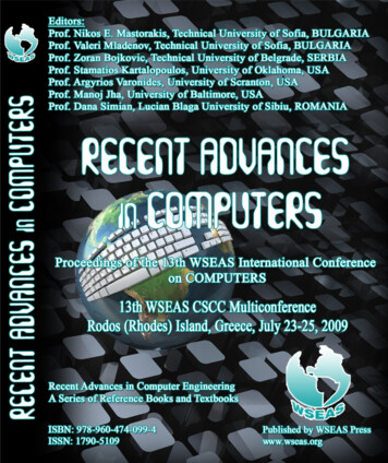
Zhao et al. J Nanobiotechnol(2021) 19:335Page 2 of 15Scheme 1 Schematic of strategies for improving selective photothermal therapysite. The concentration difference between normal tissue and tumor tissue can be generated, and the temperature of the tumor site can be selectively increased[15]. The ideal solution is to give PTAs a self-regulating photothermal conversion capability, which meansthat PTAs have a weak photothermal conversion ability in normal tissue but a strong photothermal conversion ability at the tumor site [13, 14, 16]. Theoretically,the temperature of the tumor site can be selectivelyincreased, with minimal or no damage to normal cells.In this review, we summarized strategies for improvingthe selective efficiency of PTT and discussed the viewsand challenges of PTT in the fight against cancer.Improve the enrichment of photothermal agentsat tumor sitesThe process of PTT is the delivery of PTAs to tumor tissue, which is then radiated to raise the local temperature[13, 16]. Therefore, the most straightforward strategy isto increase the concentration of PTAs at the tumor siteso that normal tissue and tumor tissue produce a concentration difference, which selectively raises the tumorsite temperature [13]. Presently, the most convenient andcommonly employed solution is intratumor injection. Alarge amount of PTAs can be enriched in the tumor tissue, and the temperature of the tumor site can be selectively increased [17, 18]. However, this method is notdirectly applied for metastatic and deep tissue tumorsin the body compared to intravenous injections [19].
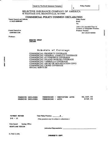
Zhao et al. J Nanobiotechnol(2021) 19:335To achieve a high abundance of intravenous drugs attumor sites, nanodelivery systems have been developedfrom nanoparticles to targeted nanoparticles, biomimetic targeting systems, and programmed targeting systems. These strategies have made significant progress inenhancing the stability of drug circulation and tumor celluptake [20].Intratumor injectionAccording to a previous study, the amount of PTAs thatcan reach the tumor site for cancer treatment is muchsmaller than the amount of intravenous injection due tothe devouring effect of the RES system after administration of the whole body and does not have a satisfactoryrole in the efficacy of the drug [21]. The most striking quality of intratumor injection is its effectiveness inregard to avoiding PTAs loss, which is more conducive toheat ablation of the tumor. Given that light absorbed inthe near-infrared second window with a range of 1000–1400 nm has excellent potential to penetrate deep tissue[22]. Haijun Yu et al. prepared ( NH4)xWO3 nanocubes,which indicated through in vivo and in vitro studiesPage 3 of 15that (NH4)xWO3 nanocubes have an excellent abilityto suppress breast cancer under the second near-infrared window (Fig. 1a, b). After injecting nanocubes andirradiating with a 1064 nm laser in the NIR-II window,they eliminated tumors and inhibited lung metastasis oftumors in mouse models [23]. Polydopamine (PDA) as amimic of the adhesive proteins found in mussels, showsexcellent biocompatibility and biodegradability and hasbeen recently utilized as an effective PTA agent in PTTresearch. The polydopamine coated Fe3O4 magnetic composite particles prepared by Shen Shun et al. have a bettereffect of avoiding interference from the endothelial reticulum system (Fig. 1c, d) [24]. Fe3O4@PDA particles wereinjected into the tumors of tumor-bearing mice and thenirradiated with a laser. The temperature of the tumor surface rapidly increased to 59.7 C, which demonstrated itsexcellent photothermal conversion capability.Via intratumoral injection, nanoparticles enter thetumor. These nanoparticles usually remain at the injection site and have poor permeability in the tumor, leadingto incomplete ablation and recurrence [25]. Thus, cellmediated delivery has great potential in cancer therapy.Fig. 1 a Infrared thermal images and b temperature rise curves of 4T1 tumors injected with PBS or ( NH4)XWO3 Nanocubes (100 μL of 5.0 mg/mL)after 100 s of 1064 nm laser irradiation (Reprinted from Ref. [23] with permission. Copyright 2015, Elsevier Ltd.). c Schematic diagram of intra-tumoralinjection of polydopamine coated magnetic composite particles into mice to enhance the photothermal therapy; d The infrared thermal imageof Fe3O4, PDA, and Fe3O4@PDA under NIR laser irradiation (λ 808 nm; 6.6 W/cm2) (Reprinted from Ref. [24] with permission. Copyright 2015,American Chemical Society)
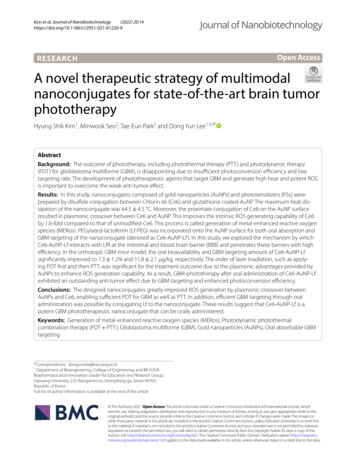
Zhao et al. J Nanobiotechnol(2021) 19:335Nanoparticles can cross nearly impermeable biologicalbarriers to reach target sites that are generally inaccessible to common drugs or nanoparticles [26, 27]. Xue-FengYu and colleagues constructed a cell-mediated deliverysystem using macrophage vehicles to transport BSAcoated Au nanorods (sAuNRs) [28]. Due to their smallsize, BSA-coated sAuNRs carried by macrophage vehiclesexhibited superior anti-phagocytosis because of the better biocompatibility of BSA. After intratumoral injection,macrophages transported, BSA-coated sAuNRs showedgreater photothermal conversion efficiency in tumors,and the tumor recurrence rate was the lowest comparedwith free BSA-coated sAuNRs. However, intratumorinjection has some limitations. PTT through intratumorinjections can easily damage the external tissue of thetumor and has the risk of spreading cancer cells to otherparts of the body. In addition, intratumor injections cannot be used for metastatic tumors and deep tumors [29].Tumor targeted enrichmentSystemic administration is utilized more widely thanintratumoral injection, especially for metastatic anddeep tumors [30]. However, due to the phagocytosis ofthe reticuloendothelial system (RES) in general systemicadministration, the amount of drugs that can reach thetumor site for cancer treatment is substantially less thanthe injection amount, and the efficacy of PTAs is hindered [31]. To achieve a high concentration of PTAs atthe tumor site, nanodelivery systems have evolved fromordinary passive targeting systems to active targetingsystems. As a common strategy in recent years, varioustargeting methods have been explored [32, 33]. Nle4-dPhe7-α-melanocyte-stimulating hormone (NDP-MSH)is an effective receptor agonist of melanocortintype-1,which is overexpressed in many melanoma cells andcombines with the melanocortintype-1 receptor withhigh affinity [34]. Chun Li and colleagues developedmelanoma-targeted hollow gold nanospheres, which stabilized with polyethylene glycol (PEG) coating and combined with NDP-MSH (NDP-MSH-PEG-HAuNS) [35](Fig. 2a). NDP-MSH-PEG-HAuNS and their aggregateswere detected in coated pits by cell uptake experiments,and many NDP-MSH-PEG-HAuNS were detected inthe cytoplasm. Moreover, the NIR laser power was 30 J/cm2 which is lower than the clinical data ( 255 J/cm2)and can avoid unnecessary damage to surrounding normal tissues. These results indicate that NDP-MSH-PEGHAuNS can be well phagocytized into cells to prolongthe treatment time in tumors and enhance the efficacy ofPTT.Yu Hu et al. designed a self-amplified drug deliverysystem for tumor PTT using multiwalled carbon nanotubes (MWNTs) as a carrier and modifying CREKAPage 4 of 15(Cys-Arg-Glu-Lys-Ala) peptides with a particular affinity to fibrin as the targeting moiety (CMWNTs-PEG)[36] (Fig. 2b). This system amplified tumor targeting by apositive feedback mechanism of the coagulation responsewhich means that fibrin is a byproduct of the coagulation reaction and can be specifically and substantiallylocated at the site of vascular damage due to the strongsignal amplification of the clotting reaction. The accumulation of CMWNTS-PEG in tumor sites was significantly higher than that of other groups, which showedan excellent tumor homing effect and realized selectivekilling of tumors. In addition to passive and active targeting, the exogenous magnetic field can also enhance thecontrolled killing of PTAs on tumors. Magnetic nanoparticles (MPSs) carrying PTAs in the blood (for example,based on superparamagnetic Fe3O4) can be redirectedand accumulate in the tumor tissue under the application of an external magnetic field, selectively destroyingthe tumor tissue while preserving normal tissue, thusimproving the selectivity and efficiency of PTT. Magneticfield-guided PTT has been successfully applied in preclinical models, truly showing its excellent clinical application prospects [37, 38].Although active nanodelivery systems that show significant efficacy in treating tumors have been developed,most of them have been affected by unanticipated highuptake of reticuloendothelial systems (RES) (e.g., liverand spleen) [39]. An endogenous, alkaline phosphatasetriggered co-assembly strategy was proposed by PengHuang et al. for the preparation of tumor-specific indocyanine green (ICG) nanofibers [21] (Fig. 2c). Tumorspecific supramolecular self-assemblies can be achievedthrough the regulation of specific enzymes. The natureof these noncovalent forces allows in situ formed nanostructures to readily incorporate drugs via the same kindof intermolecular interactions. This supramolecular system can easily avoid the undesired uptake of RES whilesustaining advantages, including high tumor accumulation rate and long tumor retention time. Phosphatasedirected co-assembly processes and their diagnosticcapabilities were carried out successfully at various levels, from in vitro experiments, cell experiments, andtissue simulations to in vivo experiments. The researchers observed that the tumor uptake of ICG significantlyincreased to 15.05 3.78% ID/g after intravenous injection for 4 h, which is 25 times higher than that of freeICG (0.59 0.24% ID/g). The resulting high signal-tonoise ratio ( 15) clearly distinguished the tumor fromthe surrounding normal tissue. Complete tumor elimination with high therapeutic accuracy was successfullyachieved by laser irradiation (0.8 W/cm2, 5 min). In thisway, this strategy successfully avoided the high uptakeof RES. Nanofibers used for PTT therapy, including
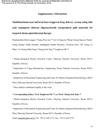
Zhao et al. J Nanobiotechnol(2021) 19:335Page 5 of 15Fig. 2 a TEM images of the B16/F10 cells incubated with NDP-MSH-PEG-HAuNS orPEG-HAuNS (Reprinted from Ref. [35] with permission. Copyright2009, Chun Li). b In vivo self-amplified accumulation of CMWNTs-PEG in tumor tissues (Reprinted from Ref. [36] with permission. Copyright2016, Elsevier Ltd.) c Scheme of in situ conversion of micelles to nanofiber after intravenous injection. Representative NIR fluorescence images ofHeLa-tumor-bearing mice after intravenous administration at different times (Reprinted from Ref. [21] with permission. Copyright 2015, AmericanChemical Society)tumor-specific ICG-doped nanofibers, have great potential to be transformed into personalized nanomedicine treatment mediums and for clinical use in cancertreatment.Biomimetic targeting strategiesAlthough nanocarrier technology has made significantprogress in cancer treatment research, the actual effectsubstantially differs from what people expect. In vivo,experimental data show that drugs collected into tumorcells are usually less than 5% of the injection amount.Most drugs are filtered from the body before enteringtumor cells [40]. Therefore, the prolonged blood circulation time of nanoparticles in the blood is a prerequisitefor targeted delivery. It is well known that specific cellscan be used as bionic targeting ligands to help target orhome drugs to tumors or other lesion sites, to increaseblood circulation time and to improve the pharmacokinetics of drugs [41]. Some stem cells, for example, arebeing applied for tumor-targeted treatment. Stem cellsalso have a crucial role in tumor metastasis. Daxiang Cuiet al. fabricated Au nanorods@SiO2@CXCR4 nanoparticles and loaded the prepared nanoparticles into humaninduced pluripotent stem cells (AuNRs-iPS) to obtainPTT nanoplatforms [42] (Fig. 3a). Due to the excellent tumor target migration capabilities of iPS cells, theresearchers discovered that the Au nanorods mediated bythe nanoplatform AuNRs-iPS had longer retention timesand even spatial distribution. Most importantly, the discovery has demonstrated that iPS can target tumor sitesand improve the efficacy of PTT to inhibit tumor growthin tumor-bearing mice.Macrophages can realize drug homing at the tumor sitethrough their excellent ability to target tumor migration[43, 44]. Macrophages containing therapeutic nanoparticles (including some magnetotactic bacteria) can actas Trojan horses, transporting the nanoparticles to thetumor site and destroying areas of low oxygen within thetumor to prevent malignant progression [45]. Jong-OhPark et al. designed a macrophage-based nanotherapeutic drug delivery system to treat solid tumors by utilizing PTT, an anticancer drug, and the tumor-infiltrating
Zhao et al. J Nanobiotechnol(2021) 19:335Page 6 of 15Fig. 3 a AuNRs@SiO2@CXCR4 loaded human iPS cells for target delivery and intratumoral homogeneous distribution of AuNRs and enhancedphotothermal therapy (Reprinted from Ref. [42] with permission. Copyright 2016, American Chemical Society.); b Tumor penetration of 4T1tumorspheres by macrophages with and without nano-loading was assessed for 12 h. Compared with LPs, AUNRS LPs@RAW showed higherpenetration and tumor coverage in 4T1 cell spheres (Reprinted from Ref. [46] with permission. Copyright 2020, The Royal Society of Chemistry). cSchematic illustration of the preparation of Cyp-MNC@RBCs for NIR fluorescence imaging and MR imaging-guided cancer PTT (Reprinted from Ref.[18] with permission. Copyright 2018, Elsevier Ltd)properties of macrophages [46] (Fig. 3b). Compared withusing nanoparticles alone, when macrophages were participating, the tumor penetration of nanoparticles wassignificantly improved. In addition, in vivo experimentsinvolving local and systemic administration in tumorbearing mice have shown that the drug delivery systemof macrophage-based nanotherapeutics can effectivelytarget and kill tumors. In addition, studies have shownthat the use of red blood cell (RBC) membranes as abionic strategy can also extend the internal blood circulation time of nanoplatforms [47]. Sheng Wang et al.prepared RBC-coated, superparamagnetic nanoclusters(MNCs); after loading with NIR cypate molecules, theirNIR absorbance was dramatically improved, and efficientphotothermal conversion efficiency was achieved [18](Fig. 3c). Cyp-MNC@RBCs had a significant tumor homing ability after intravenous administration. Moreover,the tumor-bearing mice showed higher temperatures atthe tumor site under laser irradiation.In other cases, the two membranes were merged toimprove the targeting capability, to increase circulationtime and to reduce macrophage phagocytosis. To further enhance the therapeutic efficacy of PTT, ZhiqingPang and colleagues fabricated an erythrocyte-cancer(RBC-M) hybrid membrane-camouflaged melanin nanoparticle (Melanin@RBC-M) platform by fusing the RBCmembrane with the MCF-7 cell membrane [48]. Thesehybrid membrane vesicles retained both RBC cell membraned proteins and MCF-7 cell membrane proteins;the MCF-7 membrane component can significantlyenhance the homotypic targeting function of Melanin@RBC-M; and the RBC membrane component can effectively reduce the cellular uptake of macrophages by Melanin@RBC-M and improve their circulation time, whichgreatly increases the photothermal therapeutic effect ofnanomaterials.While treating cancer with nanodelivery systems, it isoften observed that most necrosis in the center of solidtumors is caused by long-term anoxicity: the availability of oxygen and glucose is insufficient for meeting themetabolic needs of malignant cells, and the destructionof the tumor in anoxic areas, especially tumor-associated macrophages (TAMs) in these areas, can effectivelyprevent the proliferation, growth, invasion, migration,and metastasis of malignant cells, directly affecting themortality rate of patients [26, 47]. However, deliveringtherapeutic agents to the oxygen-deprived areas of thetumor is a significant challenge [25]. To solve this problem, Susan E. Clare’s team hypothesized that autonomous recruitment of tumor monocytes could be used fornanoparticle-based drug delivery and tumor therapy [44].Because monocytes have a natural phagocytosis capacity,
Zhao et al. J Nanobiotechnol(2021) 19:335they can easily carry therapeutic nanoparticles to otherwise inaccessible tumor areas. After entering the tumor,the monocytes differentiate into macrophages, and thennanoparticle-laden macrophages migrate/converge to thehypoxic region of the tumor. Once in place, nanoparticle-based therapeutic functions can be activated by NIRirradiation of the tumor to destroy TAMs. Depending onthe irradiation protocol, this therapeutic response canalso include the destruction of adjacent tumor cells andcan be combined and coordinated with other chemicalsand molecular or nanoparticle-based therapies to facilitate the destruction and remission of the tumor whilesignificantly reducing the risk of tumor regrowth andmetastasis.Programmed targeting systemsThe targeted ligand on the surface of nanoparticles canincrease the affinity between nanoparticles and target cells, thus improving the uptake efficiency of cells[49]. Nevertheless, the presence of a targeted ligand cantrigger an immune response, leading to the removal ofPage 7 of 15nanoparticles by the mononuclear phagocyte system[50]. Most of the target ligands are hydrophobic, whichcan easily cause aggregation of nanoparticles in vivo. Asa result, the blood circulation time of the nanoparticlesis shortened [51]. However, the contradiction for aggregation between tumors and blood cannot be solved byordinary nanoparticles. Programmed targeting strategiescan confer on-demand properties on target ligands to“shield” in the bloodstream and “deshield” at tumor sites,enabling them to become suitable candidates to avoidimmune system recognition and to prolong the bloodcirculation time [52, 53]. The use of shields or blockinggroups protects the ligand from being recognized by theimmune system and prolongs blood circulation. Once theshielding layer is removed at the tumor site by endogenous or exogenous stimulation, the intake of tumor cellswill increase [49, 54].By utilizing the reversible protonation of weak electrolytic groups to pH changes, Guangming Lu et al. designedlong-chain amine/carboxyl-terminated PEG decoratedgold nanostars (GNSs) for PTT [41] (Fig. 4a). WhenFig. 4 a Schematic illustration of the PEGylated mixed-charge GNSs and their pH-reversible cell affinity and photothermal therapeutic efficacy. bOptical microscopy images and cellular uptake of cells after incubation with laser for 4 h at different pH (Reprinted from Ref. [41] with permission.Copyright 2014, Wiley–VCH Verlag GmbH & Co. KGaA). c Schematic illustration of the based-gold nanostars temperature-responsive ligandreversible shielding system for combined photothermal therapy and chemotherapy. d Pharmacokinetics evaluation on Au@Pt/Re-Biotin, Au@Pt/irRe-Biotin, and cisplatin in vivo and biodistribution in the liver, spleen, and tumor (Reprinted from Ref. [49] with permission. Copyright 2018,Elsevier Ltd)
Zhao et al. J Nanobiotechnol(2021) 19:335incubated with HeLa cells, the degree of cellular uptakeof GNS-N/C at pH 6.4 was significantly higher than thatat pH 7.4 (Fig. 4b). Taking advantage of shielding nanoparticles from nonspecific interactions with normal cells/tissues before they reach tumors and after they leavetumors is crucial for the selective delivery of GNS-N/Cinto tumor cells, which provides a novel effective meansof tumor-selective therapy. They irradiated tumors withan 808 nm laser at 1 W/cm2 for 5 min 24 h postinjection.Mice treated with GNS-N/C 4 (one PEGylated mixedcharge GNS with a certain surface composition) experienced an average increase of 23 C temperature, reachingan average temperature of 56 C after 5 min of treatment,which proved the excellent PTT effect of GNS-N/C 4.Moreover, not all PTAs that arrive at the tumor wouldbe retained in the tumor tissue. Therefore, a nanosystemthat could re-shield ligands is needed to enhance thetreatment selectivity of PTT. Zhi Yuan et al. researchedthe reversible ligand shielding strategy by a reversible ligand shielding system based on a temperatureresponsive polymer [49] (Fig. 4c). The ligand biotin,cisplatin-loaded chain poly (acrylic acid)-Pt, and shielding segment thermosensitive -co-AAm))wereco-modified onto the surface of gold nanostars (Au@Pt/Re-Biotin). Among them, the lower critical solutiontemperature of P(NIPAAm-co-AAm) is approximately39 C, which helps to shield the ligand by the extensionof P(NIPAAm-co-AAm) in the blood circulation (37 C).The ligand would be deshielded through P(NIPAAmco-AAm) contraction utilizing the heat generated fromgold nanostars upon NIR irradiation when the nanoparticles arrived at the tumor site. The results indicatedthat the system could extend blood circulation (1.6-foldat 24 h), reduce immune system clearance (28% lower),and enhance tumor accumulation (37% higher) effectiveness compared with the irreversible ligand shielding system (Au@Pt/irRe-Biotin) by analysis of platinum(Fig. 4d). Photothermal imaging of tumors in vivo wasconducted to evaluate the photothermal conversion ability. Upon NIR (808 nm, 1 W/cm2, 6 min) irradiation,both Au@Pt/Re-Biotin and Au@Pt/irRe-Biotin showedapparent temperature increases, thus indicating the accumulation of gold nanostars, which possessed satisfactory photothermal conversion ability at the tumor site.In addition, this strategy showed tumor inhibition (11%higher) that was significantly superior to the irreversiblesystem.Self‑regulating photothermal conversion systemSelective killing of tumor cells means killing tumor cellswith minimal or no damage to normal cells. Althoughimproving the enrichment of photothermal materials inPage 8 of 15tumor sites can solve the problem to some extent, lowconcentrations of PTAs in normal tissue also have theability of photothermal conversion, so how to achievePTT without damaging normal tissue remains a challenge [55]. Therefore, is it possible to selectively increasetumor site temperature by intravenous injection with thesame concentration and laser radiation in tumor and normal tissue? Assume that PTAs have a weak photothermalconversion ability in normal tissues and a robust photothermal conversion ability in tumor sites. In this case, thisPTAs can selectively increase the temperature of tumorsites under inaccurate irradiation in theory, with minimalor no damage to normal cells [56]. Therefore, PTT’s otherstrategy for selective killing is to give PTAs a self-regulating photothermal conversion capability [57, 58].Self‑regulating of metal nanoparticlesTo date, many PTAs have been discovered, includingphotosensitizer polymers, metal nanoparticles, carbonnanomaterials, black phosphorus (BP) based nanomaterials, and metal sulfides [59]. However, these PTAscannot achieve a controllable photothermal conversionability. Gold nanoparticles have become one of the mostpromising drug delivery materials due to their excellent biocompatibility, surface modification, and excellent photothermal conversion efficiency. The aggregationand self-assembly of spherical gold nanoparticles givethem the photothermal conversion ability that they donot possess [60], making them the best candidate formaterials that can regulate photothermal conversion. Inthe disassembled state, the NIR absorption of sphericalgold nanoparticles is very weak and almost does not havea photothermal conversion capability. The absorptionpeak re-shifted to approximately 808 nm and generateda photothermal conversion ability in the assembled state,achieving the photothermal treatment function [61]. Tomeet the functional requirements of PTAs in differentenvironments in vivo and enable them to possess uniquecharacteristics of assembled and unassembled states,researchers have explored how to achieve responsiveaggregation of small particle-sized gold nanoparticles intumor sites.It has been reported that many pH-sensitive, sphericalgold nanoparticles can achieve responsive self-assemblyat the tumor site under the influence of the tumor microenvironment, showing strong near-infrared absorptionand thermal ablation of tumors [62]. Zhi Yuan et al. [63]used a one-pot reaction to modify lipoic acid-PEG (LAPEG), LA-PEG-Biotin, 4-mercaptobenzoic acid, andp-hydroxythiophenol on gold nanoparticles (Au@T),which can increase the hydrophobicity of the systemunder acidic conditions of pH 6.0 to agglomerate thenanoparticles (Fig. 5a). By using dynamic light scattering
Zhao et al. J Nanobiotechnol(2021) 19:335Page 9 of 15Fig. 5 a Schematic image of the tumor-microenvironment-activated self-assembled Au@T S iO2 (Reprinted from Ref. [63] with permission.Copyright 2019, Zhi Yuan). b Schematic illustration of the light-triggered assembly of dAuNPs. c Photothermal images of tumor-bearing mice forshowing the in vivo cross-linking effect of dAuNPs on tumor local temperature against irradiation time of 808 nm laser (Reprinted from Ref. [67]with permission. Copyright 2016 WILEY–VCH Verlag GmbH & Co. KGaA, Weinheim)to measure the size of Au@T, they discovered that afterbeing added to pH 6.0 PB
REVIEW Recent advances in selective photothermal therapy of tumor Liping Zhao1†, Xu Zhang2†, Xiaoxia Wang 1, Xiuwen Guan1,3,4, Weifeng Zhang 1,3,4* and Jinlong Ma1,3,4* Abstract Photothermal therapy (PTT), which converts light energy to heat energy, has become a new research hotspot in cancer treatment.