Transcription
Malaysian Journal of Microbiology, Vol 16(2) 2020, pp. 124-133DOI: http://dx.doi.org/10.21161/mjm.190512Malaysian Journal of MicrobiologyPublished by Malaysian Society for Microbiology(Insince 2011)Proteomic analysis of Pseudomonas aeruginosa biofilm treated with Chromolaenaodorata extractsWan Mohd Afifi Wan Mohd Zawawi1, Mohammad Shamil Anwar Ibrahim1, Norasfaliza Rahmad2,Umi Marshida Abdul Hamid1,3 and Mohd Fakharul Zaman Raja Yahya1,3*1Facultyof Applied Sciences, Universiti Teknologi MARA Shah Alam, 40450 Shah Alam, Selangor, Malaysia.2Agro Biotechnology Institute, Serdang, Selangor, Malaysia.3Molecular Microbial Pathogenicity Research Group, Pharmaceutical and Life Sciences Community of Research,Universiti Teknologi MARA.Email: fakharulzaman@uitm.edu.myReceived 26 June 2019; Received in revised form 6 September 2019; Accepted 31 October 2019ABSTRACTAim: Biofilm is the major causative factor of infectious diseases. Difficulty in combating biofilm-related diseases istypically due to persisters, heterogeneous microbial population and viscoelastic extracellular polymeric substances(EPS) matrix. Antibiofilm activities of Chromolaena odorata extracts have previously been demonstrated, however, theeffects of its treatment on the biofilm proteome expression remains not well understood. Thus, this study was carried outto profile changes in biofilm proteome of Pseudomonas aeruginosa following treatment with chloroform and ethanolextracts of C. odorata.Methodology and results: Biofilm was developed in 6-well microplate in the presence or absence of C. odorataextracts overnight at 37 C. Whole-cell proteome analysis was carried out by combining two-dimensional polyacrylamidegel electrophoresis and mass spectrometry. Treatment with C. odorata extracts triggered changes in two-dimensionalproteome profiles of P. aeruginosa biofilm under aerobic and anaerobic conditions. The differentially expressed proteinswere successfully identified and were assigned to various functional categories including protein metabolism,carbohydrate metabolism, peptidoglycan metabolism, electron transport and iron transport.Conclusion, significance and impact of study: The present study demonstrates differential proteome expression in P.aeruginosa biofilm following treatment with C. odorata extracts. This suggests that C. odorata extracts may targetmultiple biological processes to control P. aeruginosa biofilm. C. odorata extracts may be useful for development ofnovel antibiofilm agents.Keywords: Biofilm, Pseudomonas aeruginosa, Chromolaena odorata, proteomicINTRODUCTIONBiofilm is a conglomerate of microbial community that isencapsulated by a self-produced polymeric matrix. It iscapable of adhering to biotic or abiotic surface.Development of biofilm begins with attachment ofmicrobial cells to a particular surface which then formmicrocolonies and three-dimensional mushroom-shapedstructure (Karatan and Watnick, 2009). Over the past fewcenturies, the biofilm has caused a wide spectrum ofproblems in public health, marine and agriculture sectors.One of the important biofilm producers is P. aeruginosa,the causative factor of hospital-acquired infections. It is apathogenic Gram-negative bacterium commonly found insoil, water and skin flora. In human, it frequently infectsthe airway, wounds, urinary tract and burns. Theheterogeneity of P. aeruginosa biofilm has clearly been*Corresponding author124demonstrated by infinite focus microscope (IFM) (Mahatet al., 2012).Chromolaena odorata is a traditional medicinal plantthat is widely used for its wound healing, antibacterial,antioxidant and anticancer properties. It contains a widerange of important phytochemicals including alkaloids,flavonoids, phenolics, saponins and tannins. This fastgrowing perennial shrub is commonly found in SouthAmerica, Central America, Africa and Asia countries. Itsantibiofilm activity against P. aeruginosa has previouslybeen reported (Yahya et al., 2014), thus, highlighting thepotential use of this medicinal plant to control variousbiofilm-associated problems such as chronic humaninfection, biocorrosion, poor performance of medicaldevices and fabric damages.Proteomics is the large-scale analysis of proteinexpression of an organism that involves three major stepsISSN (print): 1823-8262, ISSN (online): 2231-7538
Malays. J. Microbiol. Vol 16(2) 2020, pp. 124-133DOI: http://dx.doi.org/10.21161/mjm.190512namely sample preparation, protein separation andprotein identification. It allows investigation of differentiallyexpressed proteins when comparing different types ofbiological samples. Indeed, there are countlessapplications of proteomics in the study of infectious andnon-infectious diseases. Since many decades ago, manyspectroscopic and proteomic studies have shed light onhow the biofilm-forming bacteria survive under diverseenvironmental challenges (Suci et al., 1994; Karatzas etal., 2008; Cho and Ahn, 2014). The inhibitory effects of C.odorata extracts against P. aeruginosa biofilm haspreviously been reported (Yahya et al., 2014). However, itremains uncertain how C. odorata extracts modulate theproteome expression in P. aeruginosa biofilm. Therefore,the present work was carried out to identify differentiallyexpressed proteins in P. aeruginosa biofilm followingtreatment with C. odorata extracts.Protein extraction and determinationThe whole-cell protein was extracted from the resultingpellet using Radio Immunoprecipitation Assay (RIPA)which consist of the following: 50 mM trisaminomethanehydrochloric acid (Tris-HCl) pH 7.4, 150 mM NaCl, 50 mMethylenediaminetetraacetic acid (EDTA), 0.2% Triton X100, 1% sodium dodecyl sulfate (SDS), 2% betamercaptoethanol (β-ME) and protease inhibitor. Celldisruption was improved by sonication on ice at 80%amplitude, 5 cycles with 45 sec burst and incubated for 2h on ice. The cell debris and intact cells were removed bycentrifugation at 8,000 rpm, 5 min. The supernatantcontaining protein fraction was stored at -20 C untilfurther use. The concentration of protein extracted wasdetermined using the standard Bradford assay.Two-dimensional polyacrylamide gel electrophoresisMATERIALS AND METHODSPlant materialThe fresh leaves of C. odorata were collected at PuncakAlam, Selangor, Malaysia (Coordinates: 3 13’42.30’’N,101 25’41.65’’E). The taxonomic identity of the plant wasauthenticated by Dr Shamsul Khamis from UniversitiPutra Malaysia. The leaves were washed with water anddried in oven for 48 h at 60 C. The dried leaves wereground into fine powder and soaked into absolute ethanolor absolute chloroform in a ratio of 1:10 (w/v) in waterbath at 50 C for 72 h. The impurities were then filteredusing muslin cloth. Rotary evaporator was used toremove excess solvents. For the proteomic study of P.aerugionsa biofilm, C. odorata extracts were tested at 200mg/mL as they exhibited the highest antibiofilm activity(Yahya et al., 2014).Test microorganismP. aeruginosa ATCC 10145 was grown in LB nutrientbroth (Difco Laboratories, USA) at 37 C. Gram-stainingwas regularly employed to assess culture purity andcolony morphology. For the purpose of each biofilmassays, the starting bacterial inoculum was adjusted to12 108 CFU/mL.Protein samples were initially precipitated using 2D-CleanUp Kit. Then, the protein samples were solubilized withrehydration buffer (7 M urea, 2 M thiourea, 2% w/v fonate(CHAPS), 0.5% immobilized pH gradient (IPG) buffer, anda trace of bromophenol blue) and left overnight torehydrate into ZOOM IPG strips (7 cm immobilized pHgradient with non-linear pH gradient range 3-10). Firstdimension isoelectric focusing (IEF) was performed in theZOOM IPGRunner Mini-Cell according to the followingprotocol: 200 V for 20 min, 450 V for 15 min, 750 V for 15min, 2,000 V for 1 h. The strips were subsequentlyequilibrated with equilibration buffer (1 NuPAGE lithiumdodecyl sulfate (LDS) Sample Buffer, NuPAGE SampleReducing Agent, 125 mM iodoacetamide (IAA)). Gelelectrophoresis was performed using 10% gels at 200 Vfor 45 min. Gels were stained using SYPRO Rubyovernight. Protein spots were visualized using UVilluminator (Alpha Imager HP) at 610 nm. Twodimensional proteome maps were analysed to identifyprotein spots which differed significantly in intensitybetween control gels and test gels using Progenesisproteomic analysis software. The volume of each spotwas normalized against the total volume of all spots in thegel, and the normalized values were expressed aspercentage spot volume. Spots with a fold-change of atleast 2.0 were excised from the gels for in-gel digestion.Microplate biofilm assayTrypsin digestionBiofilms were developed in 6-well microplate with andwithout the presence of C. odorata extracts. Following a24 h incubation at 37 C, nutrient medium was discardedand the formed biofilm fractions were rinsed twice withdistilled water, suspended in 0.9% sodium chloride (NaCl)solution (Sigma, USA) and pelleted by centrifugation at10,000 rpm for 10 min. The microplate biofilm assayprocedure was performed in three replicates under bothaerobic and anaerobic conditions. The anaerobiccondition was developed using candle jar and a strip ofpaper soaked in methylene blue dye.125The gel plugs were washed with 50 mM ammoniumcarbonate (NH4CO3) (5 min), followed by 70% acetonitrile(ACN) (15 min) and 100% ACN (5 min) at roomtemperature. The liquid were discarded following eachwashing step. The gel plugs were then dried using SpeedVac (2,000 rpm, 4 C, 15 min). Reduction and alkylationsteps were performed by incubation with 10 mMdithiothreitol (DTT)/100 mM NH4CO3 for 30 min at 60 Cand with 55 mM IAA/100 mM ammonium bicarbonate(ABC) for 20 min in the dark at room temperature.Subsequently, the gel plugs were washed twice with 50%ISSN (print): 1823-8262, ISSN (online): 2231-7538
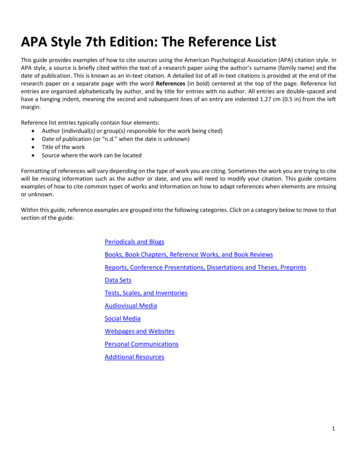
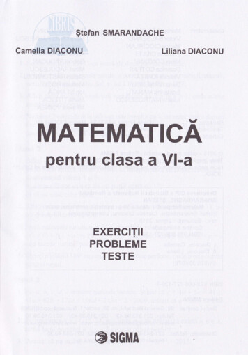
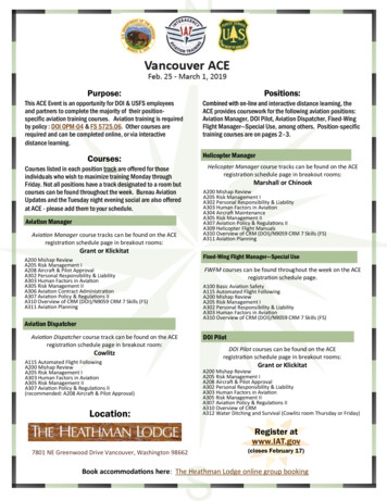
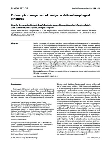
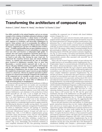
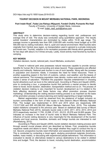
Malays. J. Microbiol. Vol 16(2) 2020, pp. 124-133DOI: http://dx.doi.org/10.21161/mjm.190512ACN/100 mM ABC (20 min each) and dehydrated with100% ACN (15 min) at room temperature beforeovernight digestion in 25 µL of 7 ng/µL of trypsin. Thedigested peptides were pooled into a clean tube and driedusing a vacuum centrifuge before MS analysis.MALDI-TOF/TOF analysisDried peptides were dissolved in 0.1% formic acid (FA)and desalted using ZipTip C18 (Millipore, Billerica, USA),according to the manufacturer’s protocol. Next, thepeptides were eluted with Elution solution (0.1%trifluoroacetic acid (TFA)/50% ACN into a newmicrocentrifuge tube. Peptides were spotted onAnchorChip Standard Targets plate in duplicates. Matrixsolubilization solution (α-cyano-4-hydroxycinnamic acid(CHCA) in 50% ACN/0.1% TFA) was then spotted oneach peptide spot and allowed to dry. Meanwhile,external calibrant spots solution was deposited ontocalibrant anchor spots on the AnchorChip target andallowed to dry. Matrix assisted laser desorption/ionisationtime of flight mass spectrometry (MALDI-TOF/TOF) wasperformed on an AB SCIEX TOF/TOF 5800 System(AB SCIEX, Framingham, MA) whilst the resulting peptidemass spectra and were submitted to the Mascot searchengine (Matrix science) to search against the nonredundant database of the National Center forBiotechnology Information (NCBIprot).BioinformaticsIdentified proteins were classified according to theirknown biological functions by database searching usingSwissProt/TrEMBL and cross-referencing with publishedreport (Hashemi et al., 2019). Prediction of functionalmotifs and subcellular localization were carried out usingScanProsite and PSORTb respectively.RESULTS AND DISCUSSIONFigures 1-4 show representative gels of P. aeruginosabiofilm proteome under aerobic and anaerobic conditions.Twelve and 18 proteins were found to be differentiallyexpressed in the biofilm following treatment with C.odorata chloroform extract (COCE) under aerobic andanaerobic conditions respectively. On the other hand,seven proteins exhibited differential expression in thebiofilm following treatment with C. odorata ethanol extract(COEE) under both aerobic and anaerobic conditions.Differentially expressed biofilm proteins weresubjected to identification by MALDI-TOF/TOF analysis(Figures 1-4). Eleven differentially expressed proteins inCOCE treated biofilm under aerobic condition and ninedifferentially expressed proteins in COCE treated biofilmunder anaerobic condition were successfully identified.On the other hand, seven differentially expressed proteinsin COEE treated biofilm under both experimentalconditions were successfully identified. Porin D,elongation factor Tu and electron transfer flavoprotein126subunit alpha appeared in more than one spots on a twodimensional polyacrylamide gel.Figures 5 and 6 show functional categories andsubcellular location of identified differentially expressedproteins respectively. Approximately 56% and 67% ofidentified differentially expressed proteins were found tobe associated with the general metabolism and cytoplasmrespectively. Porin D, elongation factor Tu and electrontransfer flavoprotein subunit alpha were analyzed forfunctional motifs associated with the post-translationalmodification. They were predicted to contain functionalmotifs of casein kinase II phosphorylation site (PS00006),protein kinase C phosphorylation site (PS00005) and Nglycosylation site (PS00001).Proteomics is a systematic and high-throughputapproach to study protein expression of living organisms.It has become a common approach to investigate themode of action of antimicrobial agents. The informationderived from this analysis is also useful to understandhow the living cells respond towards a wide range ofecological factors. In the present study, treatment with ession in P. aeruginosa biofilm. This finding is similarto Miyamoto et al. (2015) which demonstrated differentialproteome expression in Listeria monocytogenes ATCC7644 following exposure to a sub-lethal concentration ofnisin.Oxygen is reduced to water during oxidativephosphorylation in mitochondria. Changes in oxygen levelwould normally result in alteration in the amount reactiveoxygen species and expression of proteins associatedwith oxidative phosphorylation. In the present study, theoxygen level was found to affect the proteome expressionin both control and test biofilms. The bacterial proteomicstudy under aerobic and anaerobic conditions haspreviously been reported by Starck et al. (2004). Theydemonstrated that approximately 50 proteins inMycobacterium tuberculosis were expressed only underanaerobic condition but not under aerobic condition.Plant extraction using organic solvents is well knownto be useful for various analysis of plant materials. Whilemany previous works have demonstrated the effects ofdifferent extracting organic solvents on the phytochemicalprofile and biological activities of plant materials, little isknown about the effects of different extracting organicsolvents on protein expression. In the present work, theeffects of different extracting organic solvents on theproteome profile of P. aeruginosa biofilm was observed.For example, spot A5-A7 were differentially expressed inthe biofilm treated with COCE (Figure 1) but not in thebiofilm treated with COEE (Figure 3). This resultcorroborates the previous work (Yahya et al. 2014)demonstrating differential antibiofilm activities ofchloroform and ethanol extracts of C. odorata which aredue to different phytochemical profiles.The biofilm proteins are expected to be differentiallyexpressed as compared to its planktonic counterpart. Thisunderlies other physiological disparities such as metabolicrates, protection against antimicrobial attack, cell-surfaceinteraction and cell-cell communication. In the studyISSN (print): 1823-8262, ISSN (online): 2231-7538
Malays. J. Microbiol. Vol 16(2) 2020, pp. 124-133DOI: http://dx.doi.org/10.21161/mjm.190512A)B)Spot No.A1A2A3A4A5A6A7A8A9A10A11A12Accession 12J9A6V753A6UZH4P31961Q51404NDProtein identityDNA toposiomerase 4 subunit ANAD-dependent malic enzymeTryptophan-tRNA ligase carbomyltransferaseBifunctional protein GlmUCytochrome bo(3) ubiquinol oxidase subunit2Protoheme IX farnesyltransferase 2Phenazine biosynthesis protein PhzA 2Putative 4-hydroxy-4-methyl-2-oxoglutarate aldolaseElongation factor Tu 1Phosphogluconate dehydrataseFumarate hydratase class nge vs control Figure 1: Whole-cell proteome profile of P. aeruginosa biofilm following treatment with C. odorata chloroform extract(COCE) under aerobic condition. A) Differentially expressed proteins with at least two-fold change (as indicated by redcircle). Eighty µg of protein sample were focused on 13 cm, pH 3-10 non-linear IPG drystrips, followed by 12%polyacrylamide gel electrophoresis and silver staining. B) Identification of differentially expressed proteins by MALDITOF. ND, not detected by MS; , up-regulation; , down-regulation.127ISSN (print): 1823-8262, ISSN (online): 2231-7538
Malays. J. Microbiol. Vol 16(2) 2020, pp. 124-133DOI: 5C6C7C8C9C10C11C12C13C14C15C16Accession I4E1Q9HZJ8NDNDNDNDQ9HT18P57112C17C18NDNDSpot No.Protein identityFerripyoverdine receptorPolyphosphate kinaseATP synthase subunit betaPorin DProtein TolBNAD-dependent protein deacylase 2Cell division inhibitor SulAATP synthase subunit alphaSoluble pyridine 2815726Expressionchange vs control - Figure 2: Whole-cell proteome profile of P. aeruginosa biofilm following treatment with C. odorata chloroform extract(COCE) under anerobic condition. A) Differentially expressed proteins with at least two-fold change (as indicated by redcircle). Eighty µg of protein sample were focused on 13 cm, pH 3-10 non-linear IPG drystrips, followed by 12%polyacrylamide gel electrophoresis and silver staining. B) Identification of differentially expressed proteins by MALDITOF. ND, not detected by MS; , up-regulation; , down-regulation.128ISSN (print): 1823-8262, ISSN (online): 2231-7538
Malays. J. Microbiol. Vol 16(2) 2020, pp. 124-133DOI: 8Accession No.(UniProt)PORD PSEAECH60 PSEAE7EFTU PSEABATPA PSEABBRAC PSEAEA11A12PORD-PSEAEEFTU PSEABSpot No.Protein identityPorin D60kDA chaperoninElongation factor TuATP synthase subunit alphaLeucine-isoleucine-valine-threonine and alaninebinding proteinPorin DElongation factor TuMascotscore235105131663122Expressionchange vs control 118187 Figure 3: Whole-cell proteome profile of P. aeruginosa biofilm following treatment with C. odorata ethanol extract(COEE) under aerobic condition. A) Differentially expressed proteins with at least two-fold change (as indicated by redcircle). Eighty µg of protein sample were focused on 13 cm, pH 3-10 non-linear IPG drystrips, followed by 12%polyacrylamide gel electrophoresis and silver staining. B) Identification of differentially expressed proteins by MALDITOF. ND, not detected by MS; , up-regulation; , down-regulation.129ISSN (print): 1823-8262, ISSN (online): 2231-7538
Malays. J. Microbiol. Vol 16(2) 2020, pp. 124-133DOI: http://dx.doi.org/10.21161/mjm.190512A)B)Spot No.B1B7B8B9B11B12B13Accession No.(UniProt)RS1 PSEAEEFTU PSEF5HPPD PSEAEEFTA PSEAETPX PSEAEEFTU PSEABEFTA PSEAEProtein identity30s ribosomal proteinElongation factor Tu4-hydroxyphenylpyruvate dioxygenaseElectron transfer flavoprotein subunit alphaProbable thiol peroxidaseElongation factor TuElectron transfer flavoprotein subunit nge vs control Figure 4: Whole-cell proteome profile of P. aeruginosa biofilm following treatment with C. odorata ethanol extract(COEE) under anerobic condition. A) Differentially expressed proteins with at least two-fold change (as indicated by redcircle). Eighty µg of protein sample were focused on 13 cm, pH 3-10 non-linear IPG drystrips, followed by 12%polyacrylamide gel electrophoresis and silver staining. B) Identification of differentially expressed proteins by MALDITOF. ND, not detected by MS; , up-regulation; , down-regulation.130ISSN (print): 1823-8262, ISSN (online): 2231-7538
Malays. J. Microbiol. Vol 16(2) 2020, pp. 124-133DOI: http://dx.doi.org/10.21161/mjm.190512Figure 5: Functional categories of identified differentiallyexpressed proteins in P. aeruginosa biofilm according totheir known biological functions by database searchingusing SwissProt/TrEMBL.Figure 6: Subcellular locations of identified differentiallyexpressed proteins in P. aeruginosa biofilm.presented herein, the differentially expressed proteins inP. aeruginosa biofilm were successfully identified byMALDI-TOF/TOF. Expression of polyphosphate kinase(Rashid et al., 2000), elongation factor Tu (Yahya et al.,2017), ATP synthase subunit alpha (Yahya et al., 2017)and fumarate dehydratase (Schlag et al., 2007) in thebiofilms have previously been reported.The ATP synthase subunit alpha is a membranebound enzyme involved in ATP synthesis. It harnessesthe energy from the proton gradient established acrossthe inner mitochondrial membrane to drive the ATPsynthesis. In the present study, expression of ATPsynthase subunit alpha in P. aeruginosa biofilm was upregulated in all gel spots (spot C15, Figure 2A; spot A7,Figure 3A). This overexpression of ATP synthase subunitalpha is in contrast to the reduced expression of ATP131synthase subunit alpha in Salmonella typhimurium biofilmfollowing treatment with antimicrobial agent dimethylsulfoxide (Yahya et al., 2017).Porin D forms outer membrane, water filled channelsthat facilitates diffusion of small hydrophilic molecules. Itis well known to be resistant against sodium dodecylsulfate denaturation and is associated with peptidoglycanand lipopolysaccharide. The result from the present studyrevealed over expression of porin D in spot C5 (Figure2A) and spot A11 (Figure 3A), and reduced expression ofporin D in spot A2 (Figure 3A). The increased expressionof porin D following treatment with C. odorata extracts isin parallel with Kolayli et al. (2004) who demonstrated anincrease in expression of porin D in P. aeruginosafollowing treatment with carbapenem antibiotics.Elongation factor Tu is known to participate intranslation. It is a highly conserved protein in bacteria andfacilitates the binding of aminoacyl tRNA to the ribosome.The present study showed increased expression ofelongation factor Tu in spot A6 (Figure 3A), spot A12(Figure 3A) and spot B7 (Figure 4A), and reducedexpression of elongation factor Tu in spot B12 (Figure4A). Reduced expression of elongation factor Tu in S.typhimurium biofilm following treatment with antimicrobialagent dimethyl sulfoxide has previously been reported byYahya et al. (2017).Failure to determine protein sequences of excised gelspots is a common problem in a wide range of proteomicanalysis. This problem is commonly due to lowabundance proteins and post translational modification ofN termini. In the present study, appoximately 33% ofexcised gel spots failed to give sequences. This resultsupports Qi et al. (1996) who demonstrated the failure ofdetermination of cell envelope protein sequences from S.typhimurium SL1344.Spot visualization is a critical step in the gel-basedproteomics. The use of high-sensitivity fluorescent dyesuch as Sypro Ruby should be helpful in selection ofspots of interest for further mass spectrometry (MS) ortandem MS (MS/MS) analysis. In the present study, porinD, elongation factor Tu and electron transfer flavoproteinsubunit alpha were found to appear in more than onespots on a two-dimensional polyacrylamide gel andcontain multiple functional motifs associated with thepost-translational modification. The similar finding haspreviously been reported in other proteomics works (Yanet al., 2005, Tastet et al., 2006). According to Suhai(2007), the same protein may appear in different spots ona two-dimensional polyacrylamide gel due to posttranslational modification.The impact of antimicrobial treatment on variousbiological pathways in bacteria has been well established.The varying effective concentrations of antimicrobialswould normally produce differential inhibitory effects onbacterial metabolism, enzyme catalysis and proteomeexpression. In the present study, the majority ofdifferentially expressed proteins in biofilm treated with C.odorata extracts were related to pyruvate metabolism(NAD-dependent malic enzyme, putative lycanISSN (print): 1823-8262, ISSN (online): 2231-7538
Malays. J. Microbiol. Vol 16(2) 2020, pp. 124-133DOI: functionalproteinGlmU),hememetabolism (protoheme IX farnesyltransferase 2), proteinmetabolism (tryptophan-tRNA ligase carbomyltransferase,Elongation factor Tu 1, 60kDA chaperonin, ratase, fumarate hydratase class II), ATPmetabolism (ATP synthase subunit alpha, ATP synthasesubunit beta), electron transport (cytochrome bo(3)ubiquinol oxidase subunit-2, soluble pyridine nucleotidetranshydrogenase, electron transfer flavoprotein subunitalpha, etc.), iron transport (ferripyoverdine receptor) andprotein transport (porin D, leucine-isoleucine-valinethreonine and alanine binding protein). According toHamilton et al. (2009), metabolic reactions and biologicaltransport are essential for biofilm development. Thus, thisresult suggests that altered expression of these proteinsmay interrupt the sequential multistages of biofilmdevelopment and support our previous finding on theantibiofilm activities of C. odorata extracts against P.aeruginosa (Yahya et al., 2014).Porins and elongation factor Tu have become thecommon indicators for immediate response of microbialcells towards a wide range of antimicrobial agents.Investigation of their functional motifs would be useful tounderstand how they possibly interact with other bacterialproteins. In the present study, porin D, elongation factorTu and electron transfer flavoprotein subunit alpha werepredicted to contain several phosphorylations sites. Thisfinding is in parallel with our prior suggestion that the posttranslational modification may cause the appearance ofporin D, elongation factor Tu and electron transferflavoprotein subunit alpha in more than one spots on atwo-dimensional polyacrylamide gel. Treweek et al.(2015) reported that phosphorylation alters the averagesize and rate of subunit exchange of oligomeric proteinswhile Mayer et al. (2015) mentioned that the majority ofpost translational modifications affect the isoeletric pointand focusing behavior of the protein in the first dimension.Furthermore, phosphorylation of elongation factor Tu in P.aeruginosa has previously been reported by Alexander etal. (1995). They demonstrated the low ability ofphosphorylated elongation factor Tu to bind amino-acyltRNA. Phosphostaining assay and tandem massspectrometry may be useful to experimentally confirm thephosphorylation state of porin D, elongation factor Tu andelectron transfer flavoprotein subunit alpha in P.aeruginosa biofilm. In 2017, Yahya et al. identified thephosphoproteins in S. typhimurium biofilm using bothProQ diamond phosphostaining assay and electrosprayionization-QTOF (ESI-QTOF).CONCLUSIONThe present study demonstrates the effect of C. odorataextracts on the proteome expression of P. aeruginosabiofilm. Oxygen level and different extracting organicsolvents seemed to influence the outcome in terms ofprotein expression. Treatment with C. odorata extractsmay interfere with multiple biological processes in P.132aeruginosa biofilm, making this medicinal plant species aninteresting candidate for biofilm control.ACKNOWLEDGEMENTSWe would like to thank all laboratory staffs from AgroBiotechnology Institute, Serdang for providing technicaladvice on mass spectrometry. This study was supportedby Research Management Institute, UiTM and MalaysianMinistry of Higher Education under research grants,Zamalah UiTM and 600-RMI/RAGS 5/3 (11/2012),respectively.REFERENCESAlexander, C., Bilgin, N., Lindschau, C., Mesters, J. R.,Kraal, B., Hilgenfeld, R., Erdmann, V. A. andLippmann, C. (1995). Phosphorylation of elongationfactor Tu prevents ternary complex formation. TheJournal of Biological Chemistry 270, 14541-14547.Cho, Y. and Ahn, J. (2014). Physiological and molecularresponses of antibiotic resistant Salmonella entericaserovar Typhimurium to acid stress. African Journal ofMicrobiology Research 8(6), 578-589.Hamilton, S., Bongaerts, R. J. M., Mulholland, F.,Cochrane, B., Porter, J., Lucchini, S., LappinScott, H. M. and Hinton, J. C. D. (2009). Thetranscriptional programme of Salmonella entericaserovar Typhimurium reveals a key role for tryptophanmetabolism in biofilms. BMC Genomics 10, 599-620.Hashemi, M. M., Holden, B. S., Coburn, J., Taylor, M.F., Weber, S., Hilton, B., Zaugg, A. L., McEwan, C.,Carson, R., Andersen, J. L., Price, J. C., Deng, S.and Savage, P. B. (2019) Proteomic analysis ofresistance of Gram-negative bacteria to chlorhexidineand impacts on susceptibility to colistin, antimicrobialpeptides, and ceragenins. Frontier Microbiology10:210.Karatan, E. and Watnick, P. (2009) Signals, regulatorynetworks, and materials that build and break bacterialbiofilms. Microbiology and Molecular Biology Reviews73(2), 310-347.Karatzas, K. A. G., Randall, L. P., Webber, M.,Piddock, L. J. V., Humphrey, T. J., Woodward, M.J. and Coldham, N. G. (2008). Phenotypic andproteomic characterization of multiply antibioticresistant variants of Salmonella enterica serovarTyphimurium selected following exposure todisinfectants. Applied and Environmental Microbiology74(5), 1508-1516.Kolayli, F., Karadenizli, A., Savli, H., Ergen, K.,Hatirnaz, O., Balikci, E., Budak, F. and Vahaboglu,H. (2004). Effect of carbapenems on thetranscriptional expression of the oprD,
antioxidant and anticancer properties. It contains a wide range of important phytochemicals including alkaloids, flavonoids, phenolics, saponins and tannins. . Sample Buffer, NuPAGE Sample Reducing Agent, 125 mM iodoacetamide (IAA)). Gel electrophoresis was performed using 10% gels at 200 V for 45 min. Gels were stained using SYPRO Ruby .