Transcription
Theranostics 2022, Vol. 12, Issue 1IvyspringInternational PublisherResearch Paper127Theranostics2022; 12(1): 127-142. doi: 10.7150/thno.63481An MMP-degradable and conductive hydrogel tostabilize HIF-1α for recovering cardiac functionsXiaojuan Wei1#, Si Chen2,3,4#, Tian Xie3, Hongchi Chen5, Xin Jin3, Jumin Yang3, Shafaq Sahar2, HuanleiHuang6,7, Shuoji Zhu6,7, Nanbo Liu6,7, Changjiang Yu6,7, Ping Zhu6,7 , Wei Wang2,3,4 , Wei Zhang1,5 1.2.3.4.5.6.7.Institute of Microsurgery on Extremities, Shanghai Jiao Tong University Affiliated Shanghai Sixth People’s Hospital, Shanghai 200233, China.College of Chemical and Biological Engineering, Zhejiang University, Hangzhou 310027, China.School of Materials Science and Engineering, Tianjin Key Laboratory of Composite and Functional Materials, Tianjin University, Tianjin 300350, China.ZJU-Hangzhou Global Scientific and Technological Innovation Center, Hangzhou 311215, China.Department of Orthopedic Surgery, Shanghai Jiao Tong University Affiliated Shanghai Sixth People’s Hospital, Shanghai 200233, China.Guangdong Cardiovascular Institute, Guangdong Provincial People's Hospital, Guangdong Academy of Medical Sciences, Guangzhou, Guangdong 510100,China.Department of Cardiovascular Surgery, Sun Yat-sen Memorial Hospital, Sun Yat-sen University, Guangzhou, Guangdong 510100, China.# These authors contributed equally to this paper. Corresponding authors: Prof. Ping Zhu, tanganqier@163.com; Prof. Wei Wang, wwgfz@zju.edu.cn; Prof. Wei Zhang, orthozhang@163.com. The author(s). This is an open access article distributed under the terms of the Creative Commons Attribution License (https://creativecommons.org/licenses/by/4.0/).See http://ivyspring.com/terms for full terms and conditions.Received: 2021.06.03; Accepted: 2021.09.24; Published: 2022.01.01AbstractRationale: Although a few injectable hydrogels have shown a reliable biosafety and a moderate promisein treating myocardial infarction (MI), the updated hydrogel systems with an on-demand biodegradationand multi-biofunctions to deliver therapeutic drug would achieve more prominent efficacy in the futureapplications. In this report, a conductive and injectable hydrogel crosslinked by matrix metalloproteinasesensitive peptides (MMP-SP) was rationally constructed to stabilize hypoxia-inducible factor-1α (HIF-1α)to recover heart functions after MI.Methods: Firstly, tetraaniline (TA) was incorporated into partially oxidized alginate (ALG-CHO) toendow the hydrogels with conductivity. The 1,4-dihydrophenonthrolin-4-one-3-carboxylic acid (DPCA)nanodrug was manufactured with high drug loading capacity and decorated with polymerized dopamine(PDA) to achieve a stable release of the drug. Both ALG-CHO and DPCA@PDA can be cross-linked bythiolated hyaluronic acid (HA-SH) and thiolated MMP-SP to construct a MMP-degradable and conductivehydrogel. After administration in the infarcted heart of rats, echocardiographic assessments, histologicalevaluation, and RT-PCR were used to evaluate therapeutic effects of hydrogels.Results: The cell viability and the results of subcutaneous implantation verify a good cytocompatibilityand biocompatibility of the resulting hydrogels. The hydrogel shows remarkable strength in decreasingthe expression of inflammatory factors, maintaining a high level of HIF-1α to promote the vascularization,and promoting the expression of junctional protein connexin 43. Meanwhile, the multifunctionalhydrogels greatly reduce the infarcted area (by 33.8%) and improve cardiac functions dramatically withejection fraction (EF) and fractional shortening (FS) being increased by 31.3% and 19.0%, respectively.Conclusion: The as-prepared hydrogels in this report achieve a favorable therapeutic effect, offering apromising therapeutic strategy for treating heart injury.Key words: injectable hydrogel, myocardial infarction, matrix metalloproteinase, conductivity, hypoxia-inducible factor-1αIntroductionMyocardial infarction (MI) leads permanentdamages to the heart muscle, which is one of themajor causes of mortality worldwide [1, 2]. Theavailable treatments of MI include pharmaceuticaltherapy, medical device implants, and organtransplants, all of which have distinct limitations,such as high invasiveness, prolonged hospitalizationtime, re-plugging or restenosis, and scarcity ofhttps://www.thno.org
Theranostics 2022, Vol. 12, Issue 1available donors [3, 4]. Injectable biomaterials haveemerged as a promising solution for cardiac tissuerepair after MI. A large number of injectablehydrogels have been designed and delivered to thedamaged area of a beating heart in a minimallyinvasive approach [5]. IK-5001, an alginate hydrogel,demonstrates the therapeutic benefit on reducing theleft ventricular (LV) enlargement in porcine and doginfarction models and also in clinical trials(NCT01226563) [6-8]. Recently, a collagen hydrogelwith autologous stem cells was employed in anotherclinical study (NCT02635464). A 12-month studyshowed no serious adverse effects [9]. Although thesehydrogels have confirmed the reliable biosafety inpatients and are relatively promising in enhancing therepair of injured hearts, the updated hydrogel ctions to deliver therapeutic drug wouldachieve more prominent efficacy.Cell infiltration and neo-tissue formation need tofollow the pace of degradation of biomaterials,allowing the structural and functional integration ofhost tissue [10, 11]. It was reported that the bio-inert,nondegradable polyethylene glycol (PEG) hydrogelcould contribute to preserving LV wall thickness.Meanwhile, nondegradable hydrogel was insufficientto prevent post-MI remodeling, suggesting a limitedtherapy effect in rat MI model [12]. To construct acell-ingrowth matrix, the matrix metalloproteinasesensitive peptides (MMP-SP) have been employed toconstruct the hydrogel networks with a rationallycontrolled degradation behavior which is triggered l conditions after MI, there is apersistence of over-expression and activity of MMPsthat causes maladaptive changes in tissuearchitectures and functions [14-16]. A few injectablehydrogels crosslinked by MMP-SP have recently beendeveloped and applied post MI and the outcomesexhibited well-controllable deliverable therapeutics,contributing to a much better therapy effect [14,16-18].Another challenge for MI therapy is that theconduction of electrical impulse signals is interdictedbecause a fibrosis scar replaces normal myocardiumin infarcted area [19-21]. Conductive hydrogels mayre-establish synchronous contraction and relaxation ofa heart by facilitating the conduction of electricalimpulse in infarcted area, which shows great benefitsto the recovery of cardiac functions [22-24]. In ourprevious work, the hydrogel modified by tetraaniline(TA) nanoparticles (NPs) showed a similarconductivity to that of native myocardium andresulted in a remarkable upregulation of theexpression of junctional protein connexin 43 (Cx43) ion of TA into the hydrogels does not exerta cardiotoxicity and arrhythmia on rat MI hearts [25,26].It is well known that an adult mammalian heartpossesses a limited capability to regenerate itself.However, the hypoxia-inducible factor 1α (HIF-1α)has been recently explored to exert significantpro-regenerative effects by transcriptional regulationof various genes involved in metabolism,angiogenesis, and cell migration [27-30]. Hence, thepharmacological prevention of HIF-1α degradation byprolyl hydroxylase (PHD) is emerging as a promisingtarget in regenerative medicine [29, 31, 32]. Recently, asmall molecule drug, 1,4-dihydrophenonthrolin-4one-3-carboxylic acid (DPCA) has been reported to bea potent inhibitor of PHDs and a stabilizer of HIF-1αboth in vitro and in vivo [31, 33, 34]. However, becauseof potential side effects associated with systemicdelivery of hydroxylase inhibitors (includingangiogenic and erythropoietic effects), it is desirableto develop new methods to achieve a target deliveryof an efficacious and minimum amount ofhydroxylase inhibitors to specific regions [35]. On theother hand, the therapeutic applications of DPCAhave been hindered by its intrinsic poor solubility andshort half-life [33, 35]. A mass of novel approachesbased on nanoparticles has been developed as a smartdelivery system to provide a perfect drug target [36].Among them, a simple and versatile particlemodification strategy based on the famous dopaminepolymerization has been well studied. Briefly,dopamine catechol can be conveniently and rapidlyoxidized to quinone in a weak alkaline condition( pH 8.5) to shape a polymerized dopamine (PDA)layer on various and comprehensive surfaces withcomplexed morphologies. Due to the prominentadvantages of simplicity and versatility, thismethodology has widely been exploited tofunctionalize various types of substrates to stabilizethe nanodrugs and achieve a stable and controllabledrug release behavior [37-41]. Additionally, the PDAlayer will equip the modified nanoparticles with areactive oxygen species (ROS)-scavenging capability[42] and can act as a crosslinker to react with thiol andamine groups [41, 43].Calcium cross-linked alginate hydrogels arereadily erodible in an uncontrollable manner and areassociated with a high calcification risk [44-46]. Asillustrated in Figure 1, a multifunctional injectablehydrogel based on hyaluronic acid (HA) andALG-CHO, which was cross-linked by MMP-SP todeliver DPCA, was rationally constructed to treat theinfarcted rat heart. Firstly, TA was incorporated intoALG-CHO to endow the hydrogels with conductivity.https://www.thno.org
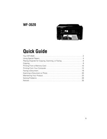
Theranostics 2022, Vol. 12, Issue 1129The hydrophobic drug DPCA was fabricated into NPsby a reprecipitation method and then coated withPDA (DPCA@PDA) to stabilize the nanodrug. BothALG-CHO and DPCA@PDA can be cross-linked bythiolated HA (HA-SH) and thiolated MMP-SP toconstruct the MMP-degradable and conductivehydrogel for restoring cardiac functions. Afteradministration in the injured rat heart, the hydrogelcould enhance the electrical conductivity andmaintain a smart and sustained release of DPCA tostabilize HIF-1α in order to reverse the harmfulmicroenvironment of infarcted zone consequentlymaintaining the heart functions in a better binant MMP-2 were purchased fromSigma-Aldrich. Thiol-modified hyaluronic acid(HA-SH, Mw 300 kDa, degree of thiol substitution 33%) was purchased from ESI BIO, USA. MMP-SP(Seq: CGPLGGGRMSMPVRDGSC where C is athiol-containing cysteine and GGRMSMPV is theMMP-cleavable sequence.) was purchased fromBankpeptide Ltd., Hefei, China. DPCA was purchasedfrom Santa Cruz Biotechnology Ltd. L929 Mousefibroblast cell line (L929) and rat cardioblasts tal Cell Bank, Beijing, China.Experimental SectionPreparation and characterization ofALG-CHO-TAMaterialsSodium alginate (ALG, low viscosity, 4-12 cP),sodium periodate (NaIO4), dopamine hydrochlorideALG-CHO-TA was synthesized according to theprevious reports [25, 26]. Briefly, ALG (5 g) wasdispersed in water (25 mL). The correspondingFigure 1. The schematic design of an MMP-degradable and conductive hydrogel to stabilize HIF-1α for recovering heart functions after MI. The hydrogel was fabricated basedon functionalized HA and ALG. The conductive TA was incorporated into ALG-CHO to endow electric conductivity on the hydrogels. DPCA nanodrug was coated with PDAto achieve a high drug loading amount and maintain a stable releasing manner. Both ALG-CHO and DPCA@PDA are crosslinked by HA-SH and thiolated MMP-SP to constructthe multifunctional hydrogels. The resulted hydrogels were administrated by intramyocardial injection into the infarcted rat hearts to evaluate therapy effects.https://www.thno.org
Theranostics 2022, Vol. 12, Issue 1amount of NaIO4 aqueous solution according to themolar ratio of ALG (sugar moiety): NaIO4 was 2:1.This reaction was carried for 4 h in dark and undercontinuous stirring at 400 r/min. The reaction mixturewas precipitated with 100% ethanol for 3 times toobtain ALG-CHO (a degree of oxidation 42.8%, Mw 4.2 105 Da). In the next step, TA (0.93 g) wasdispersed into 5mL N, N-dimethylformamide (DMF),and TA/DMF solution was added dropwise toALG-CHO solution (1g ALG-CHO in 10 mLdeionized water). The reaction was carried out for 4 hat 50 oC under nitrogen atmosphere. The resultingliquid was precipitated with 100% ethanol for 3 timesto acquire ALG-CHO-TA. ALG, ALG-CHO, TA, andALG-CHO-TA were characterized by proton nuclearmagnetic resonance (1H NMR) spectroscopy (VarianINOVA), ultraviolet-visible (UV-Vis) spectroscopy,and Fourier transform infrared (FTIR) spectrometry(PerkinElmer spectrum 100).Preparation and characterization ofDPCA@PDA NPsDPCA nanodrugs were fabricated by a typicalreprecipitation method through a sudden change ofsolvent [37-41]. Briefly, 400 μL of 25 mg/mL DPCAethanol solution was added dropwise to 10 mL ofwater with a stirring rate of 900 r/min. The DPCANPs with various sizes were prepared by adjustingthe final concentration of DPCA. Subsequently,DOPA (9 mg) was dissolved in 5 mL buffer solution(pH 8.5), then an equal volume of DPCA NPs (1mg/mL) aqueous dispersion was added, and theself-polymerization reaction was allowed to proceedunder stirring for 12 h to obtain DPCA@PDA NPs.The resulting solution was centrifuged (15 min, 10000r/min) and washed by ultrapure water for 3 times toobtain DPCA@PDA NPs. Dynamic light scattering(DLS, Malvern, Nano ZS) and transmission electronmicroscope (TEM, JEM-2100) were employed to assessthe sizes and morphology of the resulting NPs.Preparation and characterization of thedesigned hydrogelsPrecursor solution ‘A’ was obtained by mixingALG-CHO-TA and DPCA@PDA solutions. Precursorsolution ‘B’ was formed by mixing HA-SH withMMP-SP solution. Then, these two of solutions werethen mixed with the volume ratio of A: B 1: 2 toform the hydrogel. The final concentrations of variousingredients are shown in Table S1. The gelation timewas measured by an inverted vial method.Rheological measurements were performed by arheometer (MCR 302). The time scan measurementwas carried at the strain 1% and frequency of 5 Hz.The amplitude sweep was performed with a130frequency of 5 Hz. The shear thinning behavior wasconfirmed under the condition of 1% strain andfrequency of 5 Hz. Anti-fatigue assessment wasmeasured at a strain of 20% and a frequency of 5 Hz tosimulate the beating rate of a normal rat heart.The conductivities of the obtained hydrogelswith different ratios and in PBS were measured viathe four-point probe method (ST2253, China) as listedin Table S2.A classic ROS clearance experiment was carriedout to determine ROS scavenging capability. Briefly, amethanol solution of 1,1-diphenyl-2-picrylhydrazyl(DPPH) with UV absorbance at 516 nm was recordedand set as the initial state. 100 μL ALG-CHO,ALG-CHO-TA, DPCA, PDA, MMP-SP and HA-SHwater solutions corresponding to the concentration inthe hydrogel were prepared and added into 5 mLDPPH solution, and placed it in a shadingenvironment at room temperature. 1 mL of thesolution was taken to measure UV at regular timeintervals, and then put it back until the absorbance at516 nm disappeared. The ROS clearance ratio wascalculated by the change ratio to the initial absorbanceof DPPH solution.Drug release in vitro and in vivo300 μL DPCA@PDA or ALG-CHO-TA/DPCA@PDA/MMP-SP/HA-SH hydrogel was placed in adialysis bag (1000 Da). Then the dialysis bags wereimmersed into 50 mL PBS. The drug release behaviorwas carried out in a shake cultivation (300 r/min tosimulate the beating rate of rat heart) under 37 oC. Totest the drug release profile with MMP, 5 U/mLMMP-2 was added in the solution. After a specifictime interval, 3 mL solution was taken out and thesame amount of fresh PBS was added. The resultingsolution was assessed by UV-vis spectrum. The UVabsorption of 261 nm was recorded to quantify thereleasing rate of DPCA [29].A florescent model drug was employed todetermine the drug release behavior in vivo.Indocyanine green (ICG) is one of the FDA approvedphotosensitizers, which is safe and widely used inbioimaging and diagnose. In order to determine thedrug releasing behavior in vivo, the fluorescent drugICG instead of DPCA has been employed to preparenanoparticles with the same procedure, and theresulted ICG nanoparticles was coated with PDA andcrosslinked with the ALG-CHO-TA/MMP-SP/HA-SH hydrogels, and it was injected into the ratinfarcted hearts (15 rats with ALG-CHO-TA/ICG@PDA/MMP-SP/HA-SH) to test the drugreleasing behavior in vivo. The rat hearts wereharvested 1, 3, 7, 14, and 21d after the operation, andthe fluorescence intensity was measured by In Vivohttps://www.thno.org
Theranostics 2022, Vol. 12, Issue 1Imaging System (PerkinElmer, IVIS Lumina II).Subcutaneous injection and establishment ofMI model and injection of hydrogelsMale SD rats (220 20 g) were supplied byChinese Academy of Medical Science & Peking UnionMedical College Institute of Biomedical Engineering.The protocols were in accordance with the AnimalCare and Use Committee of Tianjin University and allprocedures were performed on the 3R rules. The backhair of rats was removed after the rats wereanesthetized. 15 rats were randomly divided intothree groups and were subcutaneously injected with200 μL hydrogels (n 5, III: ALG-CHO/HA-SH; IV:ALG-CHO-TA/HA-SH; V: ALG-CHO-TA/DPCA@PDA/HA-SH; VI: ALG-CHO-TA/DPCA@PDA/MMP-SP/HA-SH). In order to facilitate thedescription, the hydrogels used in subcutaneousinjection and myocardial injection are grouped as thesame. After 2, 4, and 7 d, the rats wereover-anesthetized and sacrificed. Images of the skintissue containing the hydrogel were taken to observeH&E staining.As for the rat MI model, echocardiographicmeasurements were carried out before the surgery.The ejection fraction (EF) results were employed toscreen rats. Those rats with an EF lower than 75%were gotten rid of the further research. The 114 SDrats were randomly divided into six groups (I: Sham;II: MI; III: ALG-CHO/HA-SH; IV: ALG-CHO-TA/HA-SH; V: ALG-CHO-TA/DPCA@PDA/HA-SH; VI:ALG-CHO-TA/DPCA@PDA/MMP-SP/HA-SH). Theleft artery was ligated after being anesthetized by amoderate flow of diethyl ether. The white infarctedarea could be observed in the left heart. 100 μLhydrogel was injected by a 28-gauge needle in the MIarea of two regions.Echocardiographic assessmentsEchocardiography (Vevo 2100 Imaging System,Visual Sonics, Canada) was employed to access theleft ventricular functions. The EF, fractionalshortening (FS), left ventricular internal diameter(LVID), interventricular septum thickness (IVS), andleft ventricular posterior wall thickness (LVPW) werecalculated according to the averages of fiveconsecutive cardiac cycles.Histological evaluationThe rats were euthanized at 3, 7, 14, and 28 dpost implantation. All the hearts were placed incardioplegic solution before fixation to ensure that allhearts are in a relaxed state. The hearts weredehydrated and cut into sections for Masson staining,H&E staining, Sirius red staining, and triphenyltetrazolium chloride (TTC) staining. TTC staining131sections were recorded by a digital camera. Massonstaining, H&E staining, and Sirius red stainingsections were observed by a fluorescence microscope(CKX41, Olympus, Japan). The Image J software wasemployed to calculate the ratio of infarction, fibrosis,and left ventricular wall thickness according toMasson staining sections.Immunofluorescent stainingThe paraffin sections were deparaffinized andrehydrated with gradient alcohol. After rinsing with0.01 mol/L PBS for 3 times, the slides were blocked by10 % serum in a humid box at 37 oC for 30 min. Theprimary antibody (Table S3) was added and thenincubated in a 37 oC humid box for 1 h and washedwith PBS. Then fluorescent rabbit secondary antibody(1:200, Invitrogen) was dropped onto the slide thatwas subsequently incubated in a humid box in thedarkness for 1 h. DAPI was added dropwise and thesections were incubated for 5 min in the dark. Finally,the slides were mounted with glycerol and theresulted slides were immediately observed andphotographed by a fluorescence microscope (CKX41,Olympus, Japan).RT-PCRThe removed rat hearts were immediately frozenin liquid nitrogen. The quantitative real-time RT-PCRwas carried out as previously reported [25, 26, 40].Briefly, the total RNA was extracted in strictaccordance with the HiPure Plant RNA Mini Kit(R4151-03, MAGEN). A UV spectrophotometer wasused to determine the RNA concentration. The totalRNA (1 μg) was reversely transcribed to cDNAaccording to protocol. After the reaction wascompleted, the cDNA was diluted five times withsterile deionized water and stored at -20 C. mRNAlevel of interleukin-1β (IL-1β), tumor necrosis factor(TNF-α), HIF-1α, Angiopoietin-1(ANG-1), α-Actinin,and cardiac troponin (cTnT) was detected (primersequences in Table S4).Statistical analysisThe data is shown as means standarddeviations. The statistical analysis is accessed byanalysis of variance (ANOVA). P values 0.05 wereconsidered as significant (* indicates the p 0.05 and** indicates the p 0.01).Results and DiscussionCharacterization of ALG-CHO-TATA was chosen and employed as a conductiveconstituent to graft ALG-CHO by a Shift-base reactionbetween amine and CHO groups (Figure S1). Thechemical structure of ALG, ALG-CHO, TA, andhttps://www.thno.org
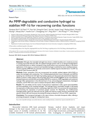
Theranostics 2022, Vol. 12, Issue 1ALG-CHO-TA was assessed by 1H-NMR. Asindicated in Figure S2, the peak at 8.3 ppm attributeto the aldehyde group and the peak at 5.6 ppmattributes to the hemiacetal group, confirmingALG-CHO was successfully synthesized. The protonpeaks appear at 6.5-7.5 ppm, representing the protonon benzene ring (Figure S2). As shown in Figure S3,the peaks of 3382 and 3030 cm-1 represent thestretching vibration of N-H and Ar-H, suggesting thesuccessful synthesis of ALG-CHO-TA. A UV-visstandard curve of TA was fabricated to determine theamount of TA in ALG-CHO-TA polymer chain. It iscalculated that the solid content of TA inALG-CHO-TA is 8.9 wt% and the grafting efficiencyof TA is 33.8%.Properties of DPCA and DPCA@PDA NPsThe DPCA@PDA NPs were fabricated as theprocedures shown in Figure 2A. As demonstrated inFigure S4, the diameters of DPCA NPs are 6.4, 40.7,and 81.2 nm when the final concentrations of DPCAare 1, 2, and 5 mg/mL, indicating a higherconcentration will lead to large sized NPs. Because asmall size is more suitable for PDA coating, the lowconcentration (1 mg/mL) of DPCA solution wasselected for further experiments. After coating withPDA, the diameter of NPs increased from 6.4 to 74.8nm (Figure 2B). The uniformly dispersed DPCA NPsare obtained in the TEM image (Figure 2C). Asindicated in Figure 2D, several DPCA nanoparticlesare encapsulated by the PDA crosslinked network,which looks like a pomegranate fruit. The sizes of132DPCA and DPCA@PDA NPs are about 3.7 and 32.8nm according to TEM images.Properties of the hydrogelsA typical thiol-aldehyde Michael addition cantake place between aldehyde group of ALG-CHO andthiol groups of HA-SH to form the hydrogel network[47]. In addition, DPCA@PDA can react with athiolated polymer chain through a thiol-benzoquinone Michael Addition reaction. Meanwhile, thiolend capped MMP-SP can participate in thecrosslinking reaction to entitle a MMP induceddegradability to the hydrogels.A parallel-plate rheometer was employed todetermine rheological properties of the resultinghydrogels. As shown in the time-sweep curve (Figure3A), G' is larger than G'' during the whole time period,revealing a crosslinked network before the sampleswere been placed on the rheometer plate. A typicalG’/G’’ crossover can hardly be observed due to therapid sol-gel transition (Table S1). The hydrogelswith various components possess a similar and softmechanical property (200-300 Pa). The gelation timecan be adjusted by the concentration of HA-SH. Asshown in Table S1, the gelation occurs within 30 swhen the concentration of HA-SH is 1.33%. Theaddition of DPCA@PDA and MMP-SP exerts fewereffects on gelation time and shows a slight increase inthe mechanical property. The injectable capability canbe clearly visualized in Figure 3B, suggesting that thehydrogel can be injected into water and maintain astable shape.Figure 2. The synthesis and characterization of DPCA@PDA NPs. (A) The synthesis process of DPCA@PDA. (B) The size distribution of DPCA and DPCA@PDA NPsassessed by DLS. TEM images of DPCA NPs (C) and DPCA@PDA NPs (D).https://www.thno.org
Theranostics 2022, Vol. 12, Issue 1133Figure 3. Properties of the as-prepared ALG-CHO-TA/DPCA@PDA/MMP-SP/HA-SH hydrogels. (A) Rheological analysis based on a time-sweep model. (B) The macro pictureto demonstrate the injectable capability of the hydrogels. (C) The shear-thinning behavior and (D) the fatigue resistance of ALG-CHO-TA/DPCA@PDA/MMP-SP/HA-SHhydrogel. (E) The macro picture indicates that the conductive hydrogel can act as a wire to light up LED. (F) The conductivity of the hydrogels. (G) The DPCA releasing behaviorin vitro. (H) The macro pictures illustrate the subcutaneous injection of hydrogels within 7 d. (I) H&E staining of the tissue around the hydrogels (black zone in the images) after7-day subcutaneous injection. (III: ALG-CHO/HA-SH; IV: ALG-CHO-TA/HA-SH; V: ALG-CHO-TA/DPCA@PDA/HA-SH; VI: ALG-CHO-TA/DPCA@PDA/MMP-SP/HA-SH.)A typical shear thinning behavior can beobserved in Figure 3C. The viscosity of the hydrogelgoes down from 3000 to 1 Pa s as the shear raterises up. Meanwhile, maintaining a stable rheologicalproperty is important for hydrogels used in acontinuous beating heart. The G' and G'' of thehydrogels remain stable at the strain range of0.1%-100% and the frequency from 0.5 to 100 rad/s(Figure S5-6). To verify a long-time stability of thehydrogel in a beating heart environment, the resultinghydrogel maintains stable after a 12000-timecontinuous shear (Figure 3D).After MI, the injury induces fibroblastproliferation to secrete extracellular matrix formingthe scar tissue, then a blockage of the transmission ofelectrical signals occurs [21, 48]. The conductive TA isreported to effectively propagate the electrical signalsin the infarcted area [25, 26]. As revealed in the UV-visspectrum of TA and ALG-CHO (Figure S7), there is asingle peak at 325 nm of TA and ALG-CHO-TA,attributing to π-π* transition of benzene ring.However, the noticeable peak at 422 nm confirms theexistence of eigenstate TA after HCl-doping, H hydrogel can light up LED asa wire (Figure 3E). As shown in Figure 3F, theaddition of TA greatly improves the conductivity ofhydrogels. The hydrogel (ALG-CHO/HA-SH)without TA possesses a conduction of 4.7 10-7 S/cm.The conductivity of hydrogel with TA is 8.8 10-5S/cm, which is consistent with the conductivity ofnative myocardium (5 10-5-1.6 10-3 S/cm) [49].Meanwhile, the TA containing hydrogels wereimmersed in PBS to test the conductivity. As shown inTable S2, the hydrogel is immersed in PBS for 24 h,the conductivity increases slightly, which mighthttps://www.thno.org
Theranostics 2022, Vol. 12, Issue 1attribute to the absorption of ions in PBS to increaseion conductivity.It is well reported that reactive oxygen species(ROS) are key signaling molecules that play animportant role in the progression of inflammatorydisorders. An enhanced ROS generation bypolymorphonuclear neutrophils (PMNs) at the site ofinflammation causes cell dysfunction and tissueinjury. A classic ROS clearance experiment wascarried out to determine the ROS scavengingcapability of the designed hydrogels. Briefly,ALG-CHO, ALG-CHO-TA, DPCA, PDA, MMP-SPand HA-SH water solutions corresponding to theconcentration in the hydrogel were prepared andadded into the ethanol solution of DPPH. Theabsorbance at 516 nm of the mixed solutions wereused to determine the ROS removal efficiency of sixcomponents. As shown in Figure S8, all the sixcomponents illustrate a different capability toscavenge ROS. PDA possesses the highest ROSclearance efficiency. 83.2% of ROS has been cleared at7 min, and the clearance rate of ROS can reach 96.7%after 48 h. In addition, compared with PDA andALG-CHO-TA, which rapidly clears ROS in the first 2h and then gradually slows down, the ROS clearancerate of HA-SH maintains stable in the first 10 h, andthe final ROS clearance efficiency is even higher thanthat of ALG-CHO-TA. This may be due to the fact thatthe concentration of HA-SH in the hydrogel is muchhigher than that of other components, which cangradually scavenge free radicals in DPPH. Theexperimental results show that each component of thehydrogel has a certain ROS scavenging effect,indicating a ROS clearance ability of the hydrogel.As illustrated in Figure 3g, DPCA inDPCA@PDA NPs can quickly diffuse to PBS solutionwithin 24 h. The DPCA drug exhibits a very fastrelease rate (90% drug released within 12 h) when it isloaded in the hydrogel without a PDA layer. On theother hand, the drug releasing maintains a stable andmuch slower speed after being loaded in thehydrogels (30% drug released after 48 h). The drugrelease behavior can be sped up by adding MMP inthe solution (50% drug released after 48 h). Theseoutcomes indicate that DPCA can be triggered to afaster release in an infarcted heart due to significantamount of over-expressing MMPs. The releasing rateof DPCA in the hydrogel without the addition ofMMP-SP was only slightly increased, which may beattributed to a higher crosslinking density due to theaddition of MMP-SP in the hydrogel. Regardless ofwhether MMP is added or not in the media, therelease rate of DPCA in the hydrogel withoutMMP-SP is basically the same. These results provethat MMP-SP confers a MMP sensitivity on the134hydrogels and both the hydrogel and PDA layer willenhance the controll
to prevent post-MI remodeling, suggesting a limited therapy effect in rat MI model [12]. To construct a cell-ingrowth matrix, the matrix metalloproteinase- sensitive peptides (MMP-SP) have been employed to construct the hydrogel networks with a rationally controlled degradation behavior which is triggered by over-expression of MMPs [13]. Under