Transcription
(2022) 22:3Saggese et al. BMC 22-9Open AccessRESEARCH ARTICLEAn antimicrobial peptide specifically activeagainst Listeria monocytogenes is secretedby Bacillus pumilus SF214Anella Saggese1 , Ylenia De Luca1, Loredana Baccigalupi2and Ezio Ricca1*AbstractBackground: Members of the Bacillus genus produce a large variety of antimicrobial peptides including linear orcyclic lipopeptides and thiopeptides, that often have a broad spectrum of action against Gram-positive and Gramnegative bacteria. We have recently reported that SF214, a marine isolated strain of Bacillus pumilus, produces twodifferent antimicrobials specifically active against either Staphylococcus aureus or Listeria monocytogenes. The antiStaphylococcus molecule has been previously characterized as a pumilacidin, a nonribosomally synthesized lipopetidecomposed of a mixture of cyclic heptapeptides linked to fatty acids of variable length.Results: Our analysis on the anti-Listeria molecule of B. pumilus SF214 indicated that it is a peptide slightly smallerthan 10 kDa, produced during the exponential phase of growth, stable at a wide range of pH conditions and resistant to various chemical treatments. The peptide showed a lytic activity against growing but not resting cells ofListeria monocytogenes and appeared extremely specific being inactive also against L. innocua, a close relative of L.monocytogenes.Conclusions: These findings indicate that the B. pumilus peptide is unusual with respect to other antimicrobials bothfor its time of synthesis and secretion and for its strict specificity against L. monocytogenes. Such specificity, togetherwith its stability, propose this new antimicrobial as a tool for potential biotechnological applications in the fightagainst the dangerous food-borne pathogen L. monocytogenes.Keywords: Bacillus, Antimicrobials, AMP, anti-Listeria, Lytic peptide, Membrane damagesBackgroundAntibiotic resistance is a major threat to global healthand is predicted to cause by 2050 millions of annualdeaths [1]. In addition to an increased incidence of infectious diseases, the lack of effective antibiotics will certainly affect many surgical procedures and treatmentsthat suppress the immune system (for example, chemotherapies) [1]. Therefore, the identification of new strategies to fight the continuous rise of resistant pathogens is*Correspondence: ericca@unina.it1Department of Biology, Federico II University, via Cinthia, ComplessoUniversitario di Monte Sant’Angelo, 80126 Naples, ItalyFull list of author information is available at the end of the articlean enormous challenge for the scientific community [1,2]. So far, alternatives to antibiotics such as vaccines andprobiotics, have been proposed together with the identification and design of new antibiotics.In this context, antimicrobial peptides (AMPs) havegained great attention as new antibiotics. They havebeen characterized from almost every organism of alldomains of life and shown to play an important role ininnate immunity, protecting the producing organismsfrom infections [1]. Many AMPs act on membrane lipidsand show a broad-spectrum of action against Gram-positive and Gram-negative bacteria, some AMPs are activeagainst fungi, viruses or other parasites [1]. AMPs fromvertebrates (defensins and cathelicidins), in addition to The Author(s) 2021. Open Access This article is licensed under a Creative Commons Attribution 4.0 International License, whichpermits use, sharing, adaptation, distribution and reproduction in any medium or format, as long as you give appropriate credit to theoriginal author(s) and the source, provide a link to the Creative Commons licence, and indicate if changes were made. The images orother third party material in this article are included in the article’s Creative Commons licence, unless indicated otherwise in a credit lineto the material. If material is not included in the article’s Creative Commons licence and your intended use is not permitted by statutoryregulation or exceeds the permitted use, you will need to obtain permission directly from the copyright holder. To view a copy of thislicence, visit http:// creat iveco mmons. org/ licen ses/ by/4. 0/. The Creative Commons Public Domain Dedication waiver (http:// creat iveco mmons. org/ publi cdoma in/ zero/1. 0/) applies to the data made available in this article, unless otherwise stated in a credit line to the data.
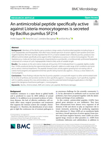
Saggese et al. BMC Microbiology(2022) 22:3the antimicrobial activity, also act as chemo-attractants,are involved in the activation of complement pathwaysand are able to modulate the immune response [1]. Over3,000 AMPs, showing an extraordinary chemical diversity and isolated from a variety of biological sources, havebeen described and are collected in specific databases(see for example: http:// aps. unmc. edu/ AP/ main. php;https:// wanga pd3. com/ main. php; https:// dbaasp. org/).Bacteria and fungi in particular have been investigatedas a source of new AMPs, and although most microorganisms produce AMPs, bacteria of the Bacillus genushave received deep attention as potential starting pointsin the search for new inhibitory substances [2]. Theserod-shaped, endospore-forming, Gram-positive bacteriaare common in many natural habitats and produce a variety of AMPs: short linear or cyclic peptides of up to 30amino acid residues; lipopeptides containing a hydrocarbon tail linked to the N-terminus of the linear or cyclicpeptide; thiopeptides, sulphur-rich peptides with thiazole, oxazole, or thiazoline rings; and 2,5-diketopiperazines (DKPs), extremely small peptides (even di-aminoacids) with structural modifications [2, 3].The best known AMPs produced by Bacilli are cycliclipopeptides of three structural categories: surfactins,iturins and fengycins. In addition, tens of linear or cycliclipopeptides not belonging to those three classes, havebeen identified and extensively reviewed by Zhao et al.[2] and Cauller et al. [3].In Bacilli AMPs biosynthesis principally occurs bytwo different biosynthetic pathways: the non-ribosomalsynthesis of peptides by large mega-enzymes, the nonribosomal peptide synthetases (NRPSs) [4, 5], and theribosomal synthesis of linear precursor peptides that aresubjected to post-translational modifications and proteolytic processing [6, 7].We have previously reported that SF214, a marine isolated strain of B. pumilus, produced two independentantimicrobials specifically active against Staphylococcusaureus or Listeria monocytogenes [8]. SF214 is known toproduce a water soluble yellow-orange pigment, essential to protect the bacterium against oxidative stresses[9, 10]. Synthesis of the still uncharacterized pigment ofSF214 is a bistable process that occurs in alternative tospore formation [10]. Pigment synthesis, sporulation andmatrix synthesis in SF214 are connected phenomena,controlled by the same master regulators [11]. The antimicrobial molecule active against S. aureus has been previously characterized as a pumilacidin, a nonribosomallysynthesized cyclic lipopeptide of the surfactin class,composed of seven amino acid residues linked to fattyacids of variable length [8]. Here is reported that the antiListeria molecule produced by SF214 is a peptide withlytic and specific activity against growing cells of ListeriaPage 2 of 11monocytogenes and that it is stable at conditions of lowpH and high temperature.Results and DiscussionExponentially growing cells of B. pumilus SF214 producea stable anti‑Listeria moleculeBacillus pumilus SF214 produces during its exponentialphase of growth an antimicrobial molecule apparentlybigger than 10 kDa active against the Listeria monocytogenes strain ATCC 7644 [8]. Production of antimicrobials during active growth is not common since mostAMPs are secondary metabolites produced during thestationary phase of growth [3]. To confirm the previous observation and further characterize the synthesisof the anti-Listeria molecule, SF214 cells were grownin S7 medium at 25 C, and samples collected at various times. For each time point, the cell-free supernatantwas size-fractionated and the 10 kDa fraction tested forthe anti-Listeria activity, as previously reported [8]. Thecell extracts of all time points were assayed for activities known to occur during the exponential or stationaryphase of growth: the glucose-6-phosphate dehydrogenase(G6PDH) and the production of the orange pigment,respectively [12, 11]. As reported in Fig. 1a, the anti-Listeria and G6PDH activities appeared during the activegrowth of SF214 cells (panel a) while the production ofthe pigment started later, at the entry into the stationaryphase of growth (panel a). Results of Fig. 1a confirmedthat actively growing cells of SF214 produce the antiListeria molecule but also indicated that the activity ofsuch molecule appeared about one hour later than theG6PDH activity. This delay may be due to the secretionand eventual maturation/processing steps involved in theactivation of some AMPs. An additional indication coming out of Fig. 1a is that the anti-Listeria activity persistedfor several hours after its appearance.This latter observation may be explained by a prolonged synthesis of the molecule, starting during theexponential growth and continuing during the stationary phase, or by the stability of the molecule that onceproduced remains active for a long time. To distinguishbetween these two possibilities, SF214 cells were grownas described above and the antibiotic chloramphenicol (5 µg/ml) added when cell reached 0.7 O D600 (redarrow in Fig. 1b), immediately blocking growth. Cell-freesupernatants, collected right before as well as 1 and 3 hafter the antibiotic treatment, were size-fractionated and 10 kDa fractions tested for the anti-Listeria activity inplate assays. An identical antimicrobial activity (mm ofinhibition halo) was observed in the three samples, indicating that the inhibition of protein synthesis and growthdid not reduce the antimicrobial activity and, therefore, suggesting that, once produced, the anti-Listeria
Saggese et al. BMC Microbiology(2022) 22:3Page 3 of 11Fig. 1 Production of the antimicrobial during growth (a) and after a treatment with chloramphenicol (b). In (a), aliquots of the cell culture werecollected at all-time points, the cell-free supernatant size-fractionated and the 10 kDa fraction tested against L. monocytogenes [8]. For all timepoints, cell extracts were analyzed for G6PD activity and pigment production as markers of of exponential and stationary phase of growth,respectively. The antimicrobial activities are reported as % of growth inhibition (mm of inhibition halo on plates) considering 100% the maximalactivity observed. In (b) the red arrow indicates the time of chloramphenicol supplementation. Grey bars indicate the % of antimicrobial activity(mm of inhibition halo on L. monocytogenes plates) considering 100% the maximal activity observed before the antibiotic treatmentTable 1 Effect of pH on the anti-Listeria activityaapH1 hour5 hours27.57.547.57.571010101010131010aDiameter (mm) of the inhibition halo in plate assaymolecule remains stable for at least three hours in thecytoplasm of resting cells (Fig. 1b).Stability of the anti-Listeria molecule was also testedover a wide range of pH and temperature conditions andafter treatments with chemicals and enzymes. The antiListeria molecule was similarly active against its targetcells after 1 or 5 h of incubation at pH values rangingfrom 7.0 to 13.0, was only slightly reduced at pH 2.0 and4.0 (Table 1). It was fully active after 15 min of incubationat 60 or 80 C (Table 2) and slightly reduced after 15 minat 100 C (Table 2). None of the organic solvents testedshowed any effect on the antimicrobial that was totallydegraded by both a trypsin or a proteinase K treatment(Table 2). Altogether, results of Tables 1 and 2 indicatethat the anti-Listeria molecule is highly stable and of proteinaceous nature.The anti‑Listeria molecule of SF214 is specifically activeagainst L. monocytogenesThe anti-Listeria molecule of SF214 has been previously found inactive against all other the Gram positive(Streptococcus faecalis, Staphylococcus aureus, BacillusTable 2 Effect of enzymes, solvents and heat on the anti-ListeriaactivityTreatmentActivity onplate assayaNone10TrypsinbProteinase Kb00DNaseb10Ribonuclease Ab10Acetonec10Ethyl alcoholc10Chloroformc10Toluenec1015 min 60 C1015 min 80 C1015 min 100 C8aDiameter (mm) of the inhibition halo in plate assaybEnzyme concentration was 100 µg/mlcA 10% (v/v) concentration was usedmegaterium, Mycobacterium smegmatis, Enterococcusfaecalis) or negative (Citrobacter freundii, Pseudomonasfluorescens, Shigella sonnei, Escherichia coli, Salmonellaenterica) bacteria tested [8]. Here, the 10 kDa fractionof the SF214 cell-free supernatant was tested and foundactive against five other strains L. monocytogenes and butinactive against of Listeria innocua (Table 3). L. innocuais a non-pathogenic species of the Listeria genus,very similar to L. monocytogenes [14]. The two specieshave a genome of very similar size (2.94 and 3.01 Mbfor L. monocytogenes and L. innocua, respectively),
Saggese et al. BMC Microbiology(2022) 22:3Page 4 of 11Table 3 List of Listeria strains used for the antimicrobial activitySpeciesStrainListeria monocytogenesATCC 7644Listeria monocytogenesATCC 19,115Listeria monocytogenesLM012018aListeria monocytogenesLM001bListeria monocytogenesATCC 13,932Listeria monocytogenesATCC 19,111Listeria innocuaATCC 33,090Listeria innocuaBUG 499 [13] cListeria innocuaDPC 1770dGenerous gifts from aM. Guida (Federico II University, Naples, Italy); bE. Ghelardi(University of Pisa, Pisa, Italy); cB. Dupuy (Inst. Pasteur, Paris, France) dD. Ercolini(Federico II University, Naples, Italy)characterized by a high number of genes coding for surface and secreted proteins, transporters, and transcriptional regulators [15]. Although the human pathogen L.monocytogenes and the non-pathogenic L. innocua arestrict relatives [14, 15], a comparative proteome analysisof the secreted and cell wall-associated proteins of thetwo bacteria revealed over 50 L. monocytogenes-specificproteins [16]. In addition to known virulence factors,this subgroup of proteins includes proteins with a putative role in cell wall metabolism, transporters, penicillinbinding proteins, cell division and motility proteins [15].The anti-Listeria molecule of SF214 is, therefore, highlyspecific and for this reason peculiar with respect to otherAMPs that have a broad spectrum of action [1–3]. Examples are the recently discovered Amyloliquecidic GF610of B. velenzensis [17] and a molecule produced by B.amyliquefaciens JFL21 [18], both active against Listeriabut also against several Gram-positives and the latter alsoagainst some fungal pathogens. For its specificity and itsproteinaceous nature the SF214 molecule will be thereinindicate as specific anti-Listeriamonocytogenespeptide(SAMP) and all further analyses performed with L.monocytogenes ATCC 7644 as the target bacterium.SAMP has a lytic activityIn order to characterize the antimicrobial activity ofSAMP, L. monocytogenes cells were grown in LB mediumat 37 C, up to 0.3 OD600 (black arrow in Fig. 2a), thensupplemented with different amounts of the 10 kDafraction of the cell-free supernatant of SF214 and growthmonitored for several hours. While 5% and 10% (vol/vol)of supernatant showed respectively a slight reduction ofgrowth and a bacteriostatic effect on L. monocytogenes,20% of it rapidly killed all cells, suggesting for the antimicrobial a lytic activity (Fig. 2a). Stationary cells of L.monocytogenes not treated with the anti-microbial werecollected at the time indicated by the red arrow in Fig. 2a,incubated with supernatant 20% (vol/vol) for 3 and 16 hand plated for CFU analysis. No CFU reduction wasobserved (not treated: T0 1.10 109; T3 0.99 109;9 T16 1.28 10 CFUs; after supernatant treatment: T0 1.10 109; T3 1.30 109; T16 1.29 109 CFUs),Fig. 2 Antimicrobial activity of the supernatant ( 10 kDa fraction of the cell-free supernatant) of SF214 on L. monocytogenes cells. (a) Differentamounts of supernatant were added during growth at the time indicated by the black arrow. The red arrow indicates the time of collection of cellsto test Antimicrobial activity on resting cells (see text). (b) MTT assay performed on L. monocytogenes cell without any supplementation (Control) orsupplemented with the 10 or 10 kDa fractions of the SF214 cell-free supernatant
Saggese et al. BMC Microbiology(2022) 22:3indicating that the antimicrobial was only active againstgrowing cells of L. monocytogenes.The lytic activity of SAMP was also confirmed by acolorimetric (MTT) assay that, by measuring the intracellular NAD(P)H-dependent oxidoreductase activity,indicates the metabolic activity of cells. As shown inFig. 2b, growing cell of L. monocytogenes (OD600 0.6)treated with 20% (vol/vol) of the 10 kDa fraction of theSF214 cell-free supernatant, showed a strong reduction ofthe enzymatic activity and therefore of their viability withrespect to the untreated cells and to cells treated with thesame amount of the 10 kDa fraction of the SF214 cellfree supernatant, inactive against L. monocytogenes.The effects of SAMP on L. monocytogenes cells werevisualized by fluorescence microscopy after co-stainingof the cells with 4,6-diamidino-2-phenylindole (DAPI)and propidium iodide (PI). The membrane-permeableDAPI stains all cells that appear blue while PI enters onlydamaged/death cells that appear red [19, 20]. As reportedin Fig. 3, only blue cells were observed when the supernatant was not used while death (red) cells appearedafter the antimicrobial treatment. Incubation for 4 h withincreasing concentrations of the antimicrobial producedan increasing number of damaged/death (red) cells andonly red cells were found at the highest concentration ofantimicrobial used in the experiment (20%) (Fig. 3). Thisassay clearly shows an increased permeability of the L.monocytogenes cells in response to the treatment, indicating that the bactericidal effect occurs through membrane permeabilization and surface damages.Page 5 of 11The morphological defects due to the supernatant werevisualized by a Scanning Electron Microscopy (SEM)analysis. L. monocytogenes cells treated for 4 h with 20%(vol/vol) supernatant showed distinct morphologicalchanges (Fig. 4). Untreated cells appeared intact (Fig. 4a)while supernatant-treated cells showed on their surfacecell debris (white arrows in Fig. 4b, c, d), an indicationof the loss of cell integrity, similar to what detected withother antimicrobials affecting the membrane integrity[20, 21]. Interestingly, the defects observed after treatment with SAMP appeared different from those causedby another anti-Listeria compound. After a treatmentwith the commercially available Linalool the Listeria cellsurface appeared by SEM analysis as a wrinkled [22] anddid not show the cell debris observed in Fig. 4, suggestinga different mechanism of action for Linalool and SAMP.To evaluate the toxicity of SAMP against eukaryoticcells, an MTT assay with human epithelial (Caco-2) cellswas performed. SF214 supernatant w (20% vol/vol) wasadded to the growth medium of the Caco-2 cells. Noeffects on cell viability were observed after 24 h of incubation suggesting that SAMP is not toxic for human cells(not shown).Characterization of SAMPTo expand the preliminary observation that SAMP has aproteinaceous nature (Tables 1 and 2), the 10 Da fraction of the SF214 supernatant was analyzed on a 18%SDS-PAGE. As shown in Fig. 5a, about a dozen proteinsranging from slightly less than 10 to over 50 kDa wasFig. 3 Viability assay by fluorescence and phase-contrast (PC) microscopy of L. monocytogenes cells stained with DAPI and PI. Cells were not treated(NT) or treated with increasing concentration of supernatant (from 5 to 20% vol/vol)
Saggese et al. BMC Microbiology(2022) 22:3Page 6 of 11Fig. 4 Scanning Electron Microscopy (SEM) analysis of L. monocytogenes cells not treated (a) or treated for 4 h with 20% (vol/vol) of supernatant(b,c,d). White arrows indicate points of cell ruptureobserved. A second gel was run in parallel, not stainedwith Coomassie-Blue but fixed and overlaid with softagar containing L. monocytogenes cells for direct detection of antimicrobial activity as previously described [23].As shown in Fig. 5b, the anti-Listeria activity was associated with a band of apparent molecular mass slightlysmaller than 10 kDa.Although smaller than 10 kDa (Fig. 5b), SAMP did notpass through filters with a 10 kDa cut-off, suggesting theformation of aggregates dissolved during the SDS PAGE.To evaluate whether such potential high molecularweight aggregates were due to hydrophobic interactions,different amounts of methanol were added to the cellfree supernatant of SF214 to obtain final methanol concentrations of 20, 50 or 75%. Methanol was selected assolvent because it is known to weaken the hydrophobicinteractions between proteins or between amino acidsof the same protein [24] and because it did not have anyinhibitory activity on L. monocytogenes at the used concentrations (not shown). After size-fractionation the 10and 10 kDa fractions of the supernatant were dilutedto the same final volume (10 ml) and aliquots tested foranti-Listeria activity. While 20% methanol did not haveeffects on SAMP and the anti-Listeria activity was foundonly in the 10 kDa fraction, in the presence of 50 and75% methanol the activity was found only in the 10 kDafraction (Table 4), suggesting that in 50 and 75% methanol SAMP did not form aggregates as in water or 20%methanol. Formation of high molecular weight aggregates and micelles by antimicrobials produced by Bacillushas been recently reported [25], supporting the conclusion based on data of Fig. 5; Table 4.Genome analysisThe genome sequence of SF214 [11] was analysed tosearch for homologs of genes coding for potential antimicrobial peptides and for NRPS/PKS, responsible forthe non-ribosomal synthesis of peptides and polyketides.The coding gene of most ribosomally-synthesized antimicrobials produced by members of the Bacillus genus hasnot been identified, therefore the search for homologswas limited to some of the AMPs whose coding geneis known. As reported in Table 5, the SF214 genomeencodes proteins with only a limited identity with knownantimicrobials produced by Bacilli. The highest identity observed was 53% found between WP 060596419.1of SF214 and Subtilosin A of B. subtilis (Table 5). However, Subtilosin A unlikely corresponds to SAMP since
Saggese et al. BMC Microbiology(2022) 22:3Page 7 of 11Fig. 5 SDS-PAGE analysis of the 10 kDa fraction of the cell-free supernatant of SF214. (a) Coomassie-Blue stained 18% polyacrylamide gel. (b) 18%polyacrylamide gel not stained but fixed and overlaid with L. monocytogenes cells. The red arrow points to the inhibition halo observed after 18 h ofincubation at 37 C. M: molecular markerTable 4 Effect of methanol on antimicrobial activityMethanol(%)020a 10 kDa 50-75-aa 10 kDa Inhibition halo in plate assay: - no halo; presence of haloit is active against members of the Bacillus and Enterococcus genus [26] that are not affected by SAMP [8]. Abioinformatic analysis, performed by using the AntiSmash(https:// antis mash. secon darym etabo lites. org)and tools available on the PKS/NRPS Analysis Website(http:// nrps. igs. umary land. edu/), identified four clusterspotentially coding for NRPS/PKS. These four clusterswere further analysed by the HMMER tool (https:// www. ebi. ac. uk/ Tools/ hmmer/) [27] to predict their potentialproducts (Table 5). Cluster SF214 0323-SF214 0328 corresponds to the srfA-sfp locus, responsible of the synthesis of the Pumilacidin active against S. aureus andpreviously characteried [8], while the other three clusters were predicted to encode products formed by one(SF214 0601), two (SF214 3715-SF214 3717) or six(SF214 0611-SF21 0615) amino acids (Table 6), all toosmall for the predicted size of SAMP. Therefore, we conclude that none of the clusters identified by our in silicoanalysis is likely to encode for the amino acids formingSAMP. Based on the results of Tables 5 and 6, we hypothesize that SAMP is produced by transcription and translation of a structural gene and that is not a previouslycharacterized molecule. Confirming such hypothesis will
Saggese et al. BMC Microbiology(2022) 22:3Page 8 of 11Table 5 Percentage of identity of known ribosomallysynthesized antimicrobials of Bacilli with proteins encoded bythe SF214 genomeAntimicrobialsHighest Identity(%) hiton the SF214translated genomeSubtilin [27]WP 060597202.1 (37)Bacthuricin F4 [28]WP 060595519.1 (31)Subtilin JS-4 [29]WP 060597425.1 (34)Cerecin [28]WP 060597533.1 (39)Antimicrobial peptide LCI [30]WP 060596412.1 (47)EricinA [27]WP 060597202.1 (36)Mersacidin [27]WP 060596204.1 (34)EricinS [27]WP 012010311.1 (45)Sublancin 168 [27]WP 050944681.1 (42)Subtilosin A [27]WP 060596419.1 (53)Clausin [30]WP 060595627.1 (42)Lichenicidin [30]WP 003217179.1 (43)Plantazolicin [30]WP 060596638.1 (38)Haloduracin [31]WP 060595537.1 (25)Amyloliquecidin GF610 [17]WP 034659961.1 (43)Coagulin A [28]WP 060596970.1 (31)Table 6 Genes potentially coding for NRPS/PKSa in the SF214genomeClusterPredicted productSF214 0323-SF214 0328glu-leu-leu-X-asp-leu-ile-valSF21 0601asnSF214 0611-SF214 0615asn-ohmal-glu-ohmal-ohmal-ohmalbSF214 3715-SF214 3717gly-tyraNRPS: non-ribosomal peptide synthase, PKS: polyketide synthase;bohmal: hydroxymalonilbe a future challenge that will need to be based on thechemical characterization of the antimicrobial, a step sofar impaired by the small amount of SAMP produced bySF214.ConclusionsThe main conclusion of this report is that B. pumilusSF214 during its exponential phase of growth producesan antimicrobial with a lytic activity specifically againstgrowing cells of L. monocytogenes. Production of anantimicrobial by a Bacillus strain is clearly not a newobservation since hundreds of them have been previously described. However, SAMP has unravelled somepeculiar properties that make this molecule unusualand worth studying: - SAMP is not a typical secondary metabolite, asmost AMPs [2, 3, 6, 7]. It is produced at the midexponential phase of growth of B. pumilus SF214 butabout one hour later than G6PDH. Such delay couldbe due to the need of post-synthesis maturationevents (for example, processing of a secretion signal),inducing the hypothesis that it is a ribosomally synthesized peptide. This hypothesis is also supportedby the in-silico analysis of the SF214 genome, that didnot show the presence of homologs of already knownAMPs or of genes coding for NRPs suited to synthesize the anti-L. monocytogenes molecule. - SAMP affects cell wall integrity of growing but notresting cells of L. monocytogenes, suggesting the cellwall synthesis machinery rather than structural components of the cell membrane as the target of action. - SAMP is specifically active against L. monocytogenes and is not active even against strains of theclose relative species L. innocua. - SAMP is not toxic for human cells.MethodsBacterial strains, growth conditions and preparationof cell‑free fractionsBacillus pumilus SF214, Listeria monocytogens and L.innucua strains reported in Table 3 were grown eitherin LB broth (8 g/L NaCl, 10 g/L tryptone, 5 g/L yeastextract), BHI (Brain Heart Infusion) or (only B. pumilus)in S7 minimal medium (50 mM morpholine-propanesulfonic acid (MOPS) (adjusted to pH 7.0 with KOH), 10mM (NH4)2SO4, 5 mM potassium phosphate (pH 7.0), 2mM MgCl2, 0.9 mM C aCl2, 50 µM M nCl2, 5 µM F eCl3, 10µM ZnCl2, 2 µM thiamine hydrochloride, 20 mM sodiumglutamate, 1% glucose, 0.1 mg/mL phenylalanine, and0.1 mg/mL tryptophan) and cells grown in aerobic conditions at 25 C. SF214 cultures were centrifuged (1000 g for 10 min at Room Temperature) and the supernatantfilter-sterilized with a 0.22-µm filter (Millipore, Bedford,MA, USA). The supernatants were size-fractionated (10kDa cutoff spin column; Centricon, Millipore) by centrifuging 11,000 g for 10 min at 4 C. The fraction 10 kDawas concentrated 5 folds by a vacuum speed concentrator (Eppendorf ).Antimicrobial activity on Plate Assay and on gelAntimicrobial activity was determined as previouslydescribed [23] with the following modifications: 10 µLof concentrated (see above) 10 or 10 kDa fractions ofthe cell-free supernatant were spotted on the surface of asterile LB agar plate and the spots air-dried. 100 µL of L.monocytogenes or L. innocua culture was mixed with 10mL of soft agar (0.7%) and poured over the plate. Fresh
Saggese et al. BMC Microbiology(2022) 22:3media and ampicillin (1 µg/mL) were used as negativeand positive controls, respectively. The plates were incubated aerobically overnight at 37 C and the inhibitionhalos were measured and reported in mm.For the direct detection of the antibacterial activityon gel, 50 ug of total proteins of the 10 Da fraction ofSF214 cell-free supernatant were split in two identicalsamples and loaded in two independent lanes of a sodiumdodecyl sulfate-polyacrylamide gel electrophoresis (SDSPAGE, 18%). After about 1.5 h, the gel was removed andcut into two vertical parts. One part of the gel, containing one sample and the molecular weight standards, wasstained with Coomassie brilliant blue R250 (Sigma). Theother part of the gel was tested for antimicrobial activityas previously described [23] with the following modifications: the gel was fixed immediately by a 2-h treatmentin 20% isopropanol-10 mM Tris-HCl (pH 7.5) and 1-hourtreatment in 20% isopropanol (pH 7.5) and 10% aceticacid, and at the end washed for 3 h in 10 mM Tris-HCl(pH 7.5). The gel was then placed into a petri dish, airdried for 10 min and overlaid with 15 ml of 0.7% agarcontaining 106 cells of the indicator strain. The dish wasthen incubated a
and show a broad-spectrum of action against Gram-pos-itive and Gram-negative bacteria, some AMPs are active against fungi, viruses or other parasites [1]. AMPs from . Grey bars indicate the % of antimicrobial activity (mm of inhibition halo on L. monocytogenes Listeria Listeria a b 10 10. of peptide monocytogenes SAMP, , 9; .