Transcription
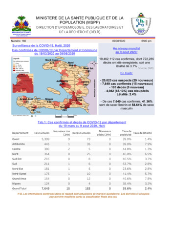
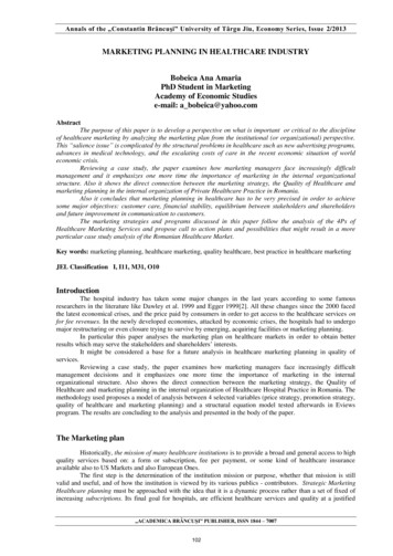
Vscan ExtendDatasheetVscan ExtendVscan Extend is a general purpose diagnostic ultrasoundimaging system that can enable qualified and trained healthcareprofessionals to visualize and measure anatomical structuresand fluid.Its pocket-sized portability and simplified user interfaceenable integration into examination and training sessions.The information can be used for basic or focused assessments.It can also be used along with other medical data for clinicaldiagnostic purposes during routine periodic monitoring andtriage assessments for adult and pediatric patients. VscanExtend can also be used for procedural guidance. It meetsrequirements of use in the home healthcare environment.Vscan Extend is available in two probe configurations – eitherwith sector probe1 or with a dual-headed probe that integratesboth sector and linear transducers.gehealthcare.comVscan Extend is offered in three connectivity configurations2: USB configuration allows the transfer of images or videos viaUSB cable connection to a PC Wi-Fi access configuration includes USB features and adds theability to export images wirelessly to shared Windows networkfolders, along with wireless access to the GE Marketplace fordownload and installation of Vscan Extend apps The DICOM configuration includes USB and Wi-Fi accessfeatures and adds the ability to wirelessly communicate withDICOM serversVscan Extend customers have access to the Vscan web portal,including online access to product and clinical reference materials.
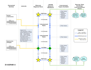
General SpecificationDimensions and WeightDisplay unit: 168 x 76 x 22 mm, 321 grams (including battery)Display: 12.7 cm, 720 x 1280 pixels resolutionSector probe: 129 x 32 x 25 mm, 85 gramsDual probe: 129 x 39 x 28 mm, 120 gramsImagingBlack-and-white mode for displaying anatomy in real-timeLinear Array Transducer for Shallow Scanning(Available with Dual Probe) (cont.)With the addition of the linear array transducer on thedual-headed probe solution, the specific clinical applicationsand exam types are expanded to include: peripheral vascularimaging (e.g. lower extremity, carotid), procedure guidancefor arterial or venous vessels (e.g. central lines, upper extremity),small organs including thyroid, musculoskeletal (long bone,hip, shoulder, elbow and knee joints), evaluation of presenceof fluid; thoracic/lung (e.g. pleural motion/sliding, line artifacts),ophthalmic,3 and pediatricsColor-coded overlay for real-time blood flow imagingTotal scan time of 60 minutes with new fully charged battery(with 80% black-and-white/20% color imaging)Universal power supply with 100 – 240 V, 50/60 Hz. Globalplugs enable recharge of battery in 75 minutes for 90%battery capacityProbe CharacteristicsVscan Extend is Available in Two Probe Configurations:Sector probe allowing deep scanningDual probe integrating both sector and linear transducers,allowing deep and shallow scanningSector Transducer Holding a Phased Array for DeepScanning (Available with Sector and with Dual Probe)User InterfaceBasic ultrasound imaging with minimized number of keys andintuitive thumb-controllable user interface for rightand left-hand users (without reconfiguration)Swiping menu to enable easy reach of advanced functionsto manage exams and dataPresets to help simplify optimized settings for imagingdifferent organs. Three presets can be set as favoritesfor quick accessProtocols to guide through series of steps during the ultrasoundexamination with automated labeling of stored imagesAutoCycle (cardiac preset) to automatically detect full heartcycle for easy and fast review or storage or reviewDuration of clip stored (non-cardiac presets) configurable toone, three or four secondsBroad-bandwidth phased array: 1.7 – 3.8 MHzField-of-view for black-and-white imaging: up to 70 degreeswith maximum depth of 24 cmColor flow sector represents blood flow within an angle ofup to 40 degreesSpecific clinical applications and exam types include: cardiac,abdominal, renal, obstetrics and gynecology, urology, fetal,evaluation of presence of fluid, imaging guidance for needle/catheter placement (e.g. paracentesis, pericardiocentesis,thoracentesis, amniocentesis), peripheral vascular imaging(e.g. arteries and veins), thoracic/lung (e.g. pleural motion/sliding, line artifacts), adult cephalic, and pediatricsLinear Array Transducer for Shallow Scanning(Available with Dual Probe)Broad-bandwidth linear array: 3.3 – 8.0 MHzField-of-view for black-and-white imaging: aperture width of2.9 cm with maximum depth of 8 cmColor flow sector represents blood flow over image with fullaperture and entire depthMeasurementsDistance measurement on-board deviceA urinary bladder volume measurement tool is availableas Vscan Extend app through GE Marketplace (optionallyavailable with Wi-Fi access or DICOM configuration)
Data StorageImage ExportData SecurityGE process todesign for privacyand securitySecured data at restSecured data onthe move Privacy Impact Assessment coversdata flows where data leaves thetrusted boundary of the device.Potential risks are identified andmitigated as part of product design Security Risk Assessment to identifysecurity risk to confidentiality andintegrity of patient data, and tomitigate risks as part of product design Images anonymized FIPS 197 compliant databaseencryption (SQL Cipher with PBKDF2based key generation and AES-256bit encryption) Password protected access topatient data on Vscan Extend device Inter-application data flow is encrypted Support of enterprise-grade wirelessencryption standards including EAPand WPA2 (PSK) Restricted administrative access tocritical settings like Wi-Fi encryption orWi-Fi certificate installationWi-Fi SpecificationIEEE 802.11 b/g/n supported4Data LabelingExams can be manually labeled with patient informationDevice synchronizes on-board DICOM Modality Worklist onrequest (option available with DICOM configuration). Suchworklist supports consistent labeling of images, videos andexams before export to DICOM PACSOn-Board of DeviceData is stored in device memory and can be backed up onMicroSD or MicroSDHC cardData is stored in generic formats: jpg for still frames, mp4for videosData is stored in examination folders that can be linked withpatient identificationAll data can be recalled for reviewData encryption is providedSelected images, videos or exams can be exported to PCwhen the device is connected via USB cable. The anonymizeddata will be seen as on a memory stickSelected images, videos, or exams can be wirelessly exported ingeneric formats (jpg, mp4) to Windows shared network folders(option available with Wi-Fi access or DICOM configuration)Selected images, videos, or exams can be wirelessly exportedin DICOM format to DICOM PACS (option available withDICOM configuration)Vscan Extend Uplink apps support easy export of selectedimages, videos, or exams to cloud-based image managementsolutions to support ultrasound case communication andcollaboration (options available with DICOM configuration)Supported DICOM Functionalities (Option Available withDICOM Configuration)VerifyModality WorklistStoreGE Marketplace (Option Available withWi-Fi Access or DICOM Configuration)Enabling the update of Vscan Extend firmware, GE Marketplace,GE Kiosk and the basic scanner application through theinternet. New versions of these software applications can beautomatically detected when connected to the GE Marketplace.Updates can then be manually downloaded and installedHost of Vscan Extend apps available over the InternetAccess to available apps with screenshots and additionalinformationNotification of newly available apps or app updates by e-mailto the customer account. App updates can beinitiated from the device itselfVscan Web PortalOnline services to enhance the Vscan experience byproviding resources from product information to clinicaland service supportSpecific application references and product education, includingan interactive basic ultrasound anatomy section, probeplacement tips and reminders, and example imagesAdditional education resources to be posted on the Vscanweb portal, including webinars, further online programs,and training opportunities
Standard ConfigurationThe following itemsare included in thestandard VscanExtend Vscan Extend device with eithersector probe or dual probe Global AC adapter withinterchangeable region-specific plugs One rechargeable battery USB cable Two MicroSD memory cards5 Soft case User manual6 Hardcopy quick card Gel (60 g bottle)7Selectable AccessoriesSafety ConformanceSafety ClassificationClassified as internally powered medical electricalequipment when not connected to the AC adapter witha type BF applied part medical electrical equipment,according to IEC 60601-1CE marked to European Medical Devices Directive(MDD) 93/42/EECCertified to CAN/CSA C22.2 No.601.1Conforms to theapplicable clauses ofthe following safetystandardsAdditional soft caseRobust case to carry complete Vscan Extend setRobust case to carry only scanner, gel, and potentialextra batteryExternal battery chargerAdditional batteryAdditional AC adapterHardcopy user manualGeneric image format (jpg, mp4) for data stored on device or exported to PCImage transfer to PC via USB cableManual labeling of exam data with patient IDFIPS-compliant data encryptionData backup capability on MicroSD cardWireless image transfer to shared network foldersEnterprise-grade wireless encryption standards including EAP and WPA2 (PSK)Access to GE Marketplace to selectively download and install Vscan Extend appsWireless image export in DICOM formatAccess to reference materials on Vscan web portalIEC 60601-18IEC 60601-1-28IEC 60601-1-48IEC 60601-1-68IEC 60601-2-378IEC 60601-1-11IEC 60601-1-12NEMA UD3ISO 10993-1EN 300 328IEC 623048IEC 623668EMC emissions group one class Brequirements per CISPR 11/EN 55011AC adapter classified as class II medical electrical equipmentaccording to IEC 60601-1Vscan Extend is Available in Three Connectivity Configurations:Wireless reading of DICOM Modality Worklist USBWi-Fi AccessDICOMConfiguration Configuration Configuration
Vscan Extend AppsApps give you the option to customize your Vscan Extend to efficiently fit the care areas you already serve,and to manage more clinical care scenarios.9 Apps are selectively downloadable from the GE Marketplace overthe internet for installation on the device.Auto OptimizeEnables automated TGC (Time Gain Compensation) with a single key stroke during live scanning. Gain will be automatically adjusted forall depths. (Auto Optimize is not available for ophthalmic scanning).Bladder VolumeProtocol-enabled measurements enable calculation of urinary bladder volume. Edge-detection algorithm suggests automaticallyplaced measurement calipers for sagittal and transverse views. Measurements can be accepted to manually adjusted beforebladder volume is calculated.Comprehensive LabelWirelessly exported .jpg files and .mpg clips can be complemented by patient identifier and scan information, as recommended byultrasound documentation standards such as AIUM and DEGUM. Linked patient name, patient ID, date of birth, exam date, examnumber, transducer name, chosen preset, transmit frequency, MI and TI, transmit focus point, and facility name will be part ofexported image or clip.Enterprise Archive UplinkThe Enterprise Archive Uplink app7 provides the interface to GE’s Centricity Enterprise Archive , a vendor neutral archive solution forboth DICOM and non-DICOM content. The Enterprise Archive Uplink app enables a secure export of Vscan Extend ultrasoundDICOM images to Centricity Enterprise Archive via the DICOMweb standard STOW-RS. (Centricity Enterprise Archive needs to bepurchased separately).Lung M-ModeThe Lung M-Mode app provides the M-Mode capability specifically to support the assessment of lung and documentation of signslike seashore.After entering this lung M-Mode tool, a centered vertical M-Mode cursor line will be applied to generate the anatomical M-Mode display. Such tool will be enabled for linear transducer with lung preset and sector transducer with cardiac preset (the recommended presetfor lung assessment with the sector transducer).Lung ProtocolThe protocol systematically guides the user to acquire and evaluate thoracic ultrasound images. A simple report summarizes findings. Temporary hints help users understand the functions of soft keys and can be deactivated if not needed. The protocol can beconfigured by number of thoracic areas and by choice between qualitative assessment and scoring.LVivo EFThe LVivo EF app enables an automated edge detection of left myocardial wall and calculates end-systolic, end-diastolic left ventricular volumes and ejection fraction using apical 4-chamber views. This app has been developed by and licensed from DiA ImagingAnalysis Ltd. (App is available for purchase).Protocol CreatorCreate a customized exam script that will help ensure exam consistency by guiding you through an ultrasound image acquisitionprotocol. These protocols can help increase consistency with exams by providing a list of scan planes and context-sensitive help.Automated presets for all steps and automated annotations for saved images are provided to help enhance productivity. Users canalso customize protocols to include ultrasound images that can be used as reference during the exam.
Vscan Extend Apps (cont.)Scan Coach FATEThe Scan Coach module for Focused Assessment Transthoracic Echo (FATE) provides a protocol of standard ultrasound imagingviews and context-based reference materials to perform a systematic FATE exam.During the exam, users will have access to a reference ultrasound image of normal anatomy and examples of common pathologies foreach scan plane.It provides 3D animations to help remind the user the relationship of probe positioning with resulting ultrasound images andannotated schematics for anatomical landmarks help acquire desired views.The steps overview page provides a checklist of views defined by the protocol to track completion during the exam.User instructions at the beginning of every step provide information related to the specific view and can be deactivated if not needed.Scan Coach FCUScan Coach module for Focused Cardiac Ultrasound (FCU) evaluation provides a protocol of standard ultrasound imaging viewsand context-based reference materials to perform a systematic evaluation of the heart.During the exam, users will have access to a reference ultrasound image of normal anatomy and examples of common pathologies foreach scan plane.It provides 3D animations to help remind the user the relationship of probe positioning with resulting ultrasound images and annotated schematics for anatomical landmarks help acquire desired views.The steps overview page provides a checklist of views defined by the protocol to track completion during the exam.User instructions at the beginning of each step provide information related to the specific view and can be deactivated if not needed.Scan Coach RHDScan Coach module for Rheumatic Heart Disease (RHD) evaluation provides a protocol of standard ultrasound imaging views andcontext-based reference materials to perform a systematic evaluation for presence of RHD.During the exam, users will have access to a reference ultrasound image of normal anatomy and examples of common pathologies foreach scan plane.It provides 3D animations to help remind the user the relationship of probe positioning with resulting ultrasound images and annotated schematics for anatomical landmarks help acquire desired views.The steps overview page provides a checklist of views defined by the protocol to track completion during the exam.User instructions at the beginning of each step provide information related to the specific view and can be deactivated if not needed.Screen MirrorMirror the display of the Vscan Extend onto a wireless display via Miracast.10 You will have the ability to share and collaborate withother healthcare providers and patients.Tricefy UplinkTricefy Uplink app7,11 supports easy export of selected images, videos or exams to Tricefy to support ultrasound case communicationand collaboration over the cloud. Tricefy is an integrated cloud-based solution from Trice Imaging.
1. Used as synonym to phased.2. All configurations may not be available in every country. Table comparing featuresfor these three configurations is provided in “Standard Configuration” section.3. Ophthalmic preset is not available in the U.S.A., China and Japan.4. Only 2.4 GHz supported (5 GHz are not supported).5. For potential service needs only (one empty card for temporary files, one with software incase of re-installation needs).6. Electronic CD or paper copy in selected countries.7. Not available in all countries.8. Including national deviations.9. All apps may not be available in every country.10. Screen Mirror app can connect displays which are Miracast-certified. For displays thatdo not have a built-in Miracast support, adapters (dongles) that plug into the HDMIports can be purchased separately. There is no need for an internet connection, as theMiracast employs peer-to-peer Wi-Fi Direct standard.11. The Vscan Extend app includes the interface to Tricefy, a cloud-based case exchangesolution which is separately provided by Trice Imaging. Customers may elect to tryTricefy via trial period by entering into an agreement with Trice Imaging. Trice Imagingbears sole responsibility for the Tricefy Uplink app and Tricefy cloud solution.Imagination at work 2018 General Electric Company – All rights reserved.GE Healthcare reserves the right to make changes in specifications and features shownherein, or discontinue the product described at any time without notice or obligation.Contact your GE Healthcare representative for the most current information. GE, the GEMonogram, imagination at work, Vscan Extend, Vscan and Centricity Enterprise Archiveare trademarks of General Electric Company. GE Healthcare, a division of General ElectricCompany. DICOM and DICOMweb are the registered trademarks of the National ElectricalManufacturers Association for its standards publications relating to digital communicationsof medical information. Windows is a registered trademark of Microsoft Corporation inthe United States and/or other countries. Tricefy trademarks are registered trademarksof Trice Imaging, Inc. GE Medical Systems, Inc., doing business as GE Healthcare.August 2018DOC1839644
The Enterprise Archive Uplink app 7 provides the interface to GE's Centricity Enterprise Archive , a vendor neutral archive solution for both DICOM and non-DICOM content. The Enterprise Archive Uplink app enables a secure export of Vscan Extend ultrasound DICOM images to Centricity Enterprise Archive via the DICOMweb standard STOW-RS .