Transcription
HindawiOxidative Medicine and Cellular LongevityVolume 2022, Article ID 7934442, 9 pageshttps://doi.org/10.1155/2022/7934442Review ArticleTargeting Oxidative Stress Mechanisms to Treat Alzheimer’s andParkinson’s Disease: A Critical ReviewAbdullahi Tunde Aborode ,1 Manas Pustake ,2,3 Wireko Andrew Awuah ,4Mariam Alwerdani ,5 Parth Shah,6 Rohan Yarlagadda,7 Shahzaib Ahmad ,8Inês F. Silva Correia ,9 Ayush Chandra ,10 Esther Patience Nansubuga ,11Toufik Abdul-Rahman,4 Aashna Mehta ,12 Omar Ali ,13 Shekinah Obinna Amaka ,4Yves Miel H. Zuñiga ,14 Anastasiia D. Shkodina ,15,16 Oko Christian Inya ,17Bairong Shen ,18 and Athanasios Alexiou 19,201Department of Chemistry, Mississippi State University, Starkville, USADepartment of Internal Medicine, Grant Government Medical College and Sir JJ Group of Hospitals, Mumbai, India3Harvard Medical School, Harvard University, Boston, MA, USA4Sumy State University, Ukraine5Mansoura Faculty of Medicine, Mansoura University, Egypt6MGM Medical College and Hospital, Aurangabad, India7Rowan University School of Osteopathic Medicine, Stratford, New Jersey, USA8Mayo Hospital Lahore, Pakistan9School of Medicine, Faculty of Health, Education, Medicine & Social Care, Anglia Ruskin University, Bishop Hall Lane,Chelmsford CM1 1SQ, UK10Tianjin Medical University, Tianjin, China11Leeds Medical School, University of Leeds, UK12University of Debrecen-Faculty of Medicine, Debrecen, Hungary13Norwich Medical School, University of East Anglia, Norwich, UK14University of the Philippines Manila, Philippines15Poltava State Medical University, Poltava, Ukraine16Municipal Enterprise “1 City Clinical Hospital of Poltava City Council”, Poltava, Ukraine17Institute of Applied Health Research, University of Birmingham, Birmingham, UK18West China School of Nursing/Institutes for Systems Genetics, Frontiers Science Center for Disease-related Molecular Network,West China Hospital, Sichuan University, 610041 Chengdu, Sichuan, China19Novel Global Community Educational Foundation, Hebersham, 2770 NSW, Australia20AFNP Med Austria, 1010 Wien, Austria2Correspondence should be addressed to Wireko Andrew Awuah; andyvans36@yahoo.comand Athanasios Alexiou; alextha@yahoo.grReceived 2 December 2021; Accepted 12 July 2022; Published 31 July 2022Academic Editor: Dragan HrnčićCopyright 2022 Abdullahi Tunde Aborode et al. This is an open access article distributed under the Creative CommonsAttribution License, which permits unrestricted use, distribution, and reproduction in any medium, provided the original workis properly cited.Neurodegenerative disorders such as Alzheimer’s disease (AD) and Parkinson’s disease (PD) are becoming more frequent as theage increases. Contemporary therapies provide symptom resolution instead of targeting underlying pathological pathways.Consequently, there is considerable heterogeneity in response to treatment. Research has elucidated multiple potential ofpathophysiological mechanisms contributing to neurodegenerative conditions, among which oxidative stress pathways appearto be suitable drug targets. The oxidative stress pathway has given rise to numerous novel pharmacological therapies that may
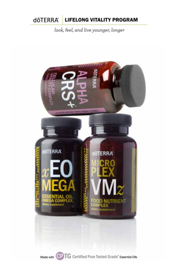
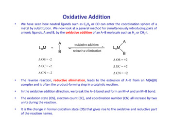
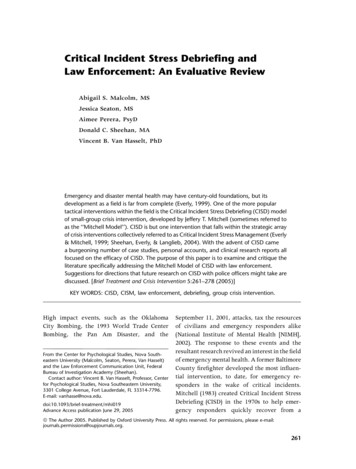
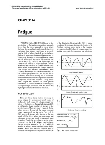
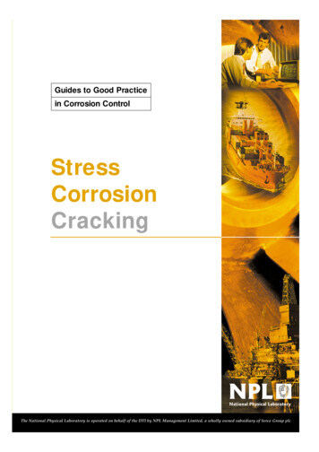
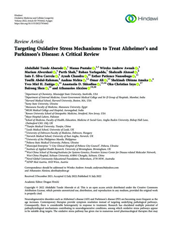
2Oxidative Medicine and Cellular Longevityprovide a new avenue for neurodegenerative diseases. For example, SKQ (plastoquinone), MitoVitE, vitamin E, SOD mimic,MitoTEMPO (SOD mimetic), and bioactive molecules like curcumin and vitamin C have indeed been examined. To betterunderstand how oxidative stress contributes to neurodegenerative diseases (such as Alzheimer’s and Parkinson’s), we analyzedthe medicinal qualities of medicines that target markers in the cellular oxidative pathways. The specific pathway by whichmitochondrial dysfunction causes neurodegeneration will require more investigation. An animal study should be carried out onmedications that tackle cellular redox mechanisms but are not currently licensed for use in the management ofneurodegenerative conditions.1. IntroductionSince they are both neurological diseases, Alzheimer’s disease (AD) and Parkinson’s disease (PD) remain untreated[1]. PD affects approximately one percent of the overall ofthe individuals over the age of 60, while Alzheimer’s disease(AD) affects approximately four percent of the overall of thepeople above the age of 65 [2, 3].Oxidative stress, which is a hallmark of aging, is themajor trigger for both disorders. Mitochondrial dysfunctionis the redox condition that results from an imbalancebetween the production and elimination of reactive oxygenspecies (ROS) (Figure 1). It is impossible to avoid ROS eventhough they metabolize all significant cellular components,especially DNA, RNA, protein, and triglycerides [4]. ROShave been used in cell signaling pathways when they arepresent in high concentrations.Neuropathologically, PD is distinguished by(a) a reduction in the amount of dopaminergic currentlyoffered for neurotransmission in the substantia nigrapars compacta (SNpc) resulting in a loss of dopamine pathways; and(b) there is a development of Lewy bodies, neurofibrillary tangles aggregates that incorporate microfibrilssynuclein [5]Disruption of circuitry that controls movement and posture is caused by deficit in the dopamine neurotransmitter inthe SNpc, which results in symptoms including slowness ofmovement and relaxation trembling. Parkinson’s diseaseis also known to cause nonmotor signs such as sleep disorders, anxiety, memory impairment, and malfunctionsof the autonomous nervous system and the senses [6, 7].The neurodegenerative disorders expression of AD is substantially more extensive, with functional decline occurringin conjunction with amyloid plaques, neurofibrillary tangles, and cerebral amyloid angiopathy [8]. The final outcome of degenerative progressions that have yet to bedefined in AD and PD is oxidative stress as a result of cellular malfunction.It is still not obvious how oxidative stress plays a role ineither disease’s onset or progression, but this is one theorythat has been floated by some researchers. Previous studieshave attempted to disseminate the role of oxidative stressin the pathophysiology of neurodegenerative diseases [9].Treatments aiming cellular oxidative pathways may bebeneficial in the management of neurodegenerative disorders, particularly those associated with endothelial dysfunc-tion recently. Oxidative stress is a major contributor toneurodegeneration in general and to the development ofboth Parkinson’s disease and Alzheimer’s disease (AD).There will also be an examination of present and upcomingtreatments for oxidative stress. For these new medicines,future outlooks are presented to discuss their possibility fordisease-modifying.2. The Role of OxidativeStress in Neurodegeneration2.1. Mechanisms of Oxidative Stress. The electrons in theoutermost electron shells of reactive species (subatomic particles, molecules, or ions) are unpaired, and this gives them ahigh degree of responsiveness. Endogenous reactive oxygenspecies (also known as ROS) have the ability to metabolizemacromolecules since they are capable of doing so. Numerous metabolic reactions, such as oxidative phosphorylation,generate ROS. Mitochondrial-derived reactive oxygen species (mtROS) (Figures 2 and 3) include singlet oxygen(O2), superoxide anion (O2 ), hydrogen peroxide (H2O2),nitric oxide (NO ), hydroxyl radical (OH ), and hydroxylion (OH-). Xanthine oxidase (XO) or mitochondrial respiratory chain complexes I (NADH dehydrogenase) and III (bc1complex) first convert oxygen to superoxide anion [3, 10](Figure 2). In both the matrix and the intermembranousregion, complex III generates a superoxide anion (Figure 2)[11].A patient’s condition, age, and hormonal status all affecthow much of these complexities are present in IMM. Inorder to produce hydrogen peroxide, the superoxide anionmust first be converted by SOD. Hydrogen peroxide can bedetoxicated to water and oxygen using glutathione peroxidase, catalase (CAT), or thioredoxin peroxidase (TPx) [12,13]. It can also be converted to hydroxyl radicals andhydroxyl ions via the Fenton reaction (Figure 3) [14].At physiological levels, ROS are sequestered by endogenous antioxidants such as glutathione peroxidase andsuperoxide dismutase. The central nervous system (CNS)is especially vulnerable to oxidative stress [15]. The rapidenergy level of cerebral cortex, the greater amount ofpolyunsaturated fatty acids (PUFA) in the biological synapses, and the native autooxidative pathways engaged inneurotransmitters are all factors that contribute to thissensitivity.Despite unsaturated fatty acids comprising 20% of thebrain’s total fatty acid content, only around 3% of the totalglutathione peroxidase in the human liver is found in thebrain. Superoxide dismutase levels in brain tissue are comparable to those found in the heart and liver. Iron, often
Oxidative Medicine and Cellular Longevity3CytoplasmOuter membranemPTPSOD1H2OGPxsH2O2CytCO2–O2IIIIVComplex IO2Inner membraneIIO2–O2Coenzyme O2Q10XanthineoxidaseFe2 O2–SOD2TPxCycloD2H2OMitochondrial matrixATPsynthaseO2–Fe3 2H2O2 CatalaseGPxsOH– OH2H2O O24H2OFigure 1: Defining the role of free radicals and antioxidants in cellular oxidative development.OxidantsAntioxidantsSuperoxideHydrogen peroxideHydroxyl radicalAlkoxy/peroxy radicalFlavonoidCarotenoidsAscorbic acidVitamin EFigure 2: Reactive oxygen species (ROS) production in mitochondria (mtROS).found as iron-neuromelanin complexes in dopaminergicneurons, under pathological conditions may act as a cofactorfor producing ROS [16]. All the above contribute to thegreater susceptibility of CNS to ROS. Locally increased levelsof ROS can precipitate mitochondrial dysfunction by damaging membrane proteins and may cause adverse mutationsin mitochondrial DNA [17, 18].The endpoint of this cascade is neuroinflammation andneuronal dysfunction as seen in PD and AD [19]. Currentlybiological studies posit that such consequences are primarilythe result of mitochondrial dysfunction, secondary from oxidative stress, leading to neurodegeneration [20–22].2.2. Reactive Species as Modulators of the NeurologicalFunction. Reactive oxygen species (ROS) modulate manyaspects of neurological function by acting as secondary messengers in several pathways (Figure 4). There are two typesof synaptic plasticity: LTP and LTD, which refer to animprovement in synaptic performance as well as a decreasein signal transduction [23].ROS are relevant in the hippocampus and spinal cordwhere they partake in LTP [24]. Secondly, ROS activatesmicroglia and astrocytes causing the ongoing release of proinflammatory cytokines and chemokines analogous to thesystemic, low-grade inflammation that occurs secondary to
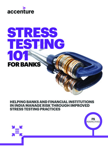
4Oxidative Medicine and Cellular LongevityOH Cl–Fe3 O2EndogenoussourcesFe2 Fe3 Fe2 MPOO2–SODsCAT2GSHNADP NADPHNOSH OHH2O2H 1/2 O2 Cl–H2O2HOCl–e–ExogenoussourcesOH Cl–O2–NADP H2OG6PH2O 1/2 O2NOL-arginineGPXL-citrulineCOCO3– NO2GRG6PDH2H2OONOO–H GSSGNADPHG6PDHNO2 OHFigure 3: Radical species development.malignancy. Thirdly, ROS play a crucial role in the differentiating of neurons by influencing the multiplication of brainprogenitor and epigenetics.Fourthly, ROS inhibits sodium currents needed foraction potentials via the oxidation of thiol groups(Figure 4). Interestingly the opening of voltage-gated calcium channels is enhanced by ROS [25, 26].Complex I: NADH dehydrogenase; II: succinate dehydrogenase; III: bc1 complex; IV: cytochrome C oxidase; V:ATP synthase; Q: Ubiquinone; Cyt C: cytochrome C; CycloD: Cyclophilin D; mPTP: mitochondrial permeability transition pore; SOD: superoxide dismutase; GPxs: glutathioneperoxidase; TPx: thioredoxin peroxidase. See text below fordetails [27].2.3. Oxidative Stress in the Pathogenesis of PD and AD. Postmortem frontal brain samples from people aged 26 to 126years showed a decline in genes linked to synapse formation,vesicular transport, and mitochondrial activity beyond theage of 40. The response to stress and antioxidants andDNA fix genes were then upregulated as a result of thesemodifications [28].PD patients’ nigral neurons, which are particularly vulnerable to mitochondrial malfunction due to high levels ofoxidative metabolism, have been the subject of extensiveinvestigation since the 1980s [2]. Regarding AD, though itspathophysiology has been explained through the amyloidand the neurofibrillary tangles, research has also implicatedother mechanisms in its development and progression;among these mechanisms is mitochondrial dysfunctionand oxidative stress [2], though, most authors agree thatmitochondrial dysfunction precedes amyloid plaque deposition and is thus not the underlying cause.In postmortem tissues, elevated levels of ATP werefound in patients with AD both in cerebral structures andin peripheral tissues, signifying the presence of mitochondrial dysfunction. Complex I, III, and IV inadequacies havebeen observed in postmortem dissections thus far. Apartfrom mitochondrial dysfunction changes, there have alsobeen changes in morphology and distribution of mitochondria, with research describing the length reduction andincrease in numbers [2].2.4. Novel Therapies Targeting Oxidative Stress Pathways.The capacity of these medications to pass the blood-brainbarrier (BBB) is a critical hurdle when developing innovativetherapeutics for neurodegenerative illness. According toresearch, the disease associated with complex 1 is the majorsource of mitochondrial dysfunction [29]; treatments thataddress this are being explored, including substances likeSKQ (plastoquinone), MitoVitE (vitamin E), MitoTEMPO(SOD mimic), MCAT (catalase), MitoPBN (CoQ), and phenyl tert butylnitrone conjugation, as well as other chemicals.Others are lipophilic cation-based tetrapeptide compounds or choline esters of glutathione and N-acetyl lcysteine that can penetrate cells. There has been evidencethat MitoQ slows down the onset of Alzheimer’s disease bydecreasing A-induced neurodegeneration in neuronal cells,reducing free radical generation [30, 31].Intracellular enzymes such as superoxide dismutase andglutathione peroxidase provide some protection from ROS.Synthetic molecules such as butylated hydroxytoluene,butylated hydroxyanisole, and ethoxyquin PAPLAL (mixture of Pd and Pt NPs) have been designed to mimic theseenzymes but may produce adverse effects resulting from systemic administration [31].
Oxidative Medicine and Cellular Longevity5Mitochondrial sourcesETCIQVIIIIICIVNonmitochondrial sourcesLOXCOXXONOXMPOCYPROSAging, pathologicalConditions: Inflammation,cancer, ischemiaRadiation, pollution,xenobioticsBIOMARKERSProtein damageLipid peroxidationAGEsALEsCDProtein damageAGEsReactive aldehydesAldehyde modifiedF(2)-isoPs; F(4)-NPsHETEHNEHODEHydroperoxideLysoPCNFS; IsoFox-LDLOxysterolsAOPPComet assayOxidative stressGGTGSSGGSSG/GSHOpht APrxsTACTQ/TQH2UQ/UQH2DNA damageCrosslinked protein5-OHC5-OHU8-OH8-OHdG8-OH-GComet assayHIF-1aCTLHydroxyperoxideLFMPOPCTyrFigure 4: Cellular redox indicators’ derivation [27].An erythropoietin (Neuro-EPO) intranasally administered shields from inflammation and neurological assaults,restoring impairments in recollection, acquisition, and recognition of new images while also reducing antioxidantactivity [32, 33].Due to the Nrf2–NF-B signaling axis, Cyclo (His-Pro)seems to have some anti-inflammatory and stress activities,as well as ability to repair neuronal functionality Antioxidantdefenses can be activated and apoptosis inducing andinflammation response can be reduced by increasing thenuclear level of Nrf2 and preventing IB degradation [34].Studies suggest Glial activation-mediated inflammationhas some role in Parkinson’s disease (PD), via the Glia maturation factor (GMF) [35]. Modified GMF articulationdirectly influences the creation of reactive oxygen speciesby 1-methyl-4-phenylpyridinium (MPP ). GMF suppression correlated with a decline in reactive oxygen speciesand resulted in downregulation of NF-kappaB-induced creation of TNFalpha and IL-1b. Thus, it decreased lipid peroxidation levels and increased levels of glutathione [35].Metal-protein attenuating compounds (MPACs) interrupt the abnormal metal-protein interaction and normalizeits distribution by competing with target proteins for metalions [36].Autopsies performed on brain tissue of PD and ADpatients show involvement of transition metals during theformation of cytotoxic tissue aggregates, and metal chelatingtherapy has demonstrated significant efficacy in certain PDmodels by preventing lipid peroxidation, although long termuse might interfere with normal physiological function [37].Although these compounds show great results, they are stillat the experimental phase, and their clinical application isbeing investigated.2.5. Oxidative Stress Drug Targets in Anti-Alzheimer’sDisease Therapy. Antioxidant properties of medications usedto treat Alzheimer’s disease show considerable differencesbased on dose and AD model. Tacrine, which was the firstanticholinesterase inhibitor approved by the Food and DrugAdministration (FDA), showed reduction in overall survival
6(OS) in an animal Alzheimer’s disease model [38] and at adose of 50–800 g/kg i.m. increased FRAP, and hence “antioxidant efficacy” [39], without raising any sign of OSassociated damage in brain tissue. Tacrine has a favorableeffect when provided in doses that improve the antioxidantsystem without increasing oxidative stress-induced damagein brain tissue [40].Donepezil, another cholinesterase inhibitor used in Alzheimer’s disease patients, resulted in dose-dependent effectson antioxidant capacity and reduced lipid peroxidationwhen administered in mice AD models at modest doses. Inthe APPswe/PS1 transgenic mice AD model, donepezil didnot decrease OS biomarkers or show significant antioxidantactivity [41]. Hence, discrepancy in results from differentstudies suggests that adaptations to its use may be the sourceof the observed discrepancy in outcomes in transgenic andnontransgenic AD mice models.Rivastigmine, another drug used to treat of Alzheimer’sdisease, does not show to decrease lipid peroxidation orreplenish GSH in an AD rat brain model [42], despite a previous report indicating antioxidant capabilities for rivastigmine when Alzheimer’s disease was produced in rats byaluminum chloride treatment [43].A single study found that another AChE inhibitor, galantamine, might lower OS, leading to decreased lipid peroxidation, nitrate, and GSSG levels, as well as increased SODactivity and lower GSH levels, while also restoring cognitiveimpairments [44].Studies performed on Alzheimer’s disease preclinicalmodels demonstrate Memantine to decrease oxidativestress induced damage to cortex and hippocampus proteins,improving age-related recognition memory in senior rats[45]. Memantine also reduced the frequency of inducibleforms of NOS in an A25–35 AD model [46] and, in addition,ROS and nitrate levels in the hippocampus and cortex in astreptozotocin AD model [47] as well as a kainic acidinduced model of dementia [47]. However, this effect wasnot observed in the striatum [45].Immunomodulatory agents such as Fingolimod orFTY720, Tanshinone I, Lenalidomide, Thalidomide, Ginsenoside Rg1, CNI-1493, Pycnogenol, and C5aR antagonistDF3016A demonstrate anti-inflammatory action, therebyreducing OS and lipid peroxidation products and decreasingmicroglial, astrocytic, and T cell activities. They might beused as a preventative measure, and there is some evidencethat they help with motor impairments and nigral dopaminergic neurotoxicity [48].Over time, innovations in the field of nanomedicine havegarnered widespread interest in the scientific community.Some compounds of interest are cerium oxide NPs, ceria/polyoxometalates hybrid, manganese tetroxide and manganese ferrite nanoparticles, yttrium oxide nanoparticles, ironoxide nanoparticles, copper nanoparticle clusters, cobaltoxide (Co3O4 NPs), and cobalt ferrite nanoparticles(CoFe2O4 NPs). Some compounds have antioxidant properties and other compounds like ceria mimic enzymes [49].Nanoceria mimics SOD and CAT activity. Due to the mixedvalency state of cerium oxide, it reacts with free radicals anddetoxifies ROS and therefore may be neuroprotective.Oxidative Medicine and Cellular LongevityApart from metal and metal oxide nanoparticlesdescribed above, inorganic nanoparticles such as mesoporous silica nanoparticles (MSNs) can have potential applications as explained by their large surface area, structuraltunability, and easy functionalization [50]. Therapeutic trialsof these drugs and therapies in animal and human modelsare discussed below [38, 41, 42, 51–65]. In patients: (i) vitamin E (α-tocopherol, 800 IU/day) vitamin C (500 mg/day) α-lipoic acid (900 mg/day) F2-isoprostane in CSF[51]; (ii) Coenzyme Q10 (400 mg 3 times/day) NOCHANGE F2-isoprostane in CSF [51]; (iii) ω-3 (3 g/day contained 675 mg DHA and 975 mg EPA) NO CHANGE F2isoprostane in urine, NO CHANGE PC in plasma [52];(iv) ω-3 α-lipoic acid (ω-3, 3 g/day contained 675 mgDHA and 975 mg EPA α-lipoic acid, 600 mg/day in onetablet) NO CHANGE F2-isoprostane in urine NOCHANGE PC in plasma [52]; (v) vitamin C (1,000 mg/day) vitamin E (400 IU/day) oxidation of CSF [53]; (vi)Curcumin (1 or 4 g/day) NO CHANGE F2-isoprostane inplasma [54]; (vii) curcuminoids (2 or 4 g/day) NO CHANGEF2-isoprostane in CSF [55]. In animal models: (i) Schisantherin A 0.1 mg/kg for 5 days i.c.v., injection started after3 days from Aβ1–42 injection ( MDA in cerebral CTX, SOD, GPx, GSH in HIP and cerebral CTX) [38]; (ii) vitamin E 150 mg/kg, p.o. for 27 days, administration began 7days before Aβ1–42 i.c.v. ( MDA, PC, Mn-SOD, Zn,Cu-SOD, GPx, Ø GR in cerebral CTX and HIP) [57]; (iii)Piperine 5 or 10 mg/kg p.o. 2 weeks before and 1 week afterAF64A ( MDA in HIP) [58]; (iv) S-allyl cysteine 30 mg/kg i.p. for 15 days pretreatment before streptozocin( TBARS, GSH, GPx, GR in HIP) [59]; (v) Imperatorin2 /day for 7 days with scopolamine injection ( MDA, SOD in CTX and HIP, GPx in CTX and HIP, GR inCTX) [42]; (vi) α-lipoic acid 30 mg/kg/day enriched dietfor 10 months ( HNE, Ø 3-NT in brain homogenates)[60]; (vii) vitamin C 125 mg/kg i.p. for 12 days (no changeMDA in HIP) [61]; (viii) vitamin C low diet content0.099 g/L of drinking water ( MDA in CTX) [62]; (ix) melatonin 5 mg/kg p.o. for 5.5 months ( MDA, PC in HIP)[63]; (x) melatonin 10 mg/kg/day for 4 months intragastrically ( TBARS, GSH, SOD in the brain homogenate) [64]2.6. Oxidative Stress Drug Targets in Anti-Parkinson’sDisease Therapy. Drugs like valproic acid, melatonin, ceftriaxone, and N-acetylcysteine have been shown in research tohave oxidative impacts on human health. Antioxidativedefense enzyme activity in mice was recovered after ceftriaxone treatment (glutathione, catalase, and SOD). Prior to taking ceftriaxone, Ropinirole considerably increased theprotective effects [66].Using 6-OHDA-induced SNpc dopaminergic neuronaldamage in rat models, serofendic acid plays a protective roleagainst 6-OHDA-induced oxidative stress parameters, suchas 3-nitrotyrosine and 4-hydroxy-2-nonenal (4-HNE).When N-acetylcysteine was administered to animals, itinduced an upsurge in the efficiency of lipid peroxylase,superoxide dismutase (SOD), and g-glutamyl transpeptidase(g-GTP) and a big decline in glutathione (GSH) levels andglutathione peroxidase function in the SNpc [67].
Oxidative Medicine and Cellular LongevityRopinirole, a dopaminergic stimulant, increased GSHlevels and CAT action, according to a new analysis. ThesePLGA nanoparticles (NPs) were developed to enhance thedrug’s effectiveness and distribution [55].Parkinson’s disease may be treated with the nootropiccentrophenoxine (CPH). Catalase and superoxide dismutase(SOD) upregulated, whereas nitric oxide (NO) and citrullinelevels decreased, according to one study [68]. These compounds seem to be promising; further studies are requiredto understand their efficacy in a natural setting because inthe above studies, to produce selective DAergic neuronaldegeneration in PD, rotenone-induced neurotoxicity wasused as a preclinical model PD in mice. Many therapeutictrials of these drugs and therapies in animal and humanmodels are conducted until date [66, 69–71]. Theseincluded (i) L-DOPA (200 mg/kg i.p. 2 injections/day for 4weeks, coadministration with MPTP (no change GSH inSN) [69], (ii) Ropinirole 1, 5, or 3 mg/kg i.p. for 14 days,after MPTP ( GSH, CAT, nitrate (only 1.5 mg/kg) inSTR and CTX) [66]; (iii) Pramipexole 1 mg/kg i.p. 2 injections/day for 4 weeks, coadministration with MPTP( GSH in SN) [69]; and (iv) Deferoxamine 50 mg/kg p.o.for 14 days, coadministration with 6-OHDA ( PC, GSH,and SOD in STR) [71].3. Conclusions and Future PerspectivesFundamentally, it is necessary to do extensive study on theinclusionary practices of ROS in the neurodegenerative process and AD, in order to analyze therapeutic targets that arenoteworthy. Current medications, though effective, shouldnot be considered optimal management of neurodegenerative disease. The drugs discussed above should undergo further evaluation for feasibly in human subjects. Theeffectiveness of pharmacological drugs that target the cellular oxidative system in reducing neurodegenerative shouldnext be thoroughly tested in medical tests. Despite the factthat mitochondrial dysfunction is an important role in thedevelopment of AD and PD, little is known about the extentof this impact, which makes it difficult to put hypotheticalpharmacological targets into reality in the real world.Conflicts of InterestThe authors of this research disclose that they have no competing interests in its manuscript.Authors’ ContributionsAbdullahi Tunde Aborode, Wireko Andrew Awuah, andManas Pustake conceptualized the concept for this review.Manas Pustake, Mariam Alwerdani, Parth Shah, Rohan Yarlagadda, Shahzaib Ahmad, Ayush Chandra, Esther PatienceNansubuga, Omar Ali, and Aashna Mehta wrote the firstdraft. Inês F Silva Correia, Toufik Abdul-Rahman, AashnaMehta, Shekinah Obinna Amaka, Yves Miel H. Zuñiga, Anastasiia D. Shkodina, Oko Christian Inya, Bairong Shen, andAthanasios Alexiou edited the second draft. AthanasiosAlexiou, Bairong Shen, Abdullahi Tunde Aborode, and Wir-7eko Andrew Awuah provided suggestions for improvementand adjustments. The final manuscript was proofread byall of the authors, and they all gave their approval.References[1] A. P. Reddy, J. Ravichandran, and N. Carkaci-Salli, “Neuralregeneration therapies for Alzheimer’s and Parkinson’sdisease-related disorders,” Biochimica et Biophysica Acta(BBA)-Molecular Basis of Disease, vol. 1866, no. 4, article165506, 2020.[2] R. Macdonald, K. Barnes, C. Hastings, and H. Mortiboys,“Mitochondrial abnormalities in Parkinson’s disease and Alzheimer’s disease: can mitochondria be targeted therapeutically?,” Biochemical Society Transactions, vol. 46, no. 4,pp. 891–909, 2018.[3] C. Qiu, M. Kivipelto, and E. Von Strauss, “Epidemiology ofAlzheimer’s disease: occurrence, determinants, and strategiestoward intervention,” Dialogues in Clinical Neuroscience,vol. 11, no. 2, pp. 111–128, 2009.[4] M. D. Ferrer, A. Sureda, A. Mestre, J. A. Tur, and A. Pons, “Thedouble edge of reactive oxygen species as damaging and signaling molecules in HL60 cell culture,” Cellular Physiology andBiochemistry, vol. 25, no. 2–3, pp. 241–252, 2010.[5] M. G. Spillantini, M. L. Schmidt, V. M. Y. Lee, J. Q. Trojanowski, R. Jakes, and M. Goedert, “α-Synuclein in Lewy bodies,” Nature, vol. 388, no. 6645, pp. 839-840, 1997.[6] F. A. Perez and R. D. Palmiter, “Parkin-deficient mice are not arobust model of parkinsonism,” Proceedings of the NationalAcademy of Sciences, vol. 102, no. 6, pp. 2174–2179, 2005.[7] W.-S. Choi, S. E. Kruse, R. D. Palmiter, and Z. Xia, “Mitochondrial complex I inhibition is not required for dopaminergicneuron death induced by rotenone, MPP , or paraquat,” Proceedings of the National Academy of Sciences, vol. 105, no. 39,pp. 15136–15141, 2008.[8] A. Serrano-Pozo, M. P. Frosch, E. Masliah, and B. T. Hyman,“Neuropathological alterations in Alzheimer disease,” ColdSpring Harbor Perspectives in Medicine, vol. 1, no. 1, articlea006189, 2011.[9] X. Chen, C. Guo, and J. Kong, “Oxidative stress in neurodegenerative diseases,” Neural Regeneration Research, vol. 7,no. 5, pp. 376–385, 2012.[10] J. St-Pierre, J. A. Buckingham, S. J. Roebuck, and M. D. Brand,“Topology of superoxide production from different sites in themitochondrial electron transport chain,” The Journal of Biological Chemistry, vol. 277, no. 47, pp. 44784–44790, 2002.[11] X. Li, P. Fang, J. Mai, E. T. Choi, H. Wang, and X. Yang, “Targeting mitochondrial reactive oxygen species as novel therapyfor inflammatory diseases and cancers,” Journal of Hematology& Oncology, vol. 6, pp. 1–19, 2013.[12] G. Barja, “Mitochondrial oxygen radical generation and leak:sites of production in states 4 and 3, organ specificity, and relation to aging and longevity,” Journal of Bioenergetics and Biomembranes, vol. 31, no. 4, pp. 347–366, 1999.[13] M. P. Murphy, “How mitochondria produce reactive oxygenspecies,” The Biochemical Journal, vol. 417, pp. 1–13, 2009.[14] U. Förstermann, “Oxidative stress in vascular disease: causes,defense mechanisms and potential therapies,” Nature ClinicalPractice Cardiovascular Medicine, vol. 5, no. 6, pp. 338–349,2008.
8[15] J. N. Cobley, M. L. Fiorello, and D. M. Bailey, “13 reasons whythe brain is susceptible to oxidative stress,” Redox Biology,vol. 15, pp. 490–503, 2018.[16] A. Suárez, M. Ramírez-Tortosa, A. Gil, and M. J. Faus, “Addition of vitamin E to long-chain polyunsaturated fatty acidenriched diets protects neonatal tissue lipids against peroxidation in rats,” European Journal of Nutrition, vol. 38, no. 4,pp. 169–176, 1999.[17] A. K. Hauck, Y. Huang, A. V. Hertzel, and D. A. Bernlohr,“Adipose oxidative stress and protein carbonylation,” TheJournal of Biological Chemistry, vol. 294, no. 4, pp. 1083–1088, 2019.[18] G. Plascencia-Villa and G. Perry, “Preventive and therapeuticstrategies in Alzheimer’s disease:
5Mansoura Faculty of Medicine, Mansoura University, Egypt 6MGM Medical College and Hospital, Aurangabad, India 7Rowan University School of Osteopathic Medicine, Stratford, New Jersey, USA 8Mayo Hospital Lahore, Pakistan 9School of Medicine, Faculty of Health, Education, Medicine & Social Care, Anglia Ruskin University, Bishop Hall Lane,